267
Erdheim-Chester disease: a two-case report
Radiol Bras. 2009 Jul/Ago;42(4):267–269
Case Report • Relato de Caso
Erdheim-Chester disease: a two-case report*
Doença de Erdheim-Chester: relato de dois casosFernando Fernandez Hexsel1, Letícia Frigo Canazaro2, Mariana Capoani3, Paula Musa Aguiar4, Eiji Suwa1, Antonio Carlos Maciel5
Erdheim-Chester disease is a rare non-Langerhans cell histiocytosis of unknown etiology, affecting multiple organ system, involving bones, central nervous system, eyes, lungs, mediastinum, kidneys and retroperitoneum. The authors report two cases that progressed with the typical presentation of the disease. Radiological findings were in agreement with literature and guided the diagnosis, confirmed by immunohistochemistry.
Keywords: Erdheim-Chester disease; Radiography; Computed tomography.
A doença de Erdheim-Chester é uma rara histiocitose de células não-Langerhans, de etiologia desconhecida, que apresenta manifestações sistêmicas, atingindo ossos, sistema nervoso central, olhos, pulmões, medias-tino, rins e retroperitônio. Relatamos dois casos que cursaram com a apresentação típica da doença. Os achados radiológicos foram concordantes com a literatura e orientaram a suspeita diagnóstica, confirmada pelo exame imuno-histoquímico.
Unitermos: Doença de Erdheim-Chester; Radiografia; Tomografia computadorizada. Abstract
Resumo
* Study developed at the Unit of Radiology – Santa Casa de Misericórdia de Porto Alegre, Porto Alegre, RS, Brazil.
1. MDs, Radiologists, Unit of Radiology – Santa Casa de Mi-sericórdia de Porto Alegre, Porto Alegre, RS, Brazil.
2. MD, Resident, Unit of Radiology – Hospital Bruno Born, Lajeado, RS, Brazil.
3. MD, Radiologist, Clínica Imagem, Florianópolis, SC, Brazil. 4. MD, Fundação Faculdade Federal de Ciências da Saúde de Porto Alegre, Porto Alegre, RS, Brazil.
5. Head for the Unit of Radiology – Santa Casa de Misericór-dia de Porto Alegre, MD, Radiologist, Unit of Radiology – Hospi-tal de Clínicas de Porto Alegre, Porto Alegre, RS, Brazil.
Mailing address: Dra. Letícia Frigo Canazaro. Rua Goiás, 37, Bairro Azenha. Porto Alegre, RS, Brazil, 90880-470. E-mail: [email protected]
Received October 19, 2007. Accepted after revision January 9, 2008.
During the hospital stay, the patient complained of decrease in visual acuity, and orbital computed tomography was per-formed and demonstrated bilateral tumes-cent lesions almost completely infiltrating the intraconal fat.
Biopsy of the right retrobulbar tumes-cent lesion demonstrated xanthogranu-loma. A subsequent immunohistochemical study demonstrated positive results for CD68 (PG-M1) and CD1a (MTB1 nega-tive), compatible with the diagnosis of Erdheim-Chester disease.
Corticosteroid therapy was introduced and the patients is currently undergoing outpatient follow-up.
Case 2 – A female, white, 37-year-old patient attended the hospital complaining of polyuria, polydipsia, and pain in the lower limbs.
Upper and lower limbs radiographies demonstrated medullary osteosclerosis and areas of symmetrical cortical hyperos-tosis.
Later during the hospital stay, the pa-tient presented exophthalmos and decrease in visual acuity. Orbital computed tomog-raphy was bilaterally performed, and iden-tified masses obliterating the intraconal fat planes involving the optic nerves with an-terior displacement of both ocular globes. Hexsel FF, Canazaro LF, Capoani M, Aguiar PM, Suwa E, Maciel AC. Erdheim-Chester disease: a two-case report. Radiol Bras. 2009;42(4):267–269.
The authors present two cases with typi-cal clinitypi-cal presentation and findings, dis-cussing clinical, radiological and prope-deutic aspects.
CASES REPORT
Case 1 – A female, white, 43-year-old patient was admitted for investigation of an abdominal mass, anemia, leukocytosis, thrombocytosis and pain in the lower limbs.
The initial chest radiography demon-strated an increase in heart volume and thickening of horizontal and oblique fis-sures of the right lung. Lower limbs radi-ography demonstrated symmetrical sclero-sis predominating in the tibial diaphyses.
Chest and abdominal computed tomog-raphy demonstrated thickening of inter-lobular septa in the pulmonary apices, le-sions presenting soft tissues density adja-cent to the descending thoracic aorta and an infiltrative lesion involving a significant extent of the abdominal aorta and internal iliac vessels, including the renal vessels and affecting both renal sinuses with a conse-quential, moderate hydronephrosis, con-tinuing as an extensive, expansive mesen-teric mass, with complete involvement of vessels in this region.
0100-3984 © Colégio Brasileiro de Radiologia e Diagnóstico por Imagem
INTRODUCTION
Erdheim-Chester disease is a rare form of non-Langerhans-cell histiocytosis, firstly described by Jakob Erdheim and William Chester in 1930(1).
The etiology of this disease still remains unknown. Histologically, it is characterized by a diffuse infiltration by foamy histio-cytes of the tissues involved, causing a lo-cal xanthomatous or xanthogranulomatous reaction(2).
268
Hexsel FF et al.
Radiol Bras. 2009 Jul/Ago;42(4):267–269
Biopsy of the conjunctival mass dem-onstrated accumulation of foamy histio-cytes and lymphocytic infiltrate whose im-munohistochemical study resulted negative for S-100 protein.
Chest computed tomography demon-strated aortic dissection with thrombosis and ulceration, bilateral pleural effusion or thickening, calcified lymphatic ganglia in the pulmonary hilum, subdiaphragmatic calcification and/or calcification in the right hepatic lobe, and a significant en-largement of both adrenal glands.
The patient underwent corticosteroid therapy and radiotherapy of the orbital le-sions followed by chemotherapy. Currently, she is under outpatient follow-up.
DISCUSSION
Most commonly, Erdheim-Chester dis-ease occurs after the fourth decade of life, with a subtle male prevalence. The disease pathogenesis is still to be elucidated, but it seems there be tropism of histiocytes to con-nective, fat and perivascular tissues, with an infiltrative pattern and local fibrosis(5).
Bone pain associated with progressive weakness, particularly in the lower limbs, is the most frequent symptom of Erdheim-Chester disease. Also, the patient
fre-quently presents fever, weight loss, exoph-thalmos, dyspnea and signs of neurologi-cal involvement, such as diabetes insipi-dus(4,6). The diagnosis is based on
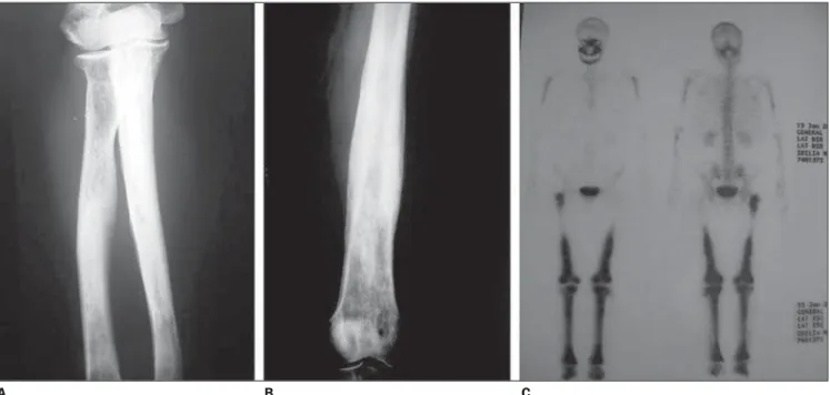
radio-logical and pathoradio-logical findings. Bone involvement present in both cases presently reported consists of symmetric sclerosis of long tubular bones, predomi-nating in metaphysis and diaphysis of lower limbs. This is the radiological abnor-mality most frequently reported in the lit-erature (60% of cases)(4–8) (Figure 1).
As for pulmonary findings, both radiog-raphy and computed tomogradiog-raphy demon-strate fissural thickening and interlobular septa thickening, besides pleural effusion, centrolobular nodules, interstitial reticular opacities, and at computed tomography multifocal areas of ground-glass attenua-tion(4,8).
Erdheim-Chester disease presents a tro-pism of histiocytes to connective, fat and perivascular tissues. This involvement may extend to the whole aorta, with invasion of the retroperitoneum and mediastinum, leading to severe complications such as cardiac failure, tamponade and renal fail-ure. Hepatic, pancreatic and mesenteric involvement is extremely rare(5).
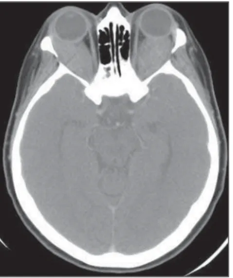
The ocular involvement observed in both case 1 and 2 may be uni- or bilateral
and is seen at images as intraorbital masses or retrobulbar tumors (Figure 2).
At computed tomography, retroperito-neal involvement is visualized as soft tis-sues masses, for example in the psoas muscle, with fat stranding, which would explain the clinical signs of renal failure and arterial hypertension resulting from the development of hydronephrosis(3,4).
Renal/perirenal involvement is ob-served in 29% of patients in a 59-case se-ries where computed tomography demon-strated hypoattenuated homogeneous peri-renal tissue infiltration with poor contrast uptake(4). The “hairy” or “onion peel”
kid-ney appearance observed in these two pa-tients is due to the symmetrical and bilat-eral infiltration of both the posterior pararenal and perirenal spaces highly sug-gestive of the diagnosis. The extension to the renal sinuses may lead to urinary tract obstruction (Figure 3).
A consensus on the management of Erdheim-Chester disease is still to be reached, but data in the literature report promising results with the utilization of corticosteroids, immunosuppressive drugs such as cyclophosphamide, chemotherapy and radiotherapy. The prognosis will de-pend on the extent of visceral involvement at the moment of the diagnosis(3,4).
Figure 1. Radiography of forearm bones (A) and tibia (B) showing symmetrical osteosclerosis of long bones typical of Erdheim-Chester disease. On C, bone scintigraphy with Tc-99m-MDP demonstrating typical contrast uptake in the distal portion of the involved bones.
269
Erdheim-Chester disease: a two-case report
Radiol Bras. 2009 Jul/Ago;42(4):267–269 REFERENCES
1. Wright R, Hermann R, Parisi J. Neurological manifestations of Erdheim-Chester disease. J Neurol Neurosurg Psychiatry. 1999;66:72–5. 2. Haroche J, Amoura Z, Dion E, et al.
Cardiovas-cular involvement, an overlooked feature of Erdheim-Chester disease: report of 6 new cases and a literature review. Medicine (Baltimore). 2004;83:371–92.
3. Haroche J, Amoura Z, Wechsler B, et al.
Erdheim-Figure 2. Orbital computed tomography showing a dense soft-tissue mass bilaterally obliterating the intraconal fat.
Figure 3. Contrast-enhanced computed tomography demonstrating bilateral, homogeneous perirenal infiltration. Also, infiltration of the descending aorta is identified.
B A
Chester disease. Presse Med. 2007;36(Pt 11): 1663–8.
4. Veyssier-Belot C, Cacoub P, Caparros-Lefebvre D, et al. Erdheim-Chester disease. Clinical and ra-diologic characteristics of 59 cases. Medicine (Baltimore). 1996;75:157–69.
5. Dion E, Graef C, Haroche J, et al. Imaging of thoracoabdominal involvement in Erdheim-Chester disease. AJR Am J Roentgenol. 2004; 183:1253–60.
6. Boissel N, Wechsler B, Leblond V. Treatment of
refractory Erdheim-Chester disease with double autologous hematopoietic stem-cell transplanta-tion. Ann Intern Med. 2001;135:844–5.
7. Gil Marculeta R, Domínguez Echávarri PD, Cano Rafart D, et al. Radiologic diagnosis of Erdheim-Chester disease. A case report. Radiologia. 2006; 48:317–20.
8. WittenbergKH, SwensenSJ, Myers JL.