231
Neointimal hyperplasia following intravascular stent implantation
Radiol Bras. 2009 Jul/Ago;42(4):231–234
Original Article • Artigo Original
Volumetric assessment of neointimal hyperplasia
in iliac arteries after metal stent implantation*
Quantificação volumétrica da hiperplasia neointimal em artérias ilíacas após implante de suporte intravascular metálico
Samuel Martins Moreira1
, Antonio Massamitsu Kambara2
, Sergio Ajzen3
, José de Ribamar Costa Junior4
OBJECTIVE: To quantify neointimal hyperplasia in iliac arteries after stent implantation, correlating clinical, arterial factors and stent material. MATERIALS AND METHODS: In the period from June/2003 to August/ 2005, 60 patients were submitted to percutaneous transluminal angioplasty and stenting. Among these patients, 30 were followed-up with intravascular ultrasonography. Data were analyzed in a laboratory of quantitative analysis by means of a specific software. RESULTS: Sixteen (53.3%) patients were men, and 14 (46.7%), women, and the mean age was 60.3 years. Arterial hypertension was observed in 22 patients (73.3%), smoking in 18 (62.1%), hyperlipidemia in 20 (66.7%), and diabetes in 9 (30%). A total of 20 nitinol stents (66.7%) and 10 stainless steel stents (33.3%) were implanted. Four patients were classified as TASC A (13.3%), 15 TASC B (50%) and 11 TASC C (36.7%). The neointimal hyperplasia volume ranged from 49.02 mm3
to 112.87 mm3
(mean, 80.33 mm3
). The rate of intrastent obstruction ranged from 18% to 47% (mean, 27.4%). The clinical outcomes achieved with stenting were sustained through the follow-up. CONCLUSION: Neointimal hyperplasia is a common finding after percutaneous transluminal angioplasty and stenting, but in the present study the stenosis rate was never higher than 50%. There was no statistically significant difference in intrastent stenosis rates in relation to stents materials, clinical and arterial risk factors.
Keywords: Stent; Iliac artery; Interventional ultrasonography; Hyperplasia.
OBJETIVO: Quantificar a hiperplasia neointimal em artérias ilíacas após stent, correlacionando fatores
clíni-cos, arteriais e materiais dos stents. MATERIAIS E MÉTODOS: De junho de 2003 a agosto de 2005, 60
pacientes realizaram angioplastia transluminal percutânea e stent. Desses, 30 foram reestudados com
ul-trassonografia intravascular. Os dados foram analisados no laboratório de análise quantitativa. RESULTA-DOS: Dezesseis pacientes eram do sexo masculino (53,3%) e 14 (46,7%), do sexo feminino. A média de idade foi de 60,3 anos. Apresentaram hipertensão arterial 22 pacientes (73,3%), tabagismo, 18 (62,1%), hiperlipidemia, 20 (66,7%), e diabetes, 9 (30%). Foram implantados 20 stents de nitinol (66,7%) e 10 de
aço inoxidável (33,3%). Quatro pacientes eram TASC A (13,3%), 15 eram TASC B (50%) e 11, TASC C (36,7%). O volume da hiperplasia variou de 49,02 mm3
a 112,87 mm3
(média de 80,33 mm3
). O percentual de obstrução intra-stent variou de 18% a 47% (média de 27,4%). Os resultados clínicos obtidos com stent
se mantiveram até o reestudo. CONCLUSÃO: A hiperplasia neointimal sempre ocorre após a angioplastia transluminal percutânea e stent, porém os percentuais de obstrução não foram superiores a 50% em
ne-nhum caso. Não houve diferença estatisticamente significante dos percentuais de obstrução intra-stent quanto
aos materiais dos stents, aos fatores clínicos e aos fatores arteriais.
Unitermos:Stent; Artéria ilíaca; Ultrassonografia de intervenção; Hiperplasia. Abstract
Resumo
* Study developed at Universidade Federal de São Paulo/Es-cola Paulista de Medicina (Unifesp/EPM), São Paulo, SP, and Instituto Dante Pazzanese de Cardiologia, São Paulo, SP, Brazil. 1. Assistant Physician, Universidade Federal de São Paulo/ Escola Paulista de Medicina (Unifesp/EPM), São Paulo, SP, Bra-zil.
2. PhD, Head for the Department of Radiology – Instituto Dante Pazzanese de Cardiologia, São Paulo, SP, Brazil.
3. Titular Professor, Department of Diagnostic Imaging – Uni-versidade Federal de São Paulo/Escola Paulista de Medicina (Unifesp/EPM), São Paulo, SP, Brazil.
4. Assistant Physician, Sector of Hemodynamics – Instituto Dante Pazzanese de Cardiologia, São Paulo, SP, Brazil.
Mailing address: Dr. Samuel Martins Moreira. Avenida Doutor Altino Arantes, 164, ap. 131, Vila Clementino. São Paulo, SP, Brazil, 04042-001. E-mail: [email protected]
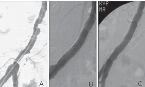
Intra-stent restenosis caused by neoin-timal hyperplasia has its occurrence peak around the sixth postoperative month (Fig-ure 1), and it has been described in other locations such as the coronary arteries(7–9) although such occurrence has not been re-ported with the same frequency as in the case of iliac arteries.
In the present study, intravascular ultra-sonography (IVUS) was performed to quantify the volume of neointimal hyper-Moreira SM, Kambara AM, Ajzen S, Costa Junior JR. Volumetric assessment of neointimal hyperplasia in iliac arteries after metal stent implantation. Radiol Bras. 2009;42(4):231–234.
INTRODUCTION
Percutaneous transluminal angioplasty with metal stents for the treatment of oc-clusive lesions of iliac arteries is a safe and effective procedure(1–3), generally accepted as the first method of choice for treatment in selected cases(4–6).
0100-3984 © Colégio Brasileiro de Radiologia e Diagnóstico por Imagem
232
Moreira SM et al.
Radiol Bras. 2009 Jul/Ago;42(4):231–234 plasia in iliac arteries treated with
percuta-neous transluminal angioplasty and stent, correlating with clinical risk factors such as smoking, arterial hypertension, hyperlipi-demia and diabetes mellitus as well as stents material and local arterial character-istics such as type of occlusion.
MATERIALS AND METHODS
Between July, 2003 and August, 2005, 60 consecutive patients were submitted to percutaneous transluminal angioplasty and stenting for treatment of occlusive lesions in the iliac arteries. Among these patients, 30 were re-studied with IVUS (In-Vision Gold®; Volcano Therapeutics, Rancho Cor-dova, USA), 16 of them men (53.3%), with ages ranging from 39 to 78 years (mean, 60.3 years).
As regards clinical risk factors, 22 pa-tients had arterial hypertension (73.3%), 18 patients were smokers (62.1%), 20 were hyperlipidemic (66.7%) and nine had dia-betes mellitus (30%) (Table 1). According to the Rutherford scale, the patients were classified into categories 2 (moderate clau-dication), 3 (severe clauclau-dication), 4 (is-chemic rest pain) and 5 (minor trophic le-sion); and regarding arterial involvement by the atheromatous plaque, the TASC I classification was utilized, and the patients were graded as A, B, and C(4).
All patients included in the present study underwent the procedure in a cath lab, with 2% lidocaine hydrochloride local
anesthetic, without vasoconstrictor. The ipsilateral retrograde femoral approach was preferably utilized, followed by the con-tralateral femoral approach, and the axillary approach as the last option. Among the enrolled patients, 20 received self-expand-able nitinol stents and 10 received balloon stainless steel stents based on criteria estab-lished in literature(10). In 24 of the arteries, significant stenosis was present (80%) and in six of the arteries, occlusion was present (20%).
Anti-platelet regimen included a load-ing dose of aspirin (200 mg) at least 2 days prior to the procedure followed by 200 mg/ day indefinitely. Additionally, patients were pretreated with ticlopidine (500 mg) or clopidogrel (75 mg) a day, two days before the intervention and maintained for 30 days(11).
The patients were followed up on an outpatient basis at one week, one month, three months, six months and eight months postoperatively, when the IVUS re-studies were then performed.
The patients who agreed in participat-ing in the present research, signed a term of free and informed consent and were sub-mitted to the re-study. In a cath lab, with the
patient under local anesthesia, a 6F valved introducer was inserted by ipsilateral retro-grade femoral approach, and 5000 UI in-tra-arterial heparin was injected. Next, a 0.014” guidewire was inserted and posi-tioned in the thoracic aorta distally to the stent. Over the guidewire, a catheter with a 20 MHz transducer on its tip, connected to a IVUS module table, reading module of the IVUS, was advanced and positioned distally to the stent location. Then, the cath-eter was connected to a pullback device (Trak Back II®; Volcano Therapeutics, Ran-cho Cordova, USA) with a constant 1 mm/s pullback rate.
The acquired images were digitized and recorded on a compact disc (CD) and later analyzed in a quantitative analysis labora-tory with (the aid of the) a dedicated soft-ware (Echoplaque®; Indec Systems, Moun-tain View, USA).
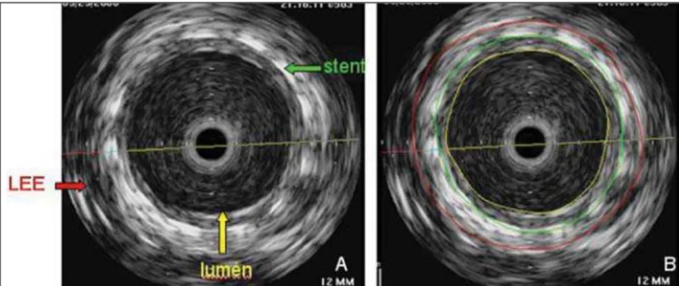
By using the IVUS, it was possible to identify and delimitate the external elastic layer, the stent and the lumen (Figure 2). With these measurements, the vessel, the stent, and lumen areas could be calculated at each millimeter within the stent; subse-quently, the volumes calculations were performed with the aid of the software. By subtracting the luminal volume the stent volume, the neointimal hyperplasia volume was determined.
A linear regression model was adjusted to evaluate the correlation between the intrastent occlusion rate, arterial hyperten-sion, smoking, hyperlipidemia, diabetes mellitus, stainless steel, stenosis and occlu-sion(12,13).
RESULTS
All patients underwent follow-up IVUS, and no complication was observed in these patients.
Eight stents were implanted in the right common iliac artery, 14 in the left common iliac artery, 5 in the right external iliac ar-tery and 3 in the left external iliac arar-tery.
Table 1 Distribution of clinical factors in the sample.
Yes
No
Total
Systemic arterial hypertension
22 (73.3%)
8 (26.7%)
30 (100%)
Diabetes mellitus
9 (30%)
21 (70%)
30 (100%)
Smoking
18 (62.1%)
12 (37.9%)
30 (100%)
Hyperlipidemia
20 (66.7%)
10 (33.3%)
233
Neointimal hyperplasia following intravascular stent implantation
Radiol Bras. 2009 Jul/Ago;42(4):231–234 The first report of the TransAtlantic Inter-Society Consensus (TASC I) was utilized to classify the anatomic arterial involve-ment by the atheromatous plaque, as fol-lows: four were TASC A (13.3%), 15 were TASC B (50%) and 11, TASC C (36.7%). With regard to the clinical status of the patients at the time of the percutaneous transluminal angioplasty and stenting, and also with follow-up purposes, the Ruther-ford scale was utilized. Thus, nine patients (30%) were in category 5 (minor trophic lesion), nine (30%) were in category 4 (rest pain), nine were in category 3 (severe lim-iting claudication) and three (10%) were in category 2 (moderate claudication) at the time of the percutaneous transluminal angioplasty and stenting. At follow-up, 15 patients (50%) were asymptomatic, 13 pre-sented long distance claudication (Ruther-ford category 1) and two presented moder-ate distance claudication. The mean neoin-timal hyperplasia volume was 766.26 mm3 (minimum of 204 mm3 and maximum of 1774 mm3). The intrastent occlusion rate caused by neointimal hyperplasia ranged from 18% to 47% (mean, 27.4%) (Figure 3).
DISCUSSION
Follow-up with IVUS demonstrated that after percutaneous transluminal angio-plasty and stenting all patients developed neointimal hyperplasia and that this is a common result from the intravascular treat-ment. However, neointimal hyperplasia was self-limited and did not cause signifi-cant restenosis among the 30 patients par-ticipating in the present study, suggest-ing that in vessels of larger caliber, like iliac
artery, only a larger volume hyperplasia could cause restenosis. However, even in cases of significant restenosis, a new cath-eter balloon or cutting balloon angioplasty can be performed, without the use of a new
stent(14).
The use of IVUS in the evaluation of intrastent restenosis has shown to be supe-rior to catheter angiography and transcuta-neous Doppler ultrasonography with high-frequency transducers, for being able to better identify the structures and also for not underestimating the lumen diameter(15). Neointimal hyperplasia remains as the main cause of failure of endovascular treat-ment with metallic stents, and much has been done do diminish this response, as follows: the use of heparin stents; the use of anti-ICAM-1 monoclonal antibodies(16), that inhibit the ICAM-1 molecular adhe-sion, hence neointimal hyperplasia; the use of probucol(17), which accelerates stents endothelialization and reduces the forma-tion of hyperplasia; hyperplasia inhibiting
drug-eluting stents(18) ; and even the oral use of neointimal hyperplasia inhibiting drugs(19). So far, the coating with polytetra-fluorethylene (Teflon) has not been able to reduce hyperplasia response(20).
Comparative studies between the pri-mary use of stents and utilization of stents in selected cases where catheter balloon angioplasty failed to show good out-comes(21) suggest that stenting should only be used in the iliac arteries, in case of fail-ure of percutaneous transluminal angio-plasty with catheter balloon. On the other hand, the type of material utilized in the composition of the stents utilized in the present study – stainless steel and nitinol – did not present differences as far as neo-intimal hyperplasia is concerned. Causal factors, such as arterial hypertension, dia-betes mellitus, smoking and hyperlipi-demia were not determinant of statistically significant differences in neointimal hyper-plasia and intrastent occlusion, and also no difference was observed in cases of arterial occlusion or stenosis. Thus, the high re-search costs and the final price of stents are not justifiable for treatment of large cali-ber arteries such as the iliac ones, even in cases where the patients present a history of diabetes mellitus, arterial hypertension, smoking, hyperlipidemia or complete ves-sels occlusion.
CONCLUSION
The results of the linear regression analysis did not demonstrate statistically significant differences for arterial hyperten-sion, smoking, hyperlipidemia, diabetes mellitus, nitinol, stainless steel, stenosis and occlusion, when correlated with the intrastent occlusion rate in the treatment of atherosclerotic lesions of iliac arteries.
REFERENCES
1. Henry M, Amor M, Ethevenot G, et al. Palmaz stent placement in iliac and femoropopliteal ar-teries: primary and secondary patency in 310 patients with 2-4 year follow-up. Radiology. 1995;197:167–74.
2. Bosch JL, Hunink MG. Meta-analysis of the re-sults of percutaneous transluminal angioplasty and stent placement for aortoiliac occlusive dis-ease. Radiology. 1997;204:87–96.
3. De Roeck A, Hendriks JMH, Delrue F, et al. Long-term results of primary stenting for long and com-plex iliac artery occlusions. Acta Chir Belg. 2006; 106:187–92.
Figure 2. Cross section of the vessel and of the stent. A: Structures identification: LEE (external elastic layer), stent, lumen. B: Construction of transverse areas.
234
Moreira SM et al.
Radiol Bras. 2009 Jul/Ago;42(4):231–234 4. Management of peripheral arterial disease (PAD).
TransAtlantic Inter-Society Consensus (TASC). Section C: acute limb ischaemia. Eur J Vasc Endovasc Surg. 2000;19 Suppl A:S115–43. 5. Norgren L, Hiatt WR, Dormandy JA, et al.
Inter-Society Consensus for the Management of Pe-ripheral Arterial Disease (TASC II). Eur J Vasc Endovasc Surg. 2007;34:411–4.
6. Bosch JL, Tetteroo E, Mali WP, et al. Iliac arte-rial occlusive disease: cost-effectiveness analysis of stent placement versus percutaneous trans-luminal angioplasty. Radiology. 1998;208:641– 8.
7. Roubin GS, King SB 3rd, Douglas JS Jr. Reste-nosis after percutaneous transluminal coronary angioplasty: the Emory University Hospital ex-perience. Am J Cardiol. 1987;60:39B–43B. 8. Palmaz JC. Intravascular stents: tissue-stent
in-teractions and design considerations. AJR Am J Roentgenol. 1993;160:613–8.
9. Caramoni PRA, Yamamoto GI, Zago AJ. Reeste-nose pós-angioplastia. Fisiopatogenia. Arq Bras Cardiol. 1997;69:141–8.
10. Leung DA, Spinosa DJ, Hagspiel KD, et al. Selec-tion of stents for treating iliac arterial occlusive
disease. J Vasc Interv Radiol. 2003;14(2 Pt 1):137– 52.
11. Leon BM, Baim DS, Popma JJ, et al. A clinical trial comparing three antithrombotic-drug regi-mens after coronary-artery stenting. Stent Anti-coagulation Restenosis Study Investigators. N Engl J Med. 1998;339:1665–71.
12. Siegel S, Castellan NJ Jr. Nonparametric statis-tics for the behavioral sciences. 2nd ed. New York: McGraw-Hill; 1988.
13. Neter J, Kutner MH, Nachtsheim CJ, et al. Ap-plied linear statistical models. 4th ed. Chicago: Times Mirror Higher Education Group, Inc.; 1996. 14. Tsetis D, Belli AM, Morgan R, et al. Preliminary experience with cutting balloon angioplasty for iliac artery in-stent restenosis. J Endovasc Ther. 2008;15:193–202.
15. Sheikh KH, Davidson CJ, Kisslo KB, et al. Com-parison of intravascular ultrasound, external ul-trasound and digital angiography for evaluation of peripheral artery dimensions and morphology. Am J Cardiol. 1991;67:817–22.
16. Kollum M, Hoefer I, Schreiber R, et al. Systemic application of anti-ICAM-1 monoclonal antibod-ies to prevent restenosis in rabbits: an
anti-inflam-matory strategy. Coron Artery Dis. 2007;18:117– 23.
17. Tanous D, Bräsen JH, Choy K, et al. Probucol inhibits in-stent thrombosis and neointimal hy-perplasia by promoting re-endotelialization. Ath-erosclerosis. 2006;189:342–9.
18. Sousa JEMR, Costa MA, Abizaid AC, et al. Lack of neointimal proliferation after implantation of sirolimus-coated stents in human coronary arter-ies: a quantitative coronary angiography and three-dimensional intravascular ultrasound study. Circulation. 2001;103:192–5.
19. Waksman R, Pakala R, Baffour R, et al. Optimal dosing and duration of oral everolimus to inhibit in-stent neointimal growth in rabbit iliac arteries. Cardiovasc Revasc Med. 2006;7:179–84. 20. Dolmatch B, Dong YH, Heeter Z. Evaluation of
three polytetrafluoroethylene stent-grafts in a model of neointimal hyperplasia. J Vasc Interv Radiol. 2007;18:527–34.