w w w . r b o . o r g . b r
Original
article
Mesenchymal
stromal
cells
from
bone
marrow
treated
with
bovine
tendon
extract
acquire
the
phenotype
of
mature
tenocytes
夽
Lívia
Maria
Mendonc¸a
Augusto
∗,
Diego
Pinheiro
Aguiar,
Danielle
Cabral
Bonfim,
Amanda
dos
Santos
Cavalcanti,
Priscila
Ladeira
Casado,
Maria
Eugênia
Leite
Duarte
InstitutoNacionaldeTraumatologiaeOrtopedia,RiodeJaneiro,RJ,Brazil
a
r
t
i
c
l
e
i
n
f
o
Articlehistory:
Received20October2014 Accepted3February2015 Availableonline6January2016
Keywords:
Tendon
Mesenchymalstromalcellsfrom bonemarrow
Tenocytes
a
b
s
t
r
a
c
t
Objective:Thisstudyevaluatedinvitrodifferentiationofmesenchymalstromalcellsisolated frombonemarrow,intenocytesaftertreatmentwithbovinetendonextract.
Methods:Bovinetendonswereusedforpreparationoftheextractandwerestoredat−80◦C. Mesenchymalstromalcellsfromthebonemarrowofthreedonorswereusedforcytotoxicity testsbymeansofMTTandcelldifferentiationbymeansofqPCR.
Results:Thedatashowedthatmesenchymalstromalcellsfrombonemarrowtreatedfor upto21daysinthepresenceofbovinetendonextractdilutedatdiminishing concentra-tions(1:10,1:50and1:250)promotedactivationofbiglycan,collagentypeIandfibromodulin expression.
Conclusion:Ourresultsshowthatbovinetendonextractiscapableofpromoting differenti-ationofbonemarrowstromalcellsintenocytes.
©2015SociedadeBrasileiradeOrtopediaeTraumatologia.PublishedbyElsevierEditora Ltda.Allrightsreserved.
Células
mesenquimais
do
estroma
da
medula
óssea
tratadas
com
extrato
de
tendão
bovino
adquirem
o
fenótipo
de
tenócitos
maduros
Palavras-chave:
Tendão
Célulasmesenquimaisdoestroma damedulaóssea
Tenócitos
r
e
s
u
m
o
Objetivo:Oestudo avaliaa diferenciac¸ãoinvitro das célulasmesenquimais isoladasdo estromadamedulaósseaemtenócitosapóstratamentocomextratodetendãobovino.
Métodos:Tendõesbovinosforamusadosparaconfecc¸ãodoextratoeestocadosa−80◦C. Célulasmesenquimaisdoestromadamedulaóssea(BMSCs)detrêsdoadoresforamusadas paraostestesdecitotoxicidadeporMTTediferenciac¸ãocelularporqPCR.
Resultados:Osdadosmostram quecélulasmesenquimais doestroma damedulaóssea tratadasporaté21diasempresenc¸adoextratodetendãobovinodiluídoemconcentrac¸ões
夽
WorkdevelopedattheInstitutoNacionaldeTraumatologiaeOrtopedia(INTO),RiodeJaneiro,RJ,Brazil. ∗ Correspondingauthor.
E-mails:[email protected],[email protected](L.M.M.Augusto). http://dx.doi.org/10.1016/j.rboe.2015.12.013
crescentes(1:10,1:50e1:250)promovemaativac¸ãodaexpressãodebiglican,colágenotipo Iefibromodulina.
Conclusão:Nossosresultadosmostramqueoextratodetendãobovinoécapazdepromover adiferenciac¸ãodasBMSCsemtenócitos.
©2015SociedadeBrasileiradeOrtopediaeTraumatologia.PublicadoporElsevier EditoraLtda.Todososdireitosreservados.
Introduction
Tendons are a specialized type of tissue composed of tenoblastsandtenocytes,whichareembeddedinan extra-cellularmatrixmostlycomposedoftypeIcollagen.Tenocytes onlyhavelimitedpotentialforproliferationandthusconfer lowregenerativecapacityontendons.1,2
Tendon injuries constitute a serious problem within orthopedicpractice and generate high costs forthe public healthcaresystem,aswellashavinganimpactonthequality oflifeofthepatientsaffected.Althoughregenerationisthe aimofthe clinicaltreatments used,the methodscurrently availablecontinueto beineffective.Thus, tendon dysfunc-tionsleadtodefinitivephysicalincapacity.3–5
Mesenchymal cells isolated from bone marrow stromal cells(BMSCs)areknowntobeapromisingtherapeuticoption withinthefieldofcelltherapyandbioengineeringof muscu-loskeletaltissues.5–8Theiruseinassociationwithsynthetic biomaterials has been proposed as an option for modern treatmentsaimingtotowardtendonreconstruction,usingan allograft,autograftorxenograft.9,10UseofautologousBMSCs biosyntheticgraftshastheaimsofimprovingtheresultsfrom conservativesurgeryandreducingthetimetakenforthe pre-injurybiomechanicalpropertiestoberestored.11Furthermore, thelowimmunogenicityofBMSCsmakesitpossibletouse themallogeneicallyandminimizestheneedfor immunosup-pressionofthereceptor.12
Despite the significant therapeutic potential of BMSCs, littleisyetknownaboutthemechanismsandsignaling path-waysinvolvedindeterminingthat BMSCswilldifferentiate toward a tenogenic route, or in relation to progression of theirdifferentiation.ConsideringthatBMSCsseemtorespond tostimuli thatarepresentinextractsfromhealthy mature tissuesandhavespecificphenotypiccharacteristics,13,14 we developedthehypothesisthattendonextractsmightinduce differentiation ofBMSCs into tenocytes. Thus, the present studyhadtheobjectiveofevaluatingtheinfluenceof treat-mentofhumanBMSCswithdifferentconcentrationsofbovine tendonextract,oninvitrodifferentiationtowardatenocytic route.
Material
and
methods
Isolationandexpansionofmesenchymalcellsfromthe stromaofhumanbonemarrow
BMSCs were isolated from surgical waste that originated fromhiparthroplastyproceduresonfivepatients(twomen and three women) aged 45–60years, who did not present
any comorbidities.Informedconsentwasobtainedfrom all of these individuals after approval of the study protocol by the institutional ethics committee. After the samples had been collectedinthe surgicalcenter,theywere stored in sterile flasks containing Iscove’s modified Dulbecco’s medium (IMDM; Sigma–Aldrich, St. Louis, MO, USA), sup-plemented with20% bovinefetal serum(BFS; Gibco,Grand Island, NY, USA),at4◦C fornotmorethan 18h. Toisolate
the total cellular fraction, the bone marrow was resus-pendedinphosphate-bufferedsaline(PBS)solutionandwas mechanicallydissociatedfromanybonefragments.Thecell suspensionsthusobtainedwerecollectedin50mLtubesand werecentrifugedat836×gand4◦Cfor5min.Thecellswere thenresuspendedin50mLofIMDMsupplementedwith20% BFSandwerecountedusingaNeubauerchamber.Following this,6×105mononuclearcellsweredistributedinculturing flasksofvolume75cm2,in10mLofIMDMwith20%BFS,and weremaintainedat37◦Cunder5%CO
2.Threedayslater,the non-adherentfractionwasremovedbymeansoflavagewith PBSandtheculturingmediumwaschanged.Afterafurther14 days,thecellswereremovedusingasolutionof0.125%trypsin and0.78mMEDTAandwereexpanded.
Preparationofbovinetendonextract
Five bovine calcaneal tendons were obtained. They were maceratedmechanicallyandthenweregroundupusingan electricblenderofpower20W,intheproportionsof1gof tis-sueto2mLofIMDM,withoutBFS.15Thetissueextractwas◦C
centrifugedat836×gand8◦Cfor5minandwasthenstored at−80◦Cforamaximumoftwomonths.
AnalysisofcellviabilityusingtheMTTmethod
BMSCs were cultured on 24-well plates, at a density of 2.5×10⁴cells/well and were treated with bovine tendon extractdilutedintheproportionsof1:10,1:50or1:250(v/v)in IMDMsupplementedwith10%BFS.Cellviabilitywasassessed 24,72,120and168hoursafterthetreatment,inthepresenceof MTT(thiazolylbluetetrazoliumbromide;Sigma–Aldrich)ata concentrationof25mg/mL.Equalconcentrationsofdimethyl sulfoxide(DMSO)wereusedasanegativecontrol. Colorimet-ricevaluationwasperformedatawavelengthof550nm,using theSIRIOSSEACreader(Burladingen,Germany).
AnalysisofgeneexpressionusingqPCR
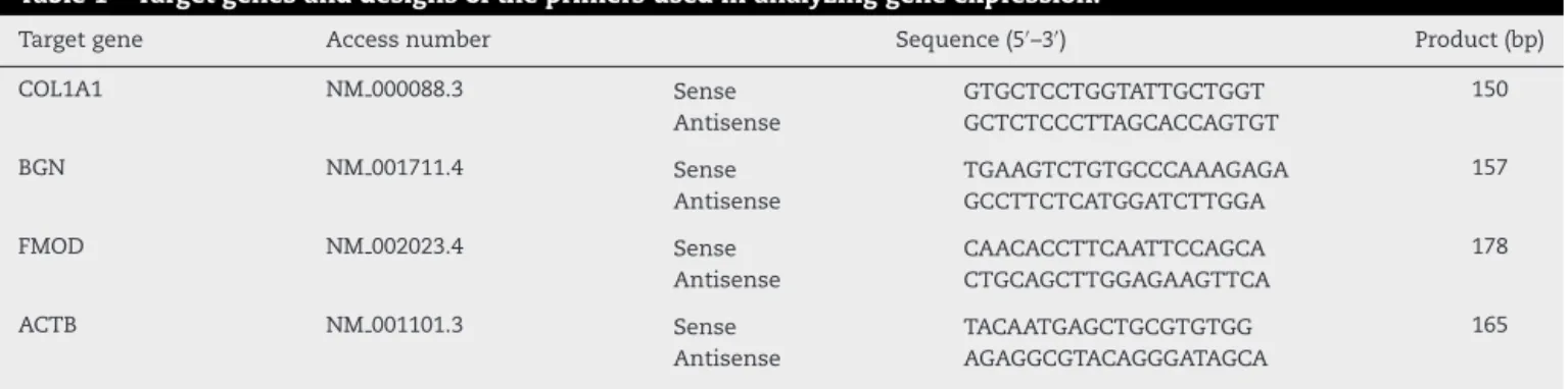
Table1–Targetgenesanddesignsoftheprimersusedinanalyzinggeneexpression.
Targetgene Accessnumber Sequence(5′–3′) Product(bp)
COL1A1 NM000088.3 Sense GTGCTCCTGGTATTGCTGGT 150
Antisense GCTCTCCCTTAGCACCAGTGT
BGN NM001711.4 Sense TGAAGTCTGTGCCCAAAGAGA 157
Antisense GCCTTCTCATGGATCTTGGA
FMOD NM002023.4 Sense CAACACCTTCAATTCCAGCA 178
Antisense CTGCAGCTTGGAGAAGTTCA
ACTB NM001101.3 Sense TACAATGAGCTGCGTGTGG 165
Antisense AGAGGCGTACAGGGATAGCA
COL1A1,type1Acollagen;BGN,biglycan;FMOD,fibromodulin;ACTB,betaactin;bp,sizeofamplifiedproductinbasepairs.
was extracted using the Trizol® method (Invitrogen Corp., Carlsbad,CA,USA)andwastreatedusingDNase(Ambion® DNA-freeTM DNase treatment; Life Technologies), in accor-dancewiththemanufacturer’sinstructions.Theintegrityand quantityoftheRNAwereevaluatedbymeansof electrophore-sison denaturing gel and bymeans ofspectrophotometry (NanodropTM 1000; Thermo Fisher Scientific, Inc.). Reverse transcription for synthesis of complementary DNA (cDNA) wasperformed induplicate, from 1.0g ofRNA, usingthe ImProm-IITMreversetranscriptionsystem(Promega),in accor-dancewiththemanufacturer’sprotocol.qPCRwasperformed using the Power Sybr Green Master MIX® detection sys-tem(AppliedBiosystems,MolecularProbes,Inc.)intheStep Oneequipment(AppliedBiosystems,MolecularProbes,Inc.). Primersfromtheconstitutivegenes(rDNA28Sandactin)were usedascontrolsforthe experiment.Theexpressionofthe genesfortypeIcollagen,biglycanandfibromodulinwas nor-malizedinrelationtotheexpressionoftheconstitutivegene for-actin(Table1).The2−CT13methodwasusedfor analyz-ingtheexpressionofthetargetgenesofthisstudyinrelation totheconstitutivegene.Thevaluesforrelativeexpressionthat havebeenpresentedtookthefixedcontrol-groupvalueof1.0 asthereference(calibrator).
24h 0
0.1 0.2 0.3 0.4 0.5 0.6 0.7
72h
Absorbance (U.A.)
120h C 1:10 1:50 1:250 B
168h
Fig.1–Evaluationofthecytotoxicityofbovinetendon extractinmesenchymalcellsofthebonemarrowstroma.C representscontrolcondition;1:10,1:50and1:250represent thedilutionsofthebovinetendonextractinIMDM;B representsthecontrolconditionforthetechniqueinwhich therewerenocells.Underallconditions,IMDMwasused withsupplementationwith1%BFS.
Results
With the aim of evaluating the toxicity of bovine tendon extract toward BMSCs, the MTT test was performed. The resultsshowedthatthebovinetendonextractdidnotalter theviabilityoftheBMSCsatanyoftheconcentrationstested (Fig.1).Thus,wecaninferthatthebovinetendonextractdid nothaveanycytotoxiceffectonthemesenchymalstemcells. Giventhattherewasnocytotoxiceffect,thepotentialofthe bovinetendonextractfordifferentiationwastestedwiththe aimofevaluatingwhetherthegrowthfactorspresentinthe extractwouldstimulatedifferentiationofthemesenchymal progenitorcells.WeobservedthatintheBMSCs,theextract was capableof activatingexpressionofthe genes fortype I collagen(Fig. 2), biglycan(Fig. 3)and fibromodulin (Fig. 4) overperiodsof7,14and21days.Theseresultsshowedthat thebovinetendonextractwasnotcytotoxicandthatitwas capable of inducingexpression ofthe genes implicatedin tenocyticdifferentiationofmesenchymalstemcells. Further-more, the tendon protein extractpromoted inductionin a
1:10 0
10 20 30 40
1:50
Relative levels of mRNA expression (type I collagen)
1:250
7 days
14 days
21 days
1:10 0
10 20 30 40
1:50
Relative levels of mRNA expression (biglycan)
1:250 7 days
14 days
21 days
Fig.3–EvaluationofbiglycanexpressionthroughqPCRon mesenchymalstromalcellstreatedwithincreasing concentrationsofbovinetendonextract(1:10,1:50and 1:250).
1:10 0
10 20 30 40 50
1:50
Relative levels of mRNA expression (fibromodulin)
1:250 7 days
14 days
21 days
Fig.4–Evaluationoffibromodulinexpressionthrough qPCRonmesenchymalstromalcellstreatedwith increasingconcentrationsofbovinetendonextract(1:10, 1:50and1:250).
dose-dependentmannerandasafunctionofthedurationof exposuretotheextract.
Discussion
Ourstudy showed that the bovine tendonextract had the potentialtoinducetenocyticdifferentiationofBMSCsandthat thisextractdidnothavecytotoxicityatany ofthe concen-trationsused.Inouranalyses,theresultsshowedthatthere was a spatial and temporal window for success regarding differentiationofBMSCsintotenocytes.Weemphasizethat fortheproceduretobeefficient,theBMSCs(at70% conflu-ence)shouldbetreatedwithtendonextractat1:50forseven days.Weobservedthatthepeakexpressionofbiglycanand
fibromodulin(whicharemarkersfortenocytes)16–21wasatthis time.ThisindicatedthattheBMSCsbecamecommittedtoa tenocyticlineage. Thetreatmentprotocolneedstoproceed withincreasedconcentrationoftheextract,whichshouldbe 1:10foranother10days.ThisallowstheBMSCstomaintain typeIcollagenexpressionandprovideanefficient extracellu-larmatrix,soasalsotomaintaincellviability.
Ourresultssuggestthatintendons,therearegrowth fac-tors that stimulate differentiation ofpluripotent cells into tendoncells.Thisopensupapossibilityforthefieldofcell therapy,fortreatingtendinopathy.Finally,thepotentialwas evaluatedinaninvitromodelandthereforethereisaneedfor validationinaninvivomodel,inordertoconfirmtheresults. Moreover,weraisethepossibilitythat,inthefuture,the poten-tial oftendonextracts originating from humantendonsin cadaverdonorsmightbeevaluated.
Conclusions
Theset ofresultsshowed thattreatment ofBMSCswitha proteinextractfrombovinetendontissuepromoted differen-tiationintotenocytes.
Conflicts
of
interest
Theauthorsdeclarenoconflictsofinterest.
r
e
f
e
r
e
n
c
e
s
1.LuoQ,SongG,SongY,XuB,QinJ,ShiY.Indirectco-culture withtenocytespromotesproliferationandmRNAexpression oftendon/ligamentrelatedgenesinratbonemarrow mesenchymalstemcells.Cytotechnology.2009;61(1–2):1–10. 2.BiY,EhirchiouD,KiltsTM,InksonCA,EmbreeMC,Sonoyama
W,etal.Identificationoftendonstem/progenitorcellsand theroleoftheextracellularmatrixintheirniche.NatMed. 2007;13(10):1219–27.
3.LinY.Fromcollagentotenocyte–howtheequinesuperficial tendonrespondstophysiologicchallengesandphysical therapy.UniversidadeUtrecht,FaculdadeDiergerneeskunde; 2005.Availablefrom:http://dspace.library.uu.nl/
bitstream/handle/1874/7411/index.htm;jsessionid= F9F0939F8C85434883E51A7B4AB1F0D6?sequence=8. 4.AndradeLR.Biomateriaisutilizadosembioengenharia
ortopédica.RevEstudBiol.2006;28(63):17–23.
5.WangT,XuZ,JiangW,MaA.Cell-to-cellcontactinduces mesenchymalstemcelltodifferentiateintocardiomyocyte andsmoothmusclecell.IntJCardiol.2006;109(1):74–81. 6.ChenWH,LaiMT,WuAT,WuCC,GelovaniJG,LinCT,etal.
Invitrostage-specificchondrogenesisofmesenchymalstem cellscommittedtochondrocytes.ArthritisRheum.
2009;60(2):450–9.
7.CsakiC,MatisU,MobasheriA,ShakibaeiM.Co-cultureof caninemesenchymalstemcellswithprimarybone-derived osteoblastspromotesosteogenicdifferentiation.Histochem CellBiol.2009;131(2):251–66.
9. Juncosa-MelvinN,BoivinGP,GoochC,GallowayMT,WestJR, DunnMG,etal.Theeffectofautologousmesenchymalstem cellsonthebiomechanicsandhistologyofgel-collagen spongeconstructsusedforrabbitpatellartendonrepair. TissueEng.2006;12(2):369–79.
10.SahooS,TohSL,GohJC.AbFGF-releasingsilk/PLGA-based biohybridscaffoldforligament/tendontissueengineering usingmesenchymalprogenitorcells.Biomaterials. 2010;31(11):2990–8.
11.BashirJ,ShermanA,LeeH,KaplanL,HareJM.Mesenchymal stemcelltherapiesinthetreatmentofmusculoskeletal diseases.PMRJ.2014;6(1):61–9.
12.LivakKJ,SchmittgenTD.Analysisofrelativegeneexpression datausingreal-timequantitativePCRandthe2(−DeltaDelta
C(T))Method.Methods.2001;25(4):402–8.
13.ChamberlainG,FoxJ,AshtonB,MiddletonJ.Concisereview: mesenchymalstemcells:theirphenotype,differentiation capacity,immunologicalfeatures,andpotentialforhoming. StemCells.2007;25(11):2739–49.
14.LeeIC,WangJH,LeeYT,YoungTH.Thedifferentiationof mesenchymalstemcellsbymechanicalstressor/and co-culturesystem.BiochemBiophysResCommun. 2007;352(1):147–52.
15.DietzFR,MukhopadhyayB,BeckerG,DanielsK,SolurshM. Peripheralnerveextracteffectsonmesenchymalcells.Iowa OrthopJ.1996;16:46–57.
16.LiuXN,YinQ,ZhangH,ZhangH,ZhuSJ,WeiYJ,etal.Tissue extractsfrominfarctedmyocardiumofratsinpromotingthe differentiationofbonemarrowstromalcellsinto
cardiomyocyte-likecells.BiomedEnvironSci. 2008;21(2):110–7.
17.SchweitzerR,ChyungJH,MurtaughLC,BrentAE,RosenV, OlsonEN,etal.Analysisofthetendoncellfateusing Scleraxis,aspecificmarkerfortendonsandligaments. Development.2001;128(19):3855–66.
18.AslanH,Kimelman-BleichN,PelledG,GazitD.Molecular targetsfortendonneoformation.JClinInvestig.
2008;118(2):439–44.
19.KuoCK,TuanRS.Mechanoactivetenogenicdifferentiationof humanmesenchymalstemcells.TissueEngA.
2008;14(10):1615–27.
20.ZhuJ,LiJ,WangB,ZhangWJ,ZhouG,CaoY,etal.The regulationofphenotypeofculturedtenocytesby microgroovedsurfacestructure.Biomaterials. 2010;31(27):6952–8.