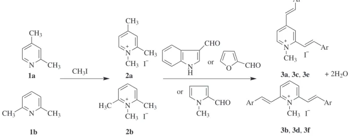
Article
J. Braz. Chem. Soc., Vol. 22, No. 1, 73-79, 2011. Printed in Brazil - ©2011 Sociedade Brasileira de Química
0103 - 5053 $6.00+0.00
A
*e-mail: [email protected]
Synthesis, Absorption and Fluorescence Spectral Characteristics of Trinucleus
Dimethine Cyanine Dyes as Fluorescent Probes for DNA Detection
Jun-Jie Su,a Lan-Ying Wang,*,a Xiang-Han Zhang,a Yi-Le Fu,a
Yi Huangb and Yong-Sheng Weib
aKey Laboratory of Synthetic and Natural Functional Molecule Chemistry (Ministry of Education), College
of Chemistry and Materials Science, Northwest University, Xi’an 710069, People’s Republic of China
bDepartment of Chemistry, Xianyang Normal University, Xianyang, Shaanxi 712000, China
Neste trabalho descreve-se a preparação de seis corantes cianina dimetina trinucleares com núcleos de piridina, obtidos a partir da condensação de iodeto de trimetilpiridínio com aldeídos aromáticos heterocíclicos. As propriedades de absorção e luorescência dos corantes foram estudadas em solventes com polaridades diferentes. O deslocamento hipsocrômico dos máximos de absorção dos corantes foi observado com o aumento da polaridade do solvente. Foram estudadas as propriedades luorescentes dos corantes em solução e na presença de DNA. Foi observado um aumento signiicativo do rendimento quântico de luorescência na presença de DNA para quatro corantes.. Em particular, um dos corantes emite luorescência fraca no tampão Tris-HCl, apresentando contudo luorescência intensa na presença de DNA.
The preparation of six trinucleus dimethine cyanine dyes with pyridine nucleus obtained by the condensation of trimethylpyridinium iodides with heterocyclic aromatic aldehyde was described. The absorption and luorescence properties of the dyes were studied in different polarity solvents. Blue shift of the maxima absorption of the dyes was observed with the increase of solvents polarity. The luorescence properties of the dyes in solution and in presence of DNA were studied. Signiicant enhancement of the luorescent quantum yield was observed in four dyes in the presence of DNA. Specially, one of six dyes emitted weak luorescence in Tris-HCl buffer, but displayed bright luorescence in the presence of DNA.
Keywords: trinucleus dimethine cyanine dyes, DNA, absorption properties, luorescence properties, luorescent dyes
Introduction
Methine cyanine dyes have been widely researched and explored as sensitizers in photography1 and as optical recording materials in laser disk.2 Recently, methine cyanine dyes have attracted attention owing to their excellent luorescence properties3-5 as well as their potential applications in probes for DNA in living cells.6-10 From the applications of cyanine dyes it is known that the optical properties of cyanine dyes are often inluenced by their conjugated systems and the solution environment. Trinucleus dimethine cyanine dyes are large conjugated systems, which contain three heteroaromatic rings joined by two vinyl chains. However the synthesis of trinucleus
dimethine cyanine dyes and their applications as luorescent probe were rarely reported.11,12 Our previous efforts have been devoted to developing series of cyanine dyes and styryl dyes.13-17
Synthesis, Absorption and Fluorescence Spectral Characteristics of Trinucleus Dimethine Cyanine Dyes J. Braz. Chem. Soc.
74
Experimental
General
All reagents were obtained from commercial sources and used without further puriication. All chemicals were of analytical grade. Melting points were taken on a XT-4 micromelting apparatus and uncorrected. IR spectra in cm-1 were recorded on Shimadzu IRPrestige-21 spectrometer and Bruker Equiox-55 spectrometer. 1H NMR spectra were recorded at 400 MHz on a Varian Inova-400 spectrometer and chemical shifts were reported relative to internal Me4Si. 13C NMR spectra were recorded at 100 MHz on a Varian Inova-400 spectrometer and chemical shifts were reported relative to internal Me4Si. Elemental analysis was performed with Vario EL-III instrument. The electron impact (EI) mass spectra were recorded at 70 eV with a GCMS-QP2010 system equipped with the solid sample direct insertion probe. The absorption spectra were recorded on a Shimadzu UV-1700 UV-Vis spectrometer. Fluorescence measurements were carried out on a Hitachi F-4500 spectroluorimeter.
Measurements of the spectral properties of the dyes in different solvents
The dye stock solutions (5.0×10⁻⁴ mol L-1 in dimethylsulfoxide (DMSO)) were diluted with different solvents and resulted in working solutions of dyes (2.0×10⁻⁵ mol L-1). The absorption spectra were examined at room temperature in different solvents and recorded using 1 cm quartz cells on a Shimadzu UV-1700 UV-Vis spectrometer. Fluorescence measurements were carried out at room temperature on a Hitachi F-4500 spectroluorimeter in 1 cm quartz cells. Fluorescence emission was excited at the maximum of the absorption. The absorption and luorescence spectral data were listed in Table 1.
Measurements of spectral properties of the dyes in the presence of DNA
Dye stock solutions (2.0×10⁻⁴ mol L-1) were prepared by dissolving the dyes in DMSO and further diluted with Tris-HCl buffer (pH 7.4) to result in working solutions of dyes (1.0×10⁻⁵ mol L-1). Stock solution of DNA was prepared by dissolving salmon sperm DNA in 0.05 mol L-1 Tris-HCl buffer. The concentration of DNA in stock solution was 3.1×10⁻³ mol L-1 base pairs (bp). The luorescence of dyes in the presence of DNA was tested by adding 0.5 mL DNA solution (3.1×10⁻³ mol L-1 bp) into 1.0 mL 1.0×10⁻⁵ mol L-1 working solutions of dyes. All working solutions were prepared immediately before the experiment.
The absorption spectra were examined at room temperature in different solvents and recorded using 1 cm quartz cells on a Shimadzu UV-1700 UV-Vis spectrometer. Fluorescence measurements were carried out at room temperature on a Hitachi F-4500 spectroluorimeter in 1 cm quartz cells. Fluorescence emission was excited at the maximum of the luorescence excitation spectrum. The absorption and luorescence spectral date were listed in Table 2 and Table 3.
Preparation of trimethyl pyridine quaternary salt 2a-2b
A mixture of 3.54 g (0.033 mol) 1a or 1b with 4.71 g (0.033 mol) CH3I was reluxed for 20 h. After cooling, the product was iltered off and puriied by recrystallization from ethanol. Pyridine quaternary salt 2a: 5.1 g, yield 62%, mp 130-131 °C. 2b: 5.4 g. Yield 66%, mp 235-236 °C.
Preparation of dyes 3a-3b
A mixture of 2a or 2b (0.10 g, 0.4 mmol), indole-3-carboxaldehyde (0.15 g, 1.0 mmol) and 5 drops of piperidine was reluxed for 24 h in 8 mL ethanol. After cooling, ether was added and the product was iltered off, washed with ether and recrystallized from ethanol:water = 9:1. Yield: 3a: 0.15 g (84%), 3b: 0.16 g (79%).
Preparation of dyes 3c-3d
The dyes 3c-3d were synthesized according to a revised literature procedure.12 A mixture of 2a or 2b (0.10 g, 0.4 mmol), 1-methylpyrrole-2-carboxaldehyde (0.20 g, 1.0 mmol) and 5 drops of piperidine was reluxed for 20 h in 5 mL ethanol. After cooling, ether was added and the product was iltered off, washed with ether and recrystallized from ethanol:water = 9:1. Yield: 3c: 0.17 g (86%), 3d: 0.15 g (76%).
Preparation of dyes 3e-3f
Quaternary salts 2a or 2b (0.2 g, 0.8 mmol) and furaldehyde (0.62 g, 6.4 mmol) were dissolved in 18 mL H2O. NaOH (20%, 4.6 mL) was added with stirring over 30 min. After 1 h, the product was iltered off, washed with cold water and recrystallized from water. Yield: 3e: 0.22 g (69%), 3f: 0.27 g (83%).
1-Methyl-2,4-bis[2-(indole-3-yl)vinyl]pyridinium iodide (3a)
Orange-red crystals with metallic luster, mp 172 oC (decomposition),1H NMR (DMSO-d
Su et al. 75 Vol. 22, No. 1, 2011
3H, N+CH
3), 7.20-7.32 (m, 6H, Ar−H, CH=CH), 7.52-7.54 (m, 2H, Ar−H), 7.89 (d, 1H, J 6.8 Hz, pyridine−H), 7.91-7.92 (m, 1H, Ar−H), 8.10-8.19 (m, 2H, CH=CH), 8.19-8.25 (m, 3H, indole-H, CH=CH), 8.48 (s, 1H, pyridine−H), 8.54 (d, 1H, J 6.8 Hz, pyridine−H), 11.84 (s, 1H, indole N−H), 11.96 (s, 1H, indole N−H). 13C NMR (DMSO-d
6, 100 MHz): d 43.6, 110.2, 112.1, 113.0, 113.1, 116.7, 117.0, 117.6, 119.7, 120.0, 120.4, 120.6, 122.3, 122.4, 124.5, 131.1, 131.3, 134.3, 136.1, 136.8, 137.0, 143.5, 152.1. IR ν
max/cm
-1 (KBr) 3400 (m, ν
=NH), 3068 (ν=C-H), 1597, 1552 (ν
C=C), 1517, 1492 1423 (s, νC=C, νC=N), 1367, 1315,1238, 1128, 1107 (dCH), 957, 744 (m, d=CH). MS: EI (70 ev) m/z (%): 361 (58 M – CH3I), 360 (100 M – CH3I – H), 142, 127. UV-Vis λ
max/nm (methanol) 465. Found: C, 59.0; H, 4.43; N, 7.56. Calc. for C26H22N3I·3/2H2O (530.3 g mol-1): C, 58.9; H, 4.72; N, 7.92%.
1-Methyl-2,6-bis[2-(indole-3-yl)vinyl]pyridinium iodide (3b)
Orange-red crystalswith metallic luster, mp 206-207 oC, 1H NMR (DMSO-d
6, 400 MHz): d 4.12 (s, 3H, N +CH
3), 7.21-7.29 (m, 6H, Ar−H), 7.51-7.54 (m, 2H, CH=CH), 7.85-7.92 (m, 2H, CH=CH), 8.10-8.24 (m, 5H, Ar−H, pyridine−H), 8.48-8.56 (m, 2H, pyridine−H), 11.86 (s, 1H, indole N−H), 11.97 (s, 1H, indole N−H). 13C NMR (DMSO-d6, 100 MHz): d 44.0, 110.7, 112.5, 113.3, 113.4, 117.2, 117.4, 118.0, 120.1, 120.4, 120.9, 121.1, 122.7, 122.8, 124.7, 124.9, 131.3, 131.5, 131.7, 134.7, 136.4, 137.1, 137.2, 137.4, 143.9, 152.5. IR ν
max/cm
-1 (KBr) 3389 (m, ν
=NH), 3071 (ν=C-H), 1598, 1551 (νC=C), 1493 1423 (s, ν
C=C, νC=N), 1313, 1273, 1237, 1189, 1128 (dCH), 956 (s, ν
=CH), 809, 745 (m, d=CH). MS: EI (70 ev) m/z (%): 361 (62 M – CH3I), 360 (100 M – CH3I – H), 142, 127. UV-Vis λ
max/nm (methanol) 464. Found: C, 61.50; H, 4.23; N, 8.13. Calc. for C26H22N3I (503.3 g mol-1): C, 61.85; H, 4.40; N, 8.32%.
1-Methyl-2,4-bis[2-(1-methylpyrrol-2-yl)vinyl]pyridinium iodide (3c)
Red needle crystals, mp 180-181oC, 1H NMR (CDCl 3, 400 MHz): d 3.70-4.01 (m, 9H, pyrrole N−CH
3, N +CH
3), 6.20 (s 1H, pyrrole−H), 6.24 (s 1H, pyrrole−H), 6.52 (d, 1H, J 15.2 Hz, CH=CH), 6.73 (s, 1H, pyrrole−H), 6.83 (s, 3H, pyrrole−H), 7.08 (d, 1H, J 16.0 Hz, CH=CH) 7.54 (d, 1H, J 16.0 Hz, CH=CH), 7.68 (d, 1H, J 6.8 Hz, pyridine−H), 8.03 (d, 1H, J 15.2 Hz, CH=CH), 8.28 (d, 1H, J 6.8 Hz, pyridine−H), 8.60 (s, 1H, pyridine−H). 13C NMR (DMSO-d
6, 100 MHz): d 34.0, 34.1, 44.2, 109.6, 109.7, 111.1, 111.3, 112.7, 117.6, 117.9, 119.2, 128.0, 128.1, 128.5, 130.2, 130.2, 130.4, 144.0, 151.6, 151.9. IR ν
max/cm
-1 (KBr) 3440 (m, ν
=NH), 3031 (ν=C-H), 1597, 1547
(ν
C=C), 1520, 1409 (s, νC=C, νC=N), 1339, 1264, 1194, 1126, 1086 (dCH), 971, 888, 814, 710 (m, d=CH). MS: EI(70 ev) m/z (%): 289 (45 M – CH3I), 288 (100 M – CH3I – H), 142, 127. UV-Vis λ
max/nm (methanol) 473. Found: C, 53.91; H, 3.57; N, 5.99. Calc. for C20H22N3I (431.09 g mol-1): C, 53.77; H, 3.61; N, 6.27%.
1-Methyl-2,6-bis[2-(1-methylpyrrol-2-yl)vinyl]pyridinium iodide (3d)
Red needle, mp 230 oC (decomposition), 1H NMR (CDCl3, 400 MHz): d 3.86 (s, 6H, pyrrole N−CH
3), 4.28 (s, 3H, N+CH
3), 6.21-6.23 (m, 2H, pyrrole−H), 6.79 (s, 2H, pyridine−H), 6.93-6.99 (m, 4H, CH=CH, pyrrole−H), 7.50 (d, 2H, J 16.0 Hz, CH=CH), 8.08-8.10 (m, 3H, pyridine−H, pyrrole−H). 13C NMR (DMSO-d
6, 100 MHz): d 33.9, 40.9, 109.5, 112.4, 112.7, 121.2, 128.2, 130.3, 141.1, 153.2. IR ν
max/cm
-1 (KBr) 3446 (m, ν
=NH), 3073 (ν=C-H), 1604, 1559 (ν
C=C), 1469, 1404 (s, νC=C, νC=N), 1335, 1307, 1228, 1189, 1144, 1090 (dCH), 948, 841, 802, 730 (m,
d=CH). MS: EI (70 ev) m/z (%): 289 (100 M – CH3I), 288 (58 M – CH3I – H), 142, 127. UV-Vis λ
max/nm (methanol) 465. Found: C, 53.88; H, 3.65; N, 6.47. Calc. for C20H22N3I (431.09 g mol-1): C, 53.77; H, 3.61; N, 6.27%.
1-Methyl-2,4-bis[2-(furan-2-yl)vinyl]pyridinium iodide (3e)
Yellow powder, mp 254-255 oC, 1H NMR (CDCl 3, 400 MHz): d 4.35 (s, 3H, N+CH
3), 6.54-6.55 (m, 2H, furan−H), 6.91-6.98 (m, 4H, furan−H, CH=CH), 7.56-7.58 (m, 2H, furan−H), 7.68-7.82 (m, 3H, CH=CH, pyridine−H), 8.14 (s, 1H, pyridine−H), 9.10 (d, 1H, J 6.8 Hz, pyridine−H). 13C NMR (DMSO-d
6, 100 MHz): d 44.4, 112.8, 115.0, 115.7, 119.8, 120.2, 126.4, 128.3, 144.9, 145.7, 146.0, 150.6, 150.8, 150.9. IR ν
max/cm
-1 (KBr) 3474 (m, ν
=NH), 3083 (ν=C-H), 1608, 1558 (νC=C), 1470, 1442 (s, ν
C=C, νC=N), 1386, 1330, 1301 (dCH) 1253(νasC−O−C), 1069(νsC−
O−C), 969, 880,751 (m, d=CH). MS: EI (70 ev) m/z (%): 264 (11 M – CH3I), 263 (53 M – CH3I – H), 142, 127. MS: EI (70 ev) m/z (%): 264 (11 M – CH3I), 263 (53 M – CH3I – H), 142, 127. UV-Vis λ
max/nm (methanol) 397. Found: C, 53.64; H, 3.60; N, 3.60. Calc. for C18H16NIO2 (405.02 g mol-1): C, 53.35; H, 3.98; N, 3.46%.
1-Methyl-2,6-bis[2-(furan-2-yl)vinyl]pyridinium iodide (3f)
Khaki-colored powder, mp 214-215 oC, 1H NMR (CDCl3, 400 MHz): d 4.37 (s, 3H, N+CH
3), 6.54-6.55 (m, 2H, CH=CH), 6.93-6.94 (m, 2H, furan−H), 7.21-7.27 (m, 2H, CH=CH), 7.50-7.56 (m, 4H, furan−H), 8.08 (d, 2H, J 8.8 Hz, pyridine−H), 8.28 (t, 1H, J 8.8 Hz, pydidine−H). 13C NMR (DMSO-d
Synthesis, Absorption and Fluorescence Spectral Characteristics of Trinucleus Dimethine Cyanine Dyes J. Braz. Chem. Soc.
76
115.5, 122.8, 128.4, 142.3, 145.7, 150.6, 152.2. IR ν max/cm
-1 (KBr) 3479 (m, ν
=NH), 3061 (ν=C-H), 1602, 1561(νC=C), 1463, 1384 (s, ν
C=C, νC=N), 1305 (dCH) 1247(νasC−O−C) 1068(νsC−O−C), 951, 982, 878, 757 (m, d=CH). MS: EI (70 ev) m/z (%): 264 (8 M – CH3I), 263 (39 M – CH3I – H), 142, 127. UV-Vis λ
max/nm (methanol) 397. Found: C, 53.31; H, 3.60; N, 3.76. Calc. for C18H16NIO2 (405.02 g mol-1): C, 53.35; H, 3.46; N, 3.98%.
Results and Discussion
Synthesis
The trinucleus dimethine cyanine dye was synthesized via the reaction of heteroaromatic aldehydes with the quaternary ammonium salt of 1,2,4-trimethyl pyridine quaternary salt or 1,2,6- trimethyl pyridine quaternary salt (Scheme 1).In all cases investigated, we found thatthe formation reactions of trinucleus dimethine cyanine dyes with pyridine nucleus proceeded eficiently under catalysis of piperidine or NaOH, and the better and purer yields were obtained in ethanol for 3a-3d andin water for 3e-3f. For example, the yield of 3d could reach 86% in ethanol, and 65% in water.12The yield of 3f could reach 83% in water, but in ethanol 3f could hardly be obtained. Because in organic solvents furaldehyde reacted with quaternary salt so quickly that the reaction could not be controlled, and there would be serious side reactions. In the synthesis of dyes 3a-3f , the required reaction time of heterocyclic aromatic aldehydes with trimethylpyridinium iodides was
furaldehyde (30 min) < 1-methylpyrrole-2-carboxaldehyde (20 h) < indole-3-carboxaldehyde (24 h), thereby the reaction activity of heterocyclic aromatic aldehydes was furaldehyde > 1-methylpyrrole-2-carboxaldehyde > indole-3-carboxaldehyde. From the structure it was also suggested that the electron-withdrawing ability of the group attached to aldehyde group was =C−O > =C−N > =C−C=, leading to the positive charge density of carbon in aldehyde group was furaldehyde > 1-methylpyrrole-2-carboxaldehyde > indole-3-carboxaldehyde. The larger the positive charge density of carbon in aldehyde group is, the higher the reaction activity of heterocyclic aromatic aldehyde with trimethylpyridinium iodides is.
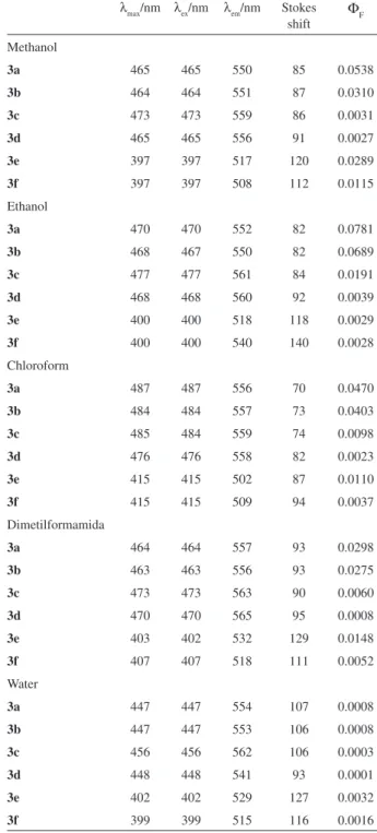
Spectral properties of the dyes in different solvents
Figure 1 gives the absorption maxima (λ
max) of six trinucleus dimethine cyanine dyes in different solvents (their dielectric constant: CHCl3 4.9, EtOH 24.6, MeOH 32.6, dimethylformamide 38.3, H2O 78.4). From Figure 1 it could be found that the λ
max of 3a, 3b, 3c and 3d was longer than the λ
max of 3e and 3f. The reason suggested here was that dyes 3a-3f were all D-π-A molecules, in dyes 3a-3d the electron donor was N of indole or pyrrole ring, and the electron donor was O of furan ring in dyes 3e-3f, and the electron acceptor of six dyes was all N+ of pyridinium. The stronger was electron-donating capability of electron donor, the longer was the λ
max of dyes under the same electron-withdrawing capability of electron acceptor. The electron-donating capability of N situated indole or pyrrole
Scheme 1. Synthesis of trinucleus dimethine cyanine dyes.
N CH3
CH3
CH3I
N CH3 CH3 CH3 N H CHO N CHO CH3 O CHO 2b I 1b or or N CH3 I
Ar Ar
N CH3 I
Ar Ar
3a, 3c, 3e
3b, 3d, 3f
Ar = indole-3-carboxaldehyde
Ar = 1-methylpyrrole-2-carboxaldehyde Ar = furaldehyde
+ 2H2O
N CH3 CH3
N CH3 CH3 H3C
I
1a 2a
Su et al. 77 Vol. 22, No. 1, 2011
ring was stronger than that of O situated furan ring, so the λ
max of 3a-3d was longer than the λmax of 3e-3f. It could be also found that with the increasing of solvent polarity the λ
max of the dyes decreased. The effect of the solvent polarity on the absorption maximum could be illustrated by interactions between the dye molecules and the solvents, as the interactions made the ground state of dye more stable by forming hydrogen bonds.18,19
The absorption, excitation maxima (λ
ex), luorescence maxima emission (λ
em) of the dyes are summarized in Table 1. The dyes exhibited luorescence properties at room temperature. Their luorescence maxima were located at 502-565 nm. Compared with the absorption maxima of the dyes, the emission spectra were shifted to the red by 69-140 nm (Stokes shift). The stokes shift of six dyes were all large, this might be attributed to an excited-state intramolecular charge transfer between the donor and acceptor in the dyes. Large stokes shift could help to reduce self-quenching and measurement error by excitation light and scattered light.20 The luorescence quantum yield of six dyes was in the region 0.0001-0.0781 in different solvents. From Table 1 it could be found that the order of luorescence quantum yield was 3a, 3b > 3c, 3d. The possible reason was that the conjugated system of 3a, 3b was larger than that of 3c, 3d. We also found the order of luorescence quantum yield was 3a > 3b and 3c > 3d. The cause lay in the fact that two D-π-A system in 3b and 3d was in opposite orientation, which made their conjugated system became small. It could be found that the luorescence performance of 3e and 3f was complex, because oxygen of furan ring was of both electron-donating and electron-acceptor effect. And there was also inluence of solvents on the luorescence quantum yield of these dyes. Further investigation of the inluence of solvents on the luorescence quantum yield of the dyes is in progressing.
Figure 1. Solvatochromic shifts for the λ
max of six trinucleus
dimethincyanine dyes in ive kinds of solvents.
Table 1. The absorption and luorescence spectral characteristics of dyes 3a-3f in different solvents
λ
max/nm λex/nm λem/nm Stokes
shift
Φ
F
Methanol
3a 465 465 550 85 0.0538
3b 464 464 551 87 0.0310
3c 473 473 559 86 0.0031
3d 465 465 556 91 0.0027
3e 397 397 517 120 0.0289
3f 397 397 508 112 0.0115
Ethanol
3a 470 470 552 82 0.0781
3b 468 467 550 82 0.0689
3c 477 477 561 84 0.0191
3d 468 468 560 92 0.0039
3e 400 400 518 118 0.0029
3f 400 400 540 140 0.0028
Chloroform
3a 487 487 556 70 0.0470
3b 484 484 557 73 0.0403
3c 485 484 559 74 0.0098
3d 476 476 558 82 0.0023
3e 415 415 502 87 0.0110
3f 415 415 509 94 0.0037
Dimetilformamida
3a 464 464 557 93 0.0298
3b 463 463 556 93 0.0275
3c 473 473 563 90 0.0060
3d 470 470 565 95 0.0008
3e 403 402 532 129 0.0148
3f 407 407 518 111 0.0052
Water
3a 447 447 554 107 0.0008
3b 447 447 553 106 0.0008
3c 456 456 562 106 0.0003
3d 448 448 541 93 0.0001
3e 402 402 529 127 0.0032
3f 399 399 515 116 0.0016
a The luorescence quantum yields of the dyes were determined by the
reference standard (rhodamine B Φ
F=0.49 in ethanol at 25 oC).21
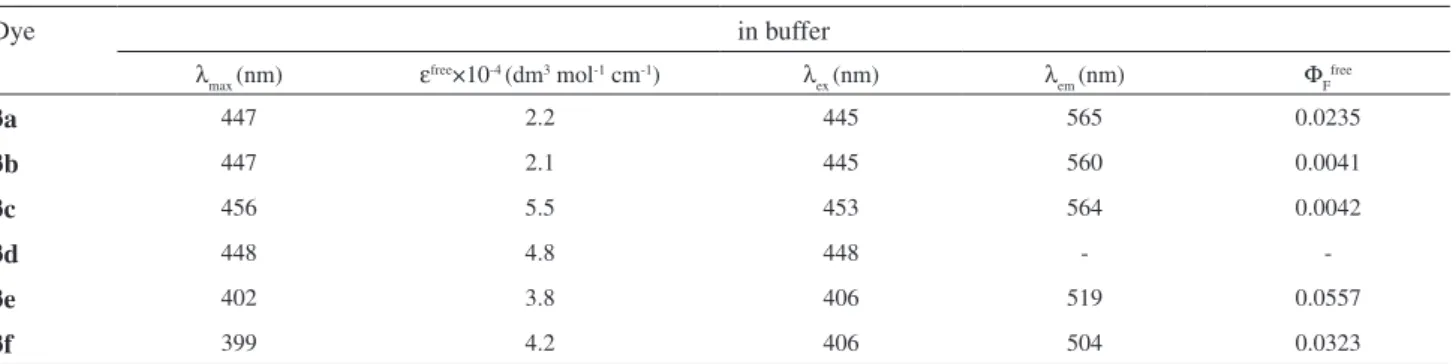
Spectral properties of the dyes in the presence of DNA
The spectral properties of the dyes in the presence of DNA are summarized in Table 2 and Table 3. The λ
Synthesis, Absorption and Fluorescence Spectral Characteristics of Trinucleus Dimethine Cyanine Dyes J. Braz. Chem. Soc.
78
free dyes in buffer. The molar extinction coeficients for dyes 3a-3d were increased in the presence of DNA, and were in the range from 3.2×10⁴ to 8.5×10⁴ L mol-1 cm-1. However, for dyes 3e and 3f the values of molar extinction coeficients were unchanged relative to free dyes, which were 3.8×10⁴ and 4.2×10⁴ L mol-1 cm-1, respectively.
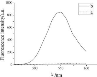
The luorescence emission maxima of DNA-dyes were located at 503-555 nm, and showed a slight blue shift relative to free dyes in solution. Stokes shift values for the DNA-dyes were in the range of 97-109 nm. Moreover, the luorescence intensity of dyes 3a-3c was greatly increased in the presence of DNA. Compared with free dyes, the quantum yields of DNA-dyes were up to 9.5 times for dye 3a, up to 31.6 times for dye 3b (Figure 2) and up to 18.9 times for dye 3c. Specially, the quantum yields of DNA-dye 3a was the highest in six dyes. It was noteworthy that free dye 3d could not be detected signiicantly luorescence in buffer, but DNA-dye 3d
showed great luorescence quantum yields. The luorescence enhancement of trinucleus dimethine cyanine dyes bound to DNA was attributable to the fact that on photoexcitation a lack of free rotation around the internuclear bridge made isomerisation around the C-C bonds of the methine chain dificult; and subsequently nonradiative deactivation of the excited state was not possible, causing the dye to luoresce.22,23 Upon binding with DNA, 3a-3d maintained
their high Stokes shift and showed a red shift, owing to a more eficient ICT in the excited state between the terminal heterocyclic aromatic and the pyridimium group. 24,25
Conclusions
Six trinucleus dimethine cyanine dyes with pyridine nucleus were synthesized and isolated in 69-86% yield with piperidine or NaOH as catalyst.
Figure 2. Fluorescence spectra of 3b in buffer, in the presence of different concentration of DNA.
Table 2. Spectral characteristics of dyes 3a-3f in buffer
Dye in buffer
λ
max (nm) ε
free×10-4 (dm3 mol-1 cm-1) λ
ex (nm) λem (nm) ΦF free
3a 447 2.2 445 565 0.0235
3b 447 2.1 445 560 0.0041
3c 456 5.5 453 564 0.0042
3d 448 4.8 448 -
-3e 402 3.8 406 519 0.0557
3f 399 4.2 406 504 0.0323
a The luorescence quantum yields of the dyes were determined by the reference standard (rhodamine B Φ
F = 0.49 in ethanol at 25 oC).21
Table 3. Spectral characteristics of dyes 3a-3f in presence of DNA
Dye in DNA presence Φ
F DNA/Φ
F free λ
max (nm) ε
DNA×10-4 (dm3 mol-1 cm-1) λ
ex (nm) λem (nm) ΦF DNA
3a 455 3.2 445 545 0.2242 9.5
3b 454 6.2 445 545 0.1291 31.6
3c 458 8.5 453 555 0.0791 18.9
3d 449 5.1 448 553 0.0187
-3e 403 3.8 406 515 0.0553 1.0
3f 401 4.2 406 503 0.0336 1.0
a The luorescence quantum yields of the dyes were determined by the reference standard (rhodamine B Φ
Su et al. 79 Vol. 22, No. 1, 2011
The absorption maxima of the dyes were located at 397-487 nm in different solvents, and with the increase of solvents polarity the maximum absorption wavelength of these dyes had a blue-shift. The luorescence maxima of the dyes in different solvents were basically located at 502-565 nm, and the luorescence quantum yield of six dyes was in the region 0.0001-0.0781 in different solvents.
The absorption maxima of the dyes in the presence of DNA were situated at 401-458 nm and showed a slight red shift relative to free dyes. The luorescence maxima of DNA-dyes were located at 503-555 nm, and showed a slight blue shift relative to free dyes. The luorescent quantum yields of the DNA-dyes 3a-3c were up to 9.5-31.6 times higher than that of free dyes. Specially, free dye 3d emitted weak luorescence in buffer, but DNA-dye 3d showed great luorescence quantum yields. Therefore dyes 3a-3d could be proposed as luorescent dyes for DNA detection.
Supplementary Information
Supplementary data are available free of charge at http://jbcs.sbq.org.br, as PDF ile.
Acknowledgements
We appreciate the inancial support for this research by a grant from the Natural Science Foundation of Shaanxi Province (No. SJ08B04), the Special Science Research Foundation of Education Committee (No. 08JK458), NWU Excellent Doctoral Dissertation Foundation (No. 08YYB04) and NWU Graduate Cross-discipline Funds (No. 09YJC20).
References
1. Karatsu, T.; Yanai, M.; Yagai, S.; Mizukami, J.; Urano, T.; Kitamura, A.; J. Photochem. Photobiol.,A2005, 170, 123. 2. Usami, T.; Asanuma, N.; Yamakawa, K.; JP Pat 265,0762000. 3. Kovalska, V.; Volkova, K.; Losytskyy, M.; Tolmachev, O.;
Balanda, A.; Yarmoluk, S.; Spectrochim. Acta, Part A 2006,
65, 271.
4. Deligeorgiev, T.; Gadjev, N.; Vasilev, A.; Maximova, V.; Timcheva, I.; Katerinopoulos, H.; Dyes Pigm. 2007, 75, 466.
5. Kovalska, V.; Kryvorotenko, D.; Balanda, A.; Losytskyy, M.; Tokar, V.; Yarmoluk, S.; Dyes Pigm. 2005, 67, 47.
6. Karlsson, H.; Bergqvist, M.;, Lincoln, P.; Westman, G.; Bioorg. Med. Chem.2004, 12, 2369.
7. Timtcheva, I.; Maximova, V.; Deligeorgiev, T.; Zaneva, D.; Ivanov, I.; J. Photochem. Photobiol., A 2000, 130, 7. 8. Hilal, H.; Taylor, J.; Dyes Pigm.2007, 75, 483.
9. Rosania, G. R.; Lee, J. W.; Ding, L. Yoon, H. S.; Chang, Y. T.;
J. Am. Chem. Soc.2003, 125, 1130.
10. Li, Q.; Kim, Y.; Namm, J.; Kulkarni, A.; Rosania, G.. R.; Ahn, Y. H.; Chang, Y. T.; Chem. Biol.2006, 13, 615.
11. Vincenza, B.; Daniele, F.; Condorelli, C.; Giuseppe, M.; Bioorg. Med. Chem.2002, 10, 2899.
12. Maria, F.; Cosimo, G. F.; Giuseppe, I.; Giuseppe, M.; Eur. J. Org. Chem.2002, 145.
13. Wang, L. Y.; Zhang, X. G.; Shi, Y. P.; Zhang, Z. X.; Dyes Pigm. 2004, 62, 21.
14. Wang, L. Y.; Zhang, X. G.; Li, F. M.; Zhang, Z. X.; Synth. Commun.2004, 34, 1.
15. Zhang, Z. X.; Zhang, Y. J.; Hao, J. X.; Li, C. E.; Sci. China, Ser. B: Chem.1995, 25, 689.
16. Zhang, X .H.; Wang,L. Y.; Zhai, G.. H.; Wen, Z. Y.; Zhang, Z. X.; Bull. Korean Chem. Soc.2007, 28, 12.
17. Huang, W.; Wang, L. Y.; Fu, Y. L.; Liu, J. Q.; Tao, Y. N.; Fan, F. L.; Zhai, G.. H.; Wen, Z. Y.; Bull. Korean Chem. Soc.2009,
30, 3.
18. Pham, W.; Lai, W. F.; Weissleder, R.; Tung, C. H.; Bioconjugate Chem.2003, 14, 1048.
19. Mishra, A.; Behera, R. K.; Behera, P. K.; Mishra, B. K.; Behera, G. B.; Chem. Rev.2000, 100, 1973; Ibrahim, A. Z.; Al-Ansari, I.; Bull. Soc. Chim. Fr.1997, 134, 593.
20. Zhang, Z.; Achilefu, S.; Org. Lett.2004, 6, 2067.
21. Casey, K. G.; Quitevis, E. L.; J. Phys. Chem.1988, 92, 6590. 22. Carlsson, C.; Larsson, A.; Jonsson, M.; Albinsson, B.; J. Phys.
Chem.1994, 98, 10313.
23. Anikovsky, M.Y.; Tatikolov, A. S.; Shvedove, L. A.; Kuzmin V. A.; Russ. Chem. Bull. 2001, 50, 1190.
24. Neto, B. A. D.; Lápis, A. A. M.; Mancilha, F. S.; Vasconcelos, I. B.; Thum, C.; Basso, L. A.; Santos, D. S.; Dupont, J.; Org. Lett. 2007, 20, 4001.
25. Neto, B. A. D.; Lápis, A. A. M.; Molecules 2009, 14, 1725.
Submitted: April 17, 2010
Supplementary Information
S
I
J. Braz. Chem. Soc., Vol. 22, No. 1, S1-S11, 2011. Printed in Brazil - ©2011 Sociedade Brasileira de Química 0103 - 5053 $6.00+0.00
*e-mail: [email protected]
Synthesis, Absorption and Fluorescence Spectral Characteristics of Trinucleus
Dimethine Cyanine Dyes as Fluorescent Probes for DNA Detection
Jun-Jie Su,a Lan-Ying Wang,*,a Xiang-Han Zhang,a Yi-Le Fu,a
Yi Huangb and Yong-Sheng Weib
aKey Laboratory of Synthetic and Natural Functional Molecule Chemistry (Ministry of Education), College
of Chemistry and Materials Science, Northwest University, Xi’an 710069, People’s Republic of China
bDepartment of Chemistry, Xianyang Normal University, Xianyang, Shaanxi 712000, China
Figure S2. Fluorescence emission of 3b in buffer, in the presence of DNA. a: 3b in buffer. b: 3b in the presence of DNA.
Figure S4. Fluorescence emission of 3d in buffer, in the presence of DNA. a: 3d in buffer. b: 3d in the presence of DNA.
Figure S3. Fluorescence emission of 3c in buffer, in the presence of DNA. a: 3c in buffer. b: 3c in the presence of DNA.
Figure S1. Fluorescence emission of 3a in buffer, in the presence of DNA. a: 3a in buffer. b: 3a in the presence of DNA.
For Fluorescence emission of 3a-3f in buffer, in the presence of DNA see Figure S1-S6. For 1H NMR, 13C NMR and
Synthesis, Absorption and Fluorescence Spectral Characteristics of Trinucleus Dimethine Cyanine Dyes J. Braz. Chem. Soc. S2
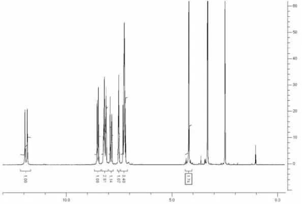
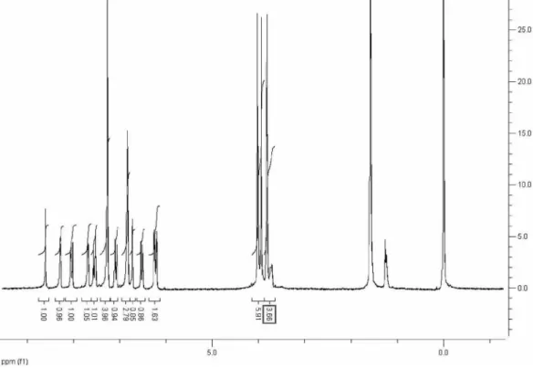
Figure S7. 1H NMR spectrum of 3a.
Figure S5. Fluorescence emission of 3e in buffer, in the presence of DNA.
a: 3e in buffer.b: 3e in the presence of DNA.
Figure S6. Fluorescence emission of 3f in buffer, in the presence of DNA. a: 3f in buffer. b: 3f in the presence of DNA.
Su et al. S3 Vol. 22, No. 1, 2011
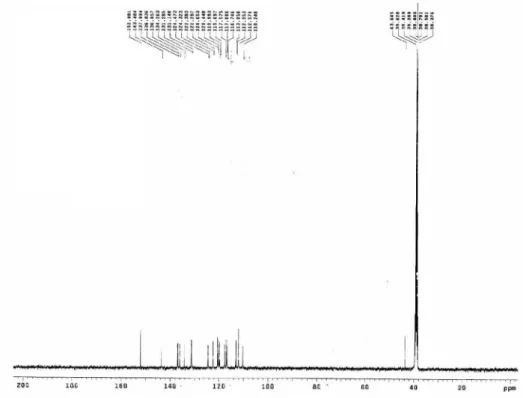
Figure S8.13C NMR spectrum of 3a.
Synthesis, Absorption and Fluorescence Spectral Characteristics of Trinucleus Dimethine Cyanine Dyes J. Braz. Chem. Soc. S4
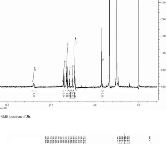
Figure S10.1H NMR spectrum of 3b.
Figure S11.13C NMR spectrum of 3b.
Su et al. S5 Vol. 22, No. 1, 2011
Figure S12. IR spectrum of 3b.
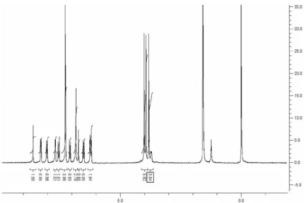
Figure S13.1H NMR spectrum of 3c.
Synthesis, Absorption and Fluorescence Spectral Characteristics of Trinucleus Dimethine Cyanine Dyes J. Braz. Chem. Soc. S6
Figure S14.13C NMR spectrum of 3c.
Su et al. S7 Vol. 22, No. 1, 2011
Figure S16.1H NMR spectrum of 3d.
Figure S17.13C NMR spectrum of 3d.
Synthesis, Absorption and Fluorescence Spectral Characteristics of Trinucleus Dimethine Cyanine Dyes J. Braz. Chem. Soc. S8
Figure S19. 1H NMR spectrum of 3e. Figure S18. IR spectrum of 3d.
Su et al. S9 Vol. 22, No. 1, 2011
Figure S21. IR spectrum of 3e.
Synthesis, Absorption and Fluorescence Spectral Characteristics of Trinucleus Dimethine Cyanine Dyes J. Braz. Chem. Soc. S10
Figure S22.1H NMR spectrum of 3f.
Figure S23. 13C NMR spectrum of 3f.
Su et al. S11 Vol. 22, No. 1, 2011