336
Pitrez EH et al.
Radiol Bras. 2010 Set/Out;43(5):336–338
Case Report • Relato de Caso
Morel-Lavallée lesion in the knee: a case report*
Lesão de Morel-Lavallée no joelho: relato de caso
Eduardo Hennemann Pitrez1, Roberto Campos Pellanda2, Mariana Eltz Silva3, Gustavo Garcia Holz3, Felipe Teixeira Hertz3, João Rubião Hoefel Filho4
Morel-Lavallée lesion is a degloving injury of the interface between the subcutaneous fat and the underlying fascia. This lesion is characterized by the development of a fluid collection that, although originally described in the thigh, it has also been described in other anatomical sites. The authors report the case of a patient with Morel-Lavallée lesion in the knee after trauma and describe the main characteristics of the lesion. Keywords: Knee trauma; Bursitis.
A lesão de Morel-Lavallée é uma lesão em cisalhamento que ocorre entre o tecido celular subcutâneo e a fáscia profunda. Caracteriza-se pela formação de uma coleção que, originalmente descrita na coxa, vem sendo observada em outros locais. Os autores relatam o caso de paciente com Morel-Lavallée no joelho e descre-vem as principais características desta lesão.
Unitermos: Traumatismos do joelho; Bursite. Abstract
Resumo
* Study developed at Clínica Radiológica Osório Lopes, Porto Alegre, RS, Brazil.
1. PhD, Radiologist at Clínica Radiológica Osório Lopes and at Hospital de Clínicas de Porto Alegre (HCPA), Porto Alegre, RS, Brazil.
2. MD, Radiologist at Hospital São Lucas da Pontifícia Univer-sidade Católica do Rio Grande do Sul (PUCRS) and at Hospital de Clínicas de Porto Alegre (HCPA), Porto Alegre, RS, Brazil.
3. MDs., Residents in Radiology at Hospital São Lucas da Pon-tifícia Universidade Católica do Rio Grande do Sul (PUCRS), Porto Alegre, RS, Brazil.
4. MD, Radiologist, Head of the Center of Imaging Diagnosis, Pontifícia Universidade Católica do Rio Grande do Sul (PUCRS), Porto Alegre, RS, Brazil.
Mailing address: Dr. Eduardo Hennemann Pitrez. Rua Ante-nor Lemos, 33, Menino Deus. Porto Alegre, RS, Brazil, 90850 100. E-mail: [email protected]
Received March 13, 2010. Accepted after revision July 13, 2010.
was admitted at the university hospital to undergo a MRI study.
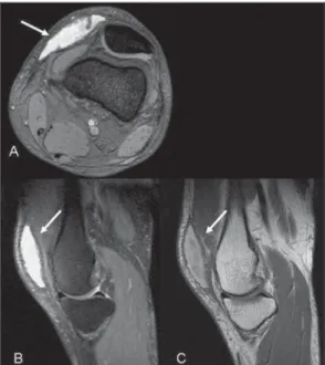
The MRI study demonstrated a lesion in the deep subcutaneous cellular tissue of the prepatellar region. The lesion was fusiform and encapsulated, with an expansile effect between the subcutaneous tissue and the underlying fascia. The lesion presented with a pseudocapsule, besides some small fat foci inside. High signal intensity in T1-weighted sequences was identified inside the lesion, because of the presence of he-matic content (Figure 1).
Pitrez EH, Pellanda RC, Silva ME, Holz GG, Hertz FT, Hoefel Filho JR. Morel-Lavallée lesion in the knee: a case report. Radiol Bras. 2010;43(5):336–338.
regions, ankles and knees have been de-scribed in the literature(3,4).
In the present article, the authors de-scribe a case of Morel-Lavallée lesion af-fecting the knee of a young patient, evalu-ated by magnetic resonance imaging (MRI).
CASE REPORT
A male, 21-year-old patient, victim of a car accident who developed pain and swell-ing in right knee after the accident. Ap-proximately 40 days after the accident, he
0100-3984 © Colégio Brasileiro de Radiologia e Diagnóstico por Imagem INTRODUCTION
Shear “degloving” Morell-Lavallée le-sions represent a traumatic separation caused by the rapid and violent avulsion of the skin over the underlying fascia, leading the skin to separate from the subcutaneous cellular tissue of the underlying muscular fascia. Such separation implies the rupture of small perforating vessels in this region, resulting in the formation of a cavity that may be filled with blood, lymph and fat foci, the latter being sometimes necrotic(1,2).
Morel-Lavallée lesion was originally described in the lateral aspect of the proxi-mal thigh, which is the most common site of this lesion, however other anatomic sites such as periscapular, lumbar and gluteal
337 Morel-Lavallée lesion in the knee
Radiol Bras. 2010 Set/Out;43(5):336–338 DISCUSSION
This lesion was described by Morel-Lavallée in 1853 and it is a hemolymphatic mass located in the deep subcutaneous cel-lular tissue, as a consequence of a shearing lesion caused by trauma. Originally de-scribed at the external aspect of the thigh, in the last few years it has been recognized and described in other anatomic regions such as the lumbar region and the scapula(1,4).
Shearing “degloving” lesions result from a traumatic separation caused by a violent and rapid avulsion of the skin over the fascia, which causes a discontinuity of the deep layer of the conjunctive tissue of the underlying muscular fascia. Such sepa-ration causes the disruption of small per-forating vessels in this region, resulting in a cavity that may be filled with blood, lymph and sometimes necrotic fat foci(1,2)
that may eventually be colonized by infec-tious agents(5). The development of
granu-lation tissue is observed, and it may orga-nize into a pseudocapsule, leading to the persistence of collections(4).
The lesion can be either painful or as-ymptomatic and up to one third of the pa-tients do not recall any significant trauma(4).
Delay in the diagnosis results in the devel-opment of collections with bulging and de-formity of the subcutaneous cellular tis-sue(5). Diagnostic delays of up to 34 years
have already been described(1,3). These
col-lections may develop rapidly when the ar-terial bed is involved, or otherwise slowly, in cases of injury to lymphatic vessels(1).
Considering that Morel-Lavallée le-sions may remain undiagnosed for long periods, it is important for the radiologist to know the characteristics of acute and chronic lesions, as well as their therapeu-tic implications. The appearance of the le-sions depends on the amount of present blood, lymph and fat, as well as the time elapsed from the trauma(4).
In the acute phase, blood clots and de-bris may be found in a hyperintense collec-tion on T2-weighted sequences. With the organization of the hematoma, resulting from the conversion of deoxyhemoglobin into metahemoglobin, such lesions are seen as iso- to hyperintense on T1-weighted im-ages. The lesion periphery becomes
hypointense on T1- and T2-weighted im-ages in the course of time, because of the presence of hemosiderin. In the course of time, the collection becomes a T2 hyperin-tense seroma with a T1 and T2 hypoinhyperin-tense pseudocapsule(4).
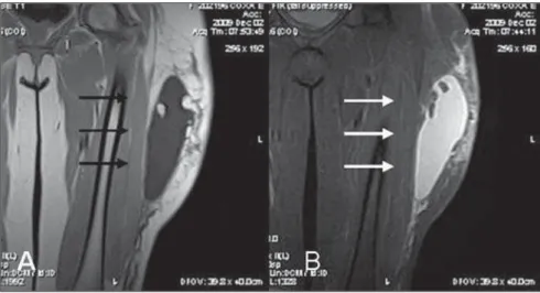
Most frequently, the lesions compro-mise the lateral aspect of the thigh (Figure 2), involving the great trochanter(6),
al-though lesions in other sites, such as the gluteal, lumbar and scapular regions, the ankles and the knees have already been de-scribed(3,4,6).
Although Morel-Lavallée lesions occur with a relatively high frequency in the knees, the radiological literature scarcely presents reports on such cases(5,7,8).
Tejwani et al. have studied 27 Morel-Lavalée lesions in the knees of 24 athletes over a 13-year period, demonstrating that the main mechanism leading to this lesion was a direct shearing force on the point of collection, and that the most relevant func-tional deficit was the restriction to flexion, affecting 41% of the patients. The authors have also demonstrated that such athletes returned to their activities even without the complete collections resolution(5).
For a long time, the classical treatment comprised surgical drainage with debride-ment and second-intention healing but, re-cently, the therapeutic alternatives have tended towards less aggressive treat-ments(5). Small lesions may be resolved by
means of a small incision and drainage, while major lesions may develop a
pseudo-capsule, becoming refractory to conserva-tive treatment(1). The lesions may also be
treated either with elastic compression or liposuction. In cases of failure, sclerosis with the administration of doxycycline is an option(5).
The most important differential diagno-sis of Morel-Lavallée knee lesions is done with prepatellar bursitis (“housemaid’s knee”). The prepatellar bursa often consists of a trilaminar bursa whose dimensions may extend a little beyond the edges of the patella measuring on average 39 × 40.5 ×
3.2 mm in diameter, in the craniocaudal, latero-medial and anteroposterior planes(5).
However, in Morel-Lavallée lesions, the collections dimensions and limits often exceed the expected proportions for the prepatellar bursa, this sign being the most reliable in the differentiation between the two entities. Collections extending above the suprapatellar region are virtually diag-nostic of Morel-Lavallée, as the bursa should not extend to this point(4,7).
Borrero et al. have studied four Morel-Lavallée collections, demonstrating in all the cases a unilocular collection whose lim-its extended beyond the prepatellar bursa, No blood or fat foci were identified within the collections(4).
Tejwani et al. have demonstrated that Morel-Lavallée collections exceeded the expected dimensions of the prepatellar bursa and reported that many of the 27 cases of Morel-Lavallée knee lesions could only be identified by clinical examination;
338
Pitrez EH et al.
Radiol Bras. 2010 Set/Out;43(5):336–338
none of these lesions were submitted to biopsy, which maintains the criterion based on dimensions still under argument in the differential diagnosis(4).
CONCLUSION
Morel-Lavallée collections have been increasingly diagnosed in the knee region, and the radiologist’s knowledge on the characteristics of the lesion at MRI can help in the diagnosis of this disease that is not always recognized, and sometimes difficult to treat. The presence of a trauma history associated with a periarticular collection in
the knee should indicate the possibility of a Morel-Lavallée lesion in the differential diagnosis, together with prepatellar bursi-tis.
REFERENCES
1. Gilbert BC, Bui-Mansfield LT, Dejong S. MRI of a Morel-Lavallée lesion. AJR Am J Roentgenol. 2004;182:1347–8.
2. Hak DJ, Olson SA, Matta JM. Diagnosis and man-agement of closed internal degloving injuries as-sociated with pelvic and acetabular fractures: the Morel-Lavallée lesion. J Trauma. 1997;42:1046– 51.
3. Mellado JM, Bencardino JT. Morel-Lavallée le-sion: review with emphasis on MR imaging. Magn Reson Imaging Clin N Am. 2005;13:775– 82.
4. Borrero CG, Maxwell N, Kavanagh E. MRI find-ings of prepatellar Morel-Lavallée effusions. Skel-etal Radiol. 2008;37:451–5.
5. Tejwani SG, Cohen SB, Bradley JP. Management of Morel-Lavallee lesion of the knee: twenty-seven cases in the national football league. Am J Sports Med. 2007;35:1162–7.
6. Moriarty JM, Borrero CG, Kavanagh EC. A rare cause of calf swelling: the Morel-Lavallee lesion. Ir J Med Sci. 2009 Jul 18. [Epub ahead of print].
7. Ciaschini M, Sundaram M. Radiologic case study. Prepatellar Morel-Lavallée lesion. Orthopedics. 2008;31:626, 719–21.