Cop
yright
© ABE&M t
odos os dir
eit
os r
eser
vados
.
486
case report
Arq Bras Endocrinol Metab. 2013;57/6
Spontaneous remission of
hypercortisolism presumed due to
asymptomatic tumor apoplexy in
ACTH-producing pituitary macroadenoma
Remissão espontânea do hipercortisolismo devido a provável apoplexia tumoral assintomática em paciente com macroadenoma produtor de ACTH
Marcio Carlos Machado1, Patricia Sampaio Gadelha2,
Marcello Delano Bronstein1, Maria Candida Barisson Vilares Fragoso1
SUMMARY
Cushing’s disease (CD) is usually caused by secretion of ACTH by a pituitary corticotroph micro-adenoma. Nevertheless, 7%-20% of patients present with ACTH-secreting macroadenomas. Our aim is to report a 36-year-old female patient with CD due to solid-cystic ACTH-macroadenoma followed up during 34 months. The patient presented spontaneous remission due to presumed asymptomatic tumor apoplexy. She showed typical signs and symptoms of Cushing’s syndrome (CS). Initial tests were consistent with ACTH-dependent CS: elevated urinary free cortisol, abnor-mal serum cortisol after low dose dexamethasone suppression test, and elevated midnight sa-livary cortisol, associated with high plasma ACTH levels. Pituitary magnetic resonance imaging (MRI) showed a sellar mass of 1.2 x 0.8 x 0.8 cm of diameter with supra-sellar extension leading to slight chiasmatic impingement, and showing hyperintensity on T2-weighted imaging, sug-gesting a cystic component. She had no visual impairment. After two months, while waiting for pituitary surgery, she presented spontaneous resolution of CS. Tests were consistent with remis-sion of hypercortisolism: normal 24-h total urinary cortisol and normal midnight salivary cortisol. Pituitary MRI showed shrinkage of the tumor with disappearance of the chiasmatic compression. She has been free from the disease for 28 months (without hypercortisolism or hypopituitarism). The hormonal and imaging data suggested that silent apoplexy of pituitary tumor led to spon-taneous remission of CS. However, recurrence of CS was described in cases following pituitary apoplexy. Therefore, careful long-term follow-up is required. Arq Bras Endocrinol Metab. 2013;57(6):486-9
SUMÁRIO
A doença de Cushing (DC) é usualmente causada por um microadenoma produtor de ACTH. Entretanto, 7%-20% dos pacientes apresentam um macroadenoma. O objetivo deste trabalho é reportar uma paciente de 36 anos, feminina, com diagnóstico de DC devido a macroadenoma hipoisário sólido-cístico com seguimento de 34 meses que apresentou remissão espontânea presumidamente em decorrência de uma apoplexia tumoral assintomática. Inicialmente, ela apresentava sinais e sintomas típicos da síndrome de Cushing (SC). Na admissão, os testes foram consistentes com o diagnóstico de SC ACTH-dependente: cortisol urinário livre de 24h elevado, não supressão do cortisol sérico após dose baixa de dexametasona e cortisol salivar noturno ele-vado, associado a concentrações elevadas do ACTH plasmático. Ressonância magnética (RM) de hipóise revelou uma massa selar de 1.2 x 0.8 x 0.8 cm com extensão suprasselar levando a uma discreta compressão do quiasma óptico e mostrando região de hipersinal na imagem ponderada em T2 sugerindo um componente cístico. A paciente não apresentava queixas visuais. Após dois meses, enquanto aguardava o tratamento cirúrgico, a paciente apresentou remissão espontânea da SC. A repetição dos exames indicou remissão do hipercortisolismo: normalização do cortisol urinário livre de 24h e normalização do cortisol salivar noturno. Nova RM de hipóise revelou redução do volume tumoral com desaparecimento da compressão quiasmática. A paciente per-manece livre da doença por 28 meses (sem hipercortisolismo ou hipopituitarismo). Os dados hor-monais e de imagem sugerem que tenha ocorrido uma apoplexia tumoral assintomática, levando à remissão espontânea da SC. Entretanto, como há relatos de recorrência após apoplexia hipoi-sária, cuidadoso seguimento a longo prazo faz-se necessário. Arq Bras Endocrinol Metab. 2013;57(6):486-9
1 Neuroendocrine Unit, Division of
Endocrinology and Metabolism, Faculdade de Medicina da Universidade de São Paulo (FMUSP), São Paulo, SP, Brazil
2 Endocrinology Service,
Universidade Federal de Pernambuco (UFPE), Recife, PE, Brazil
Correspondence to: Marcio Carlos Machado Faculdade de Medicina, Universidade de São Paulo, Unidade de Neuroendocrinologia, Divisão de Endocrinologia e Metabologia
Av. Dr. Enéas de Carvalho Aguiar, 155, 8o andar
05403-060 – São Paulo, SP, Brazil [email protected]
Cop
yright
© ABE&M t
odos os dir
eit
os r
eser
vados
.
487 Arq Bras Endocrinol Metab. 2013;57/6
IntROdUctIOn
C
ushing’s disease (CD) is generally caused byex-cessive secretion of ACTH, usually by a pituitary corticotroph microadenoma. However, 7%-20% of ca-ses are due to ACTH-producing macroadenomas (1-3).
Apoplexy is described in pituitary tumors, especially in those macroadenomas of the non-clinically func-tioning, prolactinomas, and GH-producing adenomas subtypes (4). In CD, these cases are infrequent and as-sociated with macroadenomas (5-11), although there are also descriptions in patients with ACTH-producing microadenomas (12,13). Nevertheless, the majority of CD patients with pituitary apoplexy described were symptomatic at the initial presentation.
The aim of the present study is to report a patient with 34 months of CD evolution harboring a solid-cystic ACTH-macroadenoma, who presented sponta-neous remission of hypercortisolism due to presumed asymptomatic tumor apoplexy.
cASE REPORt
A 36 year-old female patient was admitted to our Neu-roendocrine Unit in April 2009 for evaluation of CS. She had a 7-month history of weight gain, acne, hir-sutism, progressive proximal muscle weakness, arterial hypertension, and secondary amenorrhea. At admis-sion, she showed typical clinical features of CS, BP:
160 x 100 mmHg, 90 kg, BMI: 35 kg/m2 (with
trun-cal obesity), and Ferriman-Gallwey score 12. Initial hormonal data were consistent with ACTH-dependent CS: elevated urinary free cortisol (1200/1000/1180 μg/24h, reference 10-90 μg/24h), non-suppressed serum cortisol (Fs) after low-dose dexamethasone su-ppression test (Fs: 2.56 μg/dL), and elevated plasma ACTH: 89.5 pg/mL (reference < 60 pg/mL). Our lab assessment also conirmed CS diagnosis: 24-h urina-ry cortisol: 648 μg/24h (reference 30-300 μg/24h), and midnight salivary cortisol: 460 ng/dL (referen-ce < 130 ng/dL) (Table 1). The patient showed no ACTH and cortisol responses after IV administration of desmopressin (10 µg) (ACTH: 42.5 to 49.8 pg/mL, 17.2%; Fs: 26.7 to 27.1 µg/dL, 1.5%). Other hormo-nal ahormo-nalyses were normal (T3, FT4, TSH, IGF1, es-tradiol, LH, FSH, prolactin). Pituitary MRI (January 2009) revealed a 1.2 x 0.8 x 0.8 cm sellar mass with supra-sellar extension leading to minor optic chiasmatic compression, with hypointense signal in T1 and par-tial hyperintense in T2-weighted imaging, suggesting a solid and cystic component (Figure 1A and 1B).
Spontaneous remission in Cushing’s disease
Figure 1. Pituitary magnetic resonance imaging (MRI).
(A) Initial coronal post-gadolinium T1-weighted MRI pituitary scans; (B) initial coronal T2-weighted; (c) after spontaneous remission coronal post-gadolinium T1-weighted; (d) after spontaneous remission coronal T2-weighted.
A
B
c
Cop
yright
© ABE&M t
odos os dir
eit
os r
eser
vados
.
488 Arq Bras Endocrinol Metab. 2013;57/6
Figure 2. Urinary cortisol evolution during patient follow-up. ULNR: upper limit of normal range (%).
1400
1200
1000
800
600
400
200
0
Dec. 08
Dec. 08
Dec. 08
Apr. 09Jun. 09
Jun. 09
Aug. 09
Nov. 09 Jul. 10Nov. 10Mar. 11 Oct. 11
Urinar
y cortisol,
ULNR (%)
She had no visual impairment. Thoracic and abdominal computed tomography were negative for any lesions, and serum tumor markers were in the normal range. After two months, while she was waiting for pituitary surgery without any clinical treatment, she presented spontaneous resolution of CS characterized by loss of weight (6 kg), return of regular periods, improve-ment of hypertension and hirsutism, and decreased the abdominal obesity. New tests were consistent with remission of hypercortisolism: normal 24-h urinary total cortisol and midnight salivary cortisol (Table 1, Figure 2). Subsequent pituitary MRI (August 2009) showed tumor shrinkage with disappearance of optic chiasmatic compression (Figure 1C and 1D). After 28 months of follow-up, she remains in clinical remission of CS and did not develop hypopituitarism. Up to today,
she lost 22 kg (actual weight 68 kg, BMI: 27 kg/m2).
Last imaging analysis (October 2011) was similar to the August 2009 MRI. Hormonal and imaging data suggested that silent apoplexy of pituitary tumor pro-bably occurred, leading to spontaneous remission of CS. The patient remained in clinical remission until October 2011.
The Research Project and Graduate Studies Com-mission, and the Ethics Commission of our Institution approved the study, and the patient signed an informed consent form.
Spontaneous remission in Cushing’s disease
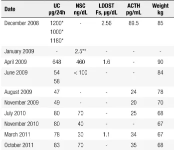
table 1. Hormonal and weight evolution during patient follow-up
date μg/24hUc ng/dLnSc Fs, μg/dLLddSt pg/mLActH Weightkg
December 2008 1200* 1000* 1180*
- 2.56 89.5 85
January 2009 - 2.5** - - -April 2009 648 460 1.6 - 90 June 2009 54
58
< 100 - - 84
August 2009 47 - - 24 78 November 2009 49 - - 20 70 July 2010 80 70 - 25 68 November 2010 80 40 - - 67 March 2011 78 30 1.1 34 67 October 2011 83 70 - 35 68
UC: urinary cortisol, reference 30-300 μg/24h. * Urinary free cortisol, reference 10-90 μg/24h; NSC: nocturnal salivary cortisol, reference < 130 ng/dL; ** Reference < 3.6 nmol/L; LDDST: low-dose dexamethasone supression test; F: serum cortisol; ACTH: reference < 60 pg/mL. To convert values of serum cortisol to nanomoles per liter, multiply by 27.59; urinary cortisol to nmol, multiply by 2.759; plasma ACTH to pmol/liter, multiply by 0.2202.
dIScUSSIOn
Spontaneous remissions in CS and CD are infrequent events that have been already reported more than ifty years ago (14,15). In the subsequent years, some other cases were published and some of them presented re-currence in the follow-up period (7,8,16). However, the cyclic aspect of cortisol secretion that account for 15% of CS patients from several etiologies, mainly CD, are well-described (17). Therefore, these two clinical conditions may overlap, and some of the cases may have been mistakenly described.
Some factors are considered to explain spontaneous remission, principally pituitary apoplexy, an event that has also been enrolled as one of the mechanisms for cyclic CS.
Pituitary apoplexy is more prevalent in other pitu-itary adenoma subtypes. This event is infrequent in CD, occurring mainly in ACTH-secreting macroadenomas (5-11). However, it may also take place in ACTH-se-creting microadenomas (12,13). Pignatta and cols. re-ported a case of CD that showed a 3-mm microadeno-ma next to the pituitary stalk that, after short period of ketoconazol use, presented a sudden clinical picture of pituitary apoplexy with remission of CS and concomi-tant disappearance of pituitary tumor in the follow-up imaging analysis (13). Commonly, pituitary apoplexy in CD patients occurs spontaneously. However, others conditions, such as pituitary radiation therapy (18) or corticotrophin-releasing hormone test (19), were pre-vious correlated with this clinical manifestation.
Cop
yright
© ABE&M t
odos os dir
eit
os r
eser
vados
.
489 Arq Bras Endocrinol Metab. 2013;57/6
experience sudden onset of symptoms that may vary in severity, from headache, with or without endocrine deiciency; visual impairment; ophthalmoplegia; coma; and even death. However, many cases are subclinical or asymptomatic (20), and are suggested by pituitary im-aging analysis, during pituitary surgeries or in necropsy studies. In CD patients, the majority of cases described was symptomatic and submitted to pituitary surgery.
The risk for pituitary infarction may be increased by provocative tests for pituitary reserve assessment, anti-coagulation, oral contraceptive agents, and clomiphene. The susceptibility to apoplexy may also be increased by head trauma, non-pituitary surgery, pregnancy, throm-bocytopenia, and increased intracranial pressure (21).
The case reported here present non-frequent aspects for CD, such as macroadenoma secreting tumor and spontaneous remission without interventions, probably due to asymptomatic pituitary apoplexy. Other similar case was previous reported by Le Nestour and cols., but in a patient with microadenoma. In the follow-up, pitu-itary MRI showed spontaneous T1-weighted hiperin-tensity suggestive of apoplexy, but without any clinical complaints. One year after that, new imaging analysis revealed an empty sella, and the patient achieved spon-taneous CS remission (12).
Natural evolution of some pituitary adenomas is known, particularly microprolactinomas. Nevertheless, this aspect is poorly established in ACTH-secreting macroadenomas.
Finally, several cases of CD that showed initial pitu-itary apoplexy or spontaneous remission presented recur-rence in the follow-up up to seven years after remission (7,8,16,22). Alarii and cols. reported a case of macroad-enoma with some two-year periods of spontaneous re-mission after clinical pituitary apoplexy events (8).
Therefore, careful long-term follow-up is required for patients with CD following remission after pituitary apoplexy, in order to detect hypercortisolism recur-rence or the development of hypopituitarism.
Disclosure: no potential conlict of interest relevant to this article was reported.
REFEREncES
1. Blevins LS Jr, Christy JH, Khajavi M, Tindall GT. Outcomes of the-rapy for Cushing’s disease due to adrenocorticotropin-secreting pituitary macroadenomas. J Clin Endocrinol Metab. 1998;83:63-7. 2. Katznelson L, Bogan JS, Trob JR, Schoenfeld DA, Hedley-Whyte
ET, Hsu DW, et al. Biochemical assessment of Cushing’s disease
in patients with corticotroph macroadenomas. J Clin Endocrinol Metab. 1998;83:1619-23.
3. Woo YS, Isidori AM, Wat WZ, Kaltsas GA, Afshar F, Sabin I, et al. Clinical and biochemical characteristics of adrenocortico-tropin-secreting macroadenomas. J Clin Endocrinol Metab. 2005;90:4963-9.
4. Randeva HS, Schoebel J, Byrne J, Esiri M, Adams CB, Wass JA. Classical pituitary apoplexy: clinical features, management and outcome. Clin Endocrinol (Oxf). 1999;51:181-8.
5. Araya V, Solís I, Lemp M, Oviedo S. Partial remission of hyper-cortisolism in Cushing disease after pituitary apoplexy. A case report. Rev Med Chil. 1998;126:1497-501.
6. Miranda M, Barros L, Knopfelmacher M, Augusto EC, Jacomossi A, Cukiert A, et al. Pituitary apoplexy followed by endocrine re-mission. Report of two cases. Arq Neuropsiquiatr. 1998;56:449-52. 7. Kamiya Y, Jin-No Y, Tomita K, Suzuki T, Ban K, Sugiyama N, et al. Recurrence of Cushing’s disease after long-term remission due to pituitary apoplexy. Endocr J. 2000;47:793-7.
8. Alarii A, Alzahrani AS, Salam SA, Ahmed M, Kanaan I. Repeated remissions of Cushing’s disease due to recurrent infarctions of an ACTH-producing pituitary macroadenoma. Pituitary. 2005;8:81-7. 9. Haboubi H, Azam I, Edavalath M, Redfern RM, Price DE,
Ste-phens JW. Apoplexy in a corticotrophin-secreting pituitary ma-croadenoma: a case report and review of the literature. QJM. 2010;103:607-9.
10. Sahin SB, Cetinkalp S, Erdogan M, Cavdar U, Duygulu G, Saygili F, et al. Pituitary apoplexy in an adrenocorticotropin-producing pituitary macroadenoma. Endocrine. 2010;38:143-6.
11. Messer CK, Fowkes ME, Gabrilove JL, Post KD, Son H, Levine AC. ACTH-producing remnants following apoplexy of an ACTH-secre-ting pituitary macroadenoma. Pituitary. 2012;15 Suppl 1:S6-9. 12. Le Nestour E, Abécassis JP, Bertagna X, Bonnin A, Luton JP.
Si-lent necrosis of a pituitary corticotroph adenoma revealed by timely magnetic resonance imaging: a cause of spontaneous re-mission of Cushing’s disease. Eur J Endocrinol. 1994;130:469-71. 13. Pignatta AB, Díaz AG, Gómez RM, Bruno OD. Spontaneous remis-sion of Cushing’s disease after disappearance of a microadeno-ma attached to the pituitary stalk. Pituitary. 2004;7:45-9. 14. Pasqualini RQ, Gurevich N. Spontaneous remission in a case of
Cushing’s syndrome. J Clin Endocrinol Metab. 1956;16:406-11. 15. Hayslett JP, Cohn GL. Spontaneous remission of Cushing’s
disea-se. Report of a cadisea-se. N Engl J Med. 1967;276:968-70.
16. Ishibashi M, Shimada K, Abe K, Furue H, Yamaji T. Spontaneous remission in Cushing’s disease. Arch Intern Med. 1993;153:251-5. 17. Alexandraki KI, Kaltsas GA, Isidori AM, Akker SA, Drake WM,
Chew SL, et al. The prevalence and characteristic features of cyclicity and variability in Cushing’s disease. Eur J Endocrinol. 2009;160:1011-8.
18. Ohtsuka T, Hanew K, Sugawara A, Goh M, Sato S, Shimizu Y, et al. A case of Cushing’s disease resulting in remission due to pituitary apoplexy during radiation therapy. Nippon Naika Gakkai Zasshi. 1988;77:1095-6.
19. Rotman-Pikielny P, Patronas N, Papanicolaou DA. Pituitary apo-plexy induced by corticotrophin-releasing hormone in a patient with Cushing’s disease. Clin Endocrinol (Oxf). 2003;58:545-9. 20. Onesti ST, Wisniewski T, Post KD. Clinical versus subclinical
pitui-tary apoplexy: presentation, surgical management, and outcome in 21 patients. Neurosurgery. 1990;26:980-6.
21. Vella A, Young W. Pituitary apoplexy. Endocrinologist. 2001;11:282-8. 22. Mercado-Asis LB, Oldield EH, Cutler GB Jr. Pituitary tumor he-morrhage in Cushing disease. Ann Intern Med. 1995;122:189-90.