Cop
yright
© ABE&M t
odos os dir
eit
os r
eser
vados
.
483
case report
Arq Bras Endocrinol Metab. 2013;57/6
Horner syndrome as a
manifestation of thyroid
carcinoma: a rare association
Síndrome de Horner como manifestação clínica de carcinoma da tireoide: uma associação rara
Bernardo Pereira1, Tiago Silva1, Henrique Luiz1, Isabel Manita1,
Luísa Raimundo1, Jorge Portugal1
SUMMARY
An 82-year-old patient presented a progressively growing hard thyroid nodule, and left pto-sis. Additionally, ophthalmologic evaluation revealed ipsilateral miosis, diagnostic indings of Horner syndrome. Computerized tomography revealed a 7.5-cm thyroid mass iniltrating the main neck vessels. Although clinical and imaging data were suggestive of poorly differentiated thyroid carcinoma, ine-needle aspiration led to the diagnosis of papillary carcinoma. Paliative care was proposed to the patient due to the advanced stage of the neoplasm and to signiicant comorbidities. Horner syndrome is an infrequent manifestation of thyroid disorders and be-nign etiologies are more often implied. Malignant thyroid neoplasms represent a rare cause of Horner syndrome. However, an appropriate and prompt diagnosis is paramount for timely treatment of rare thyroid malignancies. Arq Bras Endocrinol Metab. 2013;57(6):483-5
SUMÁRIO
Paciente de 82 anos apresentando-se com nódulo tireoidiano de crescimento progressivo e pto-se palpebral esquerda. O exame oftalmológico revelou ainda miopto-se ipsilateral e achados diag-nósticos de síndrome de Horner. A tomograia computadorizada mostrou massa tireoidiana de 7,5 cm iniltrando os grandes vasos do pescoço. Apesar dos dados clínicos e imagiológicos su-gestivos de um carcinoma pouco diferenciado da tireoide, a citologia aspirativa foi diagnóstica de carcinoma papilar. Em função do estádio avançado da neoplasia e das comorbilidades sig-niicativas, foi proposta para terapêutica paliativa. A síndrome de Horner é uma manifestação clínica infrequente em tumores tireoidianos, estando as condições benignas maioritariamente implicadas. As neoplasias malignas da tireoide representam uma causa rara de síndrome de Horner. Contudo, um diagnóstico adequado e expedito é fundamental para o tratamento atem-pado nos raros casos de malignidade da tireoide. Arq Bras Endocrinol Metab. 2013;57(6):483-5
1 Hospital Garcia de Orta, E.
P. E., Almada, Portugal
Site where the study was carried out: Serviço de Endocrinologia e Diabetologia, Hospital Garcia de Orta, E. P. E., Almada, Portugal
Correspondence to:
Bernardo Pereira
Serviço de Endocrinologia, 8º piso Hospital Garcia de Orta, E. P. E. Av. Torrado da Silva
2801-951 – Almada, Portugal [email protected]
Received on Feb/14/2013 Accepted on Mar/5/2013
INTRODUCTION
H
orner syndrome (HS) is a clinical entity classi-cally characterized by ipsilateral ptosis, miosis, and anhydrosis, being induced by lesions in the ocu-losympathetic tract. Although several etiologies may cause HS, neoplasms are most commonly implicated. In rare circumstances, thyroid disorders may be asso-ciated with HS, and benign diseases predominate (1).We report here a case of HS as the presenting symp-tom of a locally advanced thyroid carcinoma.
CLINICAL CASE
Cop
yright
© ABE&M t
odos os dir
eit
os r
eser
vados
.
484 Arq Bras Endocrinol Metab. 2013;57/6
At physical examination, a hard left cervical nodule ixed to deep structures was noted close to the thoracic inlet. Ophthalmologic evaluation revealed 3.11-mm left ptosis (Figure 1), and right and left pupillary diam-eters measuring 4.9 and 7 mm, respectively, diagnostic indings of HS.
Analytic surveys revealed normocytic normochro-mic anemia (10.8 g/dL, reference: 11.5-18), elevated serum creatinin (2.7 mg/dL, reference: 0.7-1.2) and erythrocyte sedimentation rate (61 mm/s, reference: < 30). She had low free T4 (0.74 ng/dL; reference: 0.93-1.7) but normal free T3 (3.38 pg/mL; reference: 2.57-4.43) and TSH (0.2 mUI/L; reference: 0,1-4), as well as other pituitary hormones, cortisol and IGF-1 (data not shown). Cervical ultrasonography (US) re-vealed a hypoecoic mass in the left thyroid lobe with central vascularization and margins indistinct from posterior structures. US-guided ine needle aspiration (FNA) showed hypercellularity with nuclear overlap-ping, intranuclear inclusions and grooves, diagnostic features of papillary thyroid carcinoma. Cervical com-puted tomography (CT) revealed a 7.5-cm heteroge-neous left thyroid mass with focal necrosis and mac-rocalciications, laterally displacing the trachea (Figure 2), and compressing main neck vessels posteriorly, with margins indistinct from these structures (Figure 3). Left cervical (segments III and IV) and inferior para-tracheal adenopathies were also noted, with no other potential metastasis in thoracic and abdominal regions. Brain magnetic resonance showed recent and past vas-cular ischemic lesions in the frontal cortex that were otherwise unremarkable.
A multidisciplinary team discussion concluded that, due to the locally advanced agressive thyroid carcinoma and the overall clinical context (advanced age, stage 4 chronic renal disease – MDRD formula – and cerebrovascular disease), the patient was not it for any surgical or medical curative approach, and supportive care was proposed.
Figure 1. Left ptosis in Horner syndrome associated with thyroid carcinoma.
Figure 2. CT coronal section: cervical mass with lateral displacement of the trachea and extension to mediastinum.
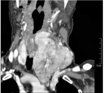
Figure 3. CT axial section: cervical mass displacing laterally the internal jugular vein (white arrow), without distinct margins (focal invasion) from common carotid artery (black arrow).
Horner syndrome and thyroid carcinoma
DISCUSSION
Cop
yright
© ABE&M t
odos os dir
eit
os r
eser
vados
.
485 Arq Bras Endocrinol Metab. 2013;57/6
in superior cervical ganglion, close to the carotid bifur-cation. The third neuron enters the skull in association with the internal carotid artery, passes through the ca-vernous sinus to the orbit and the eye, where it enerva-tes the dilator muscle of the pupil, Mueller´s muscle of eyelids and the lacrimal sac (4).
As in the case of our patient, oculosympathetic tract injuries most often occur in the pregaglionic region (21-67%), with neoplastic disruption being the most common etiology (1). Infrequently, thyroid disorders can induce HS (3,5). Benign etiologies are most commonly implicated (83%), with multinodular goiter being the most frequent reported lesion (1). Thyroid malignancy is a rare cause of HS, with a fre-quency of 0.4-0.9% in the two largest reviews of its etiology (3,5). Although the type of thyroid malig-nancy is often unreported in HS (1,3,5), anaplastic carcinoma has the potential to be more commonly in-volved due to its local aggressiveness (2,6). In our pa-tient, given the clinical and imagiologic data sugges-tive of an aggressive behavior, poorly differentiated thyroid carcinoma was at least likely the cause of neu-rological damage. Failure to diagnose it by US-FNA could be related to aspiration in well-differentiated neoplastic areas.
Physical examination is essential for the diagnosis of HS and pharmacological tests can be performed to con-irm (cocaine and apraclonidine) (7) and localize (hi-droxianphetamine) (3) the lesion. This initial approach identify the level of the lesion in 60-62% of cases (3,8). Cervical CT is a recommended imaging method when investigating HS, and except when obvious causes are suspected (e.g. trauma, surgery), it should always be performed, given the possibility of life-threatening con-ditions, such as occult thyroid carcinoma (9).
Preoperatively, FNA is essential not only for diag-nostic purposes but also for therapeutic decisions. As an example, cytological diagnosis of thyroid lymphoma as the cause of HS should lead to chemotherapy and radiotherapy as irst line approaches (10).
The recognition of thyroid disorders in association with HS should lead to short-term surgical interven-tion, not only to increase the likelihood of neurologic recovery (2,6,11), but also to improve survival, as it is the case of anaplastic thyroid carcinoma (12).
In conclusion, we present a rare case of HS in asso-ciation with a thyroid carcinoma of aggressive behavior.
Horner syndrome and thyroid carcinoma
Although the cause of HS in thyroid disease is fairly of-ten benign, appropriate investigations and timely treat-ment approach are paramount due to the possibility of a rare life-threatening thyroid malignancy.
Acknowledgements: Valeriano Horta Leite, MD, PhD (Depar-tamento de Medicina, Serviço de Endocrinologia, Instituto Por-tuguês de Oncologia de Lisboa, Francisco Gentil, E. P. E.) and Jorge Rosa Santos, MD (Departamento de Cirurgia, Serviço de Cirurgia de Cabeça e Pescoço, Instituto Português de Oncologia de Lisboa, Francisco Gentil, E. P. E.) for providing their extensive clinical experience in Thyroid Oncology. António Alves de Ma-tos, MD (Serviço de Radiologia, Hospital Garcia de Orta, E. P. E.) and Carlos David Santos, MD (Serviço de Anatomia Patoló-gica, Hospital das Clínicas da Universidade Federal do Triângulo Mineiro, Uberaba, MG) for their valuable contribution.
Disclosure: no potential conlict of interest relevant to this article was reported.
REFERENCES
1. Leuchter I, Becker M, Mickel R, Dulguerov P. Horner’s syndrome and thyroid neoplasms. ORL. 2002;64(1):49-52.
2. Harding J, Sywak M, Sidhu S, Delbridge L. Horner’s syndrome in association with thyroid and parathyroid disease. ANZ J Surg. 2004;74(6):442-5.
3. Maloney W, Younge B, Moyer N. Evaluation of the causes and ac-curacy of pharmacologic localisation in Horner’s syndrome. Am J Ophthalmol. 1980;90(3):394-402.
4. Kardon R. Anatomy and physiology of the autonomic nervous system. In: Miller NR, Newman NJ, Biousse V, Kerrison JB, edi-tors. Walsh and Hoyt Clinical Neuro-ophthalmology. 6th ed. Balti-more: Williams & Wilkins; 2005. p. 674-6.
5. Giles C, Henderson J. Horner’s syndrome: an analysis of 216 ca-ses. Am J Ophthalmol. 1958;46(3 part 1):289-96.
6. Yip D, Drachtman R, Amorosa L, Trooskin S. Papillary thyroid cancer presenting as horner syndrome. Pediatr Blood Cancer. 2010;55(4):739-41.
7. Kedar S, Biousse V, Newman N. Horner’s syndrome. In: UpToDate, Brazis P (Ed), UpToDate, Waltham, MA, 2012. Available from: http:// www.uptodate.com/contents/horners-syndrome?source=search_res ult&searc=horner&selectedTitle=1%7E99. Accessed on: Oct 15, 2012. 8. Almog Y, Gepstein R, Kesler A. Diagnostic value of imaging in
Horner syndrome in adults. J Neuro-Ophthalmol. 2010;30(1):7-11. 9. Lee J, Lee H, Lee D, Choi C, Kim S, Suh D. Neuroimaging strate-gies for three types of Horner syndrome with emphasis on ana-tomic location. Am J Roentgenol [serial on the internet] 2007Jan [cited 2012 Dec 29]; 188 (1):w74-w81. Available from: http://www. ajronline.org/content/188/1/W74.long. Accessed on: Nov 7, 2012. 10. Billie J, Wetzel W, Suen J. Thyroid lymphoma with adjacent nerve
paralysis. Arch Otolaryngol. 1982;108(8):517-9.
11. Broome J, Gauger P, Miller B, Doherty G. Anaplastic thyroid can-cer manifesting has new-onset Horner’s syndrome. Endocr Pract. 2009;15(6):563-6.