Identification and analysis of novel virulence-defective
Rhodococcus fascians mutants
Adriana Sofia Silva Cabecinhas
Thesis submitted in partial fulfillment of the requirements for the degree of Master of Applied Biotechnology
Promotor: Doctor Danny Vereecke Co-Promotor: Doctor Américo Rodrigues
ii
Title: Identification and analysis of novel virulence-defective Rhodococcus fascians mutants
Copyright © Adriana Sofia Silva Cabecinhas
A Escola Superior de Turismo e Tecnologia do Mar e o Instituto Politécnico de Leiria têm o direito, perpétuo e sem limites geográficos, de arquivar e publicar esta dissertação através de exemplares impressos reproduzidos em papel ou de forma digital, ou por qualquer outro meio conhecido ou que venha a ser inventado, e de a divulgar através de repositórios científicos e de admitir a sua cópia e distribuição com objectivos educacionais ou de investigação, não comerciais, desde que seja dado crédito ao autor e editor.
This work was conducted at the Laboratory of Applied in vitro Plant Biotechnology at the Campus Schoonmeersen of the Ghent University.
Part of the work was also conducted at the department of Plant Systems Biology (PSB) of the Flanders Institute for Biotechnology (Vlaams Instituut voor Biotechnologie - VIB).
iii
Acknowledgments
First of all, thanks a lot to Danny Vereecke, my scientific mum, to be not only a spectacular promotor, that guided and supported me through all the different stages of the thesis, but also a remarkable touristic guide, gastronomic advisor and a big friend. Thanks a lot for everything that you taught me, for all the input that you have asked from me and, specially, for all the effort that you have put into my thesis and to always believe in me. Thanks to Katrien to always have a big smile, to annoy me when I was trying to work and to try to put everyone speaking in English and encourage me to give it a try on the Dutch. Thanks to Joris to improve the quality of my photos and to bake cakes (or at least promise to). Thanks Nina that was in Belgium for a short time but was a big support for me. Thanks to the girls (Halima, Salima, Nandini and Mirni) for the support when I was really busy and tired, the company, the funny moments and to allow me to try a bit of your culture. Thanks Stefaan to let me working in your lab and for the relaxing effect that you have by always saying that “it is ok” even when it is not.
Thanks to Stephen to allow me work at your bench in VIB and be always available to help. Thanks a lot to Annick for the advice in the clonings, to take care of me when I had to work with the radioactive compounds and to help me at any time. Thanks Belén, not only for the company at weird hours at VIB but also to be always with a smile and able to help me even in the most busy days. Thanks to Pieter for the “ai, ai, ai…” and the nice conversations in the lab. Thanks to Hans and Stephanie because the party is also an important part of the work. Thanks to the Portuguese people that I met here. You made me feel at home in a country that is not ours. Thanks to Maria’s (Fonseca and Fuller, my adopted mum), my two pillars that kept me straight and never allowed me to fall. Thanks for the awesome company, the nice food and all the support. Thanks to my Erasmus friends (José, Inês, Sónia, Vicky, Olívia), it would definitely not have been the same without you. A special thank to Vicky for being the exception.
Obrigada aos Doutores Américo Rodrigues, pelo apoio prestado ao longo deste trabalho, e Marco Lemos, pela permanente disponibilidade. Por último, mas não menos importante, obrigada aos meus pais pois sem eles não estaria aqui. Obrigada mãe por não ser preciso uma palavra para me entenderes. Obrigada pai pelo apoio. Obrigada aos dois por me terem
iv
dado fortes raízes mas também por não me terem cortado as asas. Um grande obrigada ao João, meu amigo, companheiro e confidente. Obrigada por seres o meu porto de abrigo, me dares todo o apoio, acreditares em mim e teres sempre uma palavra positiva para me dizer. Obrigada manas por, cada uma à sua maneira, me darem força todos os dias para continuar. Obrigada Nádia pelas estrelinhas que me guiaram ao longo de todo o caminho e por estares sempre disposta a ouvir-me. Obrigada padrinhos pela ajuda, obrigada avó pelos miminhos e obrigada tios e primos pelo amparo. Obrigada amigos (Serguei, Dani, SaSilva, Rute, Tilui, Rita Pascoal, Joana Patriarca e Sarah Azinheiro) por terem sempre disponibilidade para mim e por nada mudar mesmo após meses de ausência.
Words do not come easy to me and it is impossible to say all the names that were important in this process. I will always be grateful to all of you.
Dank u wel Muito obrigada
v
Abstract
The phytopathogenic actinomycete Rhodococcus fascians induces a disease, known as leafy gall, characterized by the induction of multiple shoots, on a broad variety of dicotyledonous herbaceous plants. The main pathogenicity factor of the bacterium is the production of a mixture of 6 cytokinins encoded by the genes of the fas operon which is located on a linear virulence plasmid pFiD188. This research aimed to analyze two novel virulence loci of the linear plasmid: GT1 encoding a glycosyltransferase and the highly homologous mtr1 and mtr2 genes encoding SAM-dependent methyltransferases.
Previous data showed that the glycosyltransferase mutant 21D5 had a modified colony morphology and formed aggregates in liquid cultures. We established that these characteristics did not affect growth on rich media, but lead to the inability to proliferate under nutrient deprivation, strongly impacting epiphytic fitness. The latter was illustrated by the strongly attenuated virulence of 21D5, which was accompanied by a defective expression of the essential virulence genes fas and att, and the consequent reduced capacity to invade plant tissues and produce virulence factors. GT1 expression proved to be induced by compounds that accumulate in plants very early upon infection, placing the GT1 function at the onset of the interaction.
Just like R. fascians, Streptomyces turgidiscabies carries a fas operon and two associated mtr genes. Mutants in these mtr’s in R. fascians have lost all ability to provoke symptoms, but still produce 2MeS-cytokinins, implying that other methylated cytokinins are imperative for disease induction. By analysing the cytokinin profiles via TLC of a set of R. fascians strains grown under fas-inducing conditions and of S. turgidiscabies fed with 14
C-labelled SAM and adenine we tried to uncover the elusive reaction products of the MTRs. Unfortunately, no fas- or mtr-dependent compounds were detected in the supernatants. By determining the expression pattern of the mtr genes in vitro and in planta it became clear that the regulatory aspects of these genes are very complex, limiting their expression to R. fascians cells that reside on the host. To open the path to the identification of the MTR reaction products, we succeeded in developing an assay in which the in planta expression can be brought in vitro so that the labelling experiments can be repeated.
vii
Resumo
Rhodococcus fascians é uma actinomiceta fitopatogénica que induz uma doença, conhecida como irritação frondosa, caracterizada pela indução de múltiplos rebentos, numa vasta gama de plantas herbáceas dicotiledóneas. O principal factor de patogenicidade da bactéria é a produção de uma mistura de 6 citoquininas codificadas pelos genes do operão fas que está localizado num plasmídeo linear associado à virulência, pFiD188. Este trabalho teve como objectivo a análise de dois novos loci deste plasmídeo associados à virulência: GT1 que codifica uma glicosiltransferase e os genes mtr1 e mtr2, grandemente homólogos, que codificam metiltransferases dependentes de SAM.
Trabalhos prévios com 21D5, mutante na glicosiltransferase, mostraram que possui uma morfologia de colónia modificada e forma agregados em culturas líquidas. Neste trabalho demonstrou-se que essas características não afectam o crescimento em meio de cultura rico, mas levam à incapacidade de proliferação em condições de privação de nutrientes, tendo um impacto forte na competência epifítica. Este impacto foi demostrado pela atenuação severa da virulência em 21D5, que foi acompanhada de uma expressão alterada dos genes fas e att, essenciais para a virulência, e consequente redução da capacidade de invasão dos tecidos da planta e de produção dos factores de virulência. Demonstrou-se também que a expressão de GT1 é induzida por compostos que são acumulados em plantas nas fases iniciais da infecção, colocando a função de GT1 no começo da interacção.
Tal como R. fascians, Streptomyces turgidiscabies possui um operão fas e dois genes mtr associados. R. fascians mutantes nestes genes mtr’s perderam a capacidade de provocar sintomas, mas produziram 2MeS-citoquininas, implicando que outras citoquininas metiladas são cruciais para a indução da doença. De modo a identificar os produtos de reacção das MTRs procedeu-se à análise do perfil de citoquininas de R. fascians em condições que induzem a expressão dos genes do operão fas e de S. turgidiscabies alimentados com SAM e adenina marcadas com 14C em TLC. No entanto, não foi possível a identificação de compostos dependentes de fas ou mtr nos sobrenadantes. Pela determinação do perfil de expressão dos genes mtr in vitro e in planta tornou-se claro que a regulação destes genes é muito complexa, sendo a sua expressão limitada a células de R. fascians que colonizam o hospedeiro. Para possibilitar a identificação dos produtos de recção de MTR, desenvolveu-se um protocolo que permite a expressão in planta em condições in vitro, o que permitirá a repetição dos ensaios de marcação com 14C.
viii Part of this work is published in:
2014 - Adriana Cabecinhas, Petr Tarkowski, Ine Pertry, Stefaan Werbrouck, and Danny Vereecke “Methylated cytokinins: the secret weapon of the phytopathogen Rhodococcus fascians?” – Auxins and cytokinins in plant development, International Symposium
ix
Table of contents
Abstract ... v
Resumo ... vii
1. Introduction ... 1
A. Rhodococcus fascians, causative agent of the leafy gall syndrome ... 1
B. The molecular basis of the phytopathogenicity of R. fascians strain D188 ... 4
C. Glycosyltransferase (GT1), a novel player in the pathology of R. fascians ... 9
D. Two methyltransferases, intriguing members of the fas locus ... 13
2. Aim of this work ... 17
3. Materials and methods... 19
3.1 Bacterial strains and growth conditions ... 19
3.2 Determination of the colony morphology ... 21
3.3 Plant material and growth conditions... 21
3.4 Infection procedures ... 22
3.5 SEM analysis ... 23
3.6 Preparation of plant extracts ... 23
3.7 Transformation procedures and plasmid preparation ... 23
3.8 In vitro expression analysis ... 24
3.8.1 att expression ... 24
3.8.2 fas, GT1, mtr1 and mtr2 expression ... 25
3.9 In planta expression analysis ... 26
3.9.1 Quantitative in planta expression analysis ... 26
3.9.2 Histochemical in planta expression analysis ... 26
3.10 In planta survival ... 27
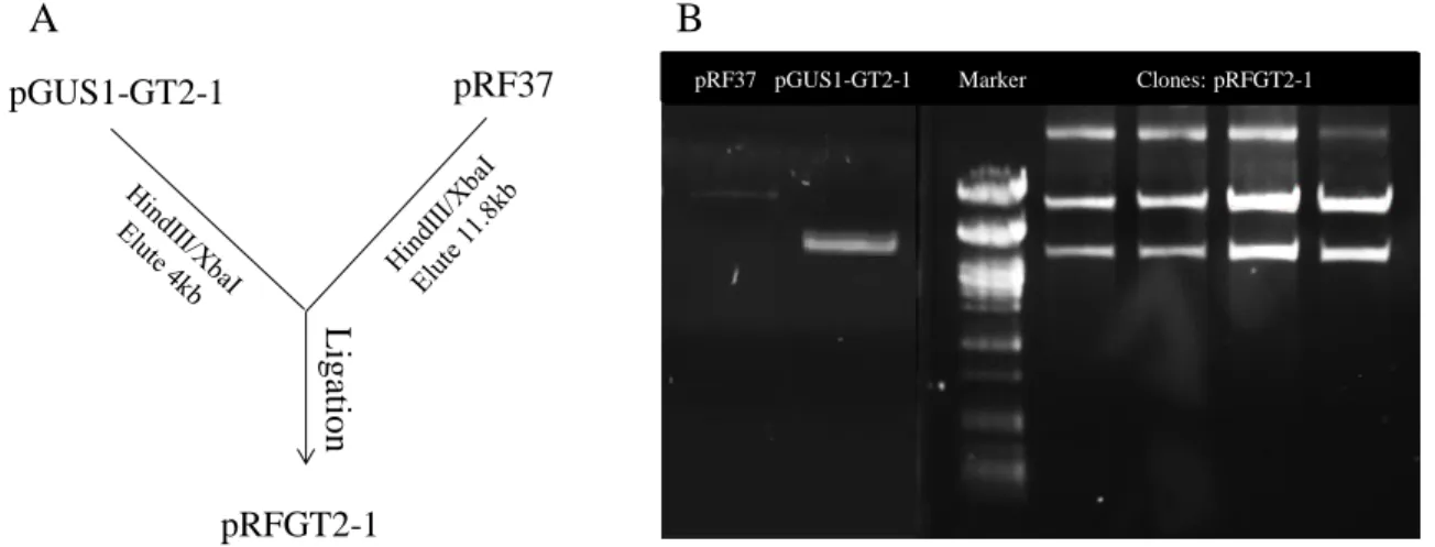
3.11 Cloning of GT1:GUS fusions ... 27
3.12 TLC analysis of bacterial cytokinins ... 28
4. Results ... 31
A. Functional analysis of the glycosyltransferase GT1 ... 31
4.A.1. The colony morphology of R. fascians is defined by the medium composition and modified in the GT1 mutant 21D5 ... 31
4.A.2. Growth capacity of different R. fascians strains on different media ... 34
4.A.3. Mutations in GT1 seriously impair the virulence of R. fascians ... 35
4.A.4. The regulation of fas gene expression is impaired in vitro and in planta affecting the level of cytokinin production by 21D5 ... 41
4.A.5. Impaired att gene expression affects the in planta production of the Att compound by 21D5 .... 43
4.A.6. 21D5 is unable to persist on the plant host... 45
4.A.7. Expression of GT1 is possibly induced by carbon sources that accumulate in infected plants and might be controlled by AttR ... 48
B. Methylated cytokinins: the secret weapon of R. fascians? ... 50
5. Discussion ... 57
6. Summary and perspectives ... 61
7. References ... 65
xi
List of figures
Figure 1.1 Typical growth of R. fascians on YEB and symptoms on Nicotiana tabacum infected
with the wild-type strain D188 at 28 dpi ... 2
Figure 1.2 Representation of the linear plasmid of R. fascians strain D188 ... 5 Figure 1.3 Organization and homologies of the fas and att loci located in region U1 of pFiD188,
the linear plasmid of R. fascians strain D188 ... 5
Figure 1.4 Representation of the molecular processes occurring after the infection of a host plant
by R. fascians ... 7
Figure 1.5 Representation of the cytokinin biosynthetic pathway encoded by the fas operon of
R. fascians D188 ... 8
Figure 1.6 Organization and homologies of the GT locus genes in R. fascians ... 10
Figure 1.7 The protein structure of Escherichia coli MurG, a typical B-fold GT, in complex with
its donor substrate, UDP-GlcNAc ... 11
Figure 1.8 Sequence alignment of selected GT-B superfamily members ... 12 Figure 1.9 Organization of the fas loci and induced symptoms by R. fascians strain D188 and S.
turgidiscabies ... 14
Figure 3.1 Cloning strategy for the generation of a construct carrying a transcriptional fusion
between GT1 and GUS in pRF37, a bifunctional vector for E. coli and R. fascians ... 28
Figure 4.1 Colony morphology of the wild-type R. fascians strain D188, its plasmid-free derivative
D188-5 and the transposon mutant 21D5 grown on different media ... 33
Figure 4.2 Overview of the growth of the wild-type R. fascians strain D188 and the transposon
mutant 21D5 ... 34
Figure 4.3 Symptoms on A. thaliana plants inoculated with different R. fascians strains ... 36 Figure 4.4 Axillary activation in A. thaliana plants inoculated with different R. fascians strains . 37 Figure 4.5 Kinetics of symptom development on A. thaliana plants inoculated with different
R. fascians strains ... 38
Figure 4.6 Symptom formation on decapitated N. tabacum xanthi plants when they were 4-week
old inoculated with different R. fascians strains ... 39
Figure 4.7 Symptoms on wounded or intact excised leaves of 4-week old N. tabacum xanthi plants
inoculated with different R. fascians strains ... 40
Figure 4.8 Expression of fasA and cytokinin production in different R. fascians strains ... 42 Figure 4.9 Expression of the attA gene and Att compound production in different R. fascians
xii
Figure 4.10 Colonization capacities of R. fascians strains D188-5, 21D5 and D188 ... 45 Figure 4.11 Epiphytic colonization of excised tobacco leaves by R. fascians strain D188 and 21D5
5 weeks after infection ... 47
Figure 4.12 Effect of different conditions on in vitro GT1 expression measured in
D188(pRFGT2-1) with MUG as a substrate ... 49
Figure 4.13 Effect of different regulators on in vitro GT1 expression measured from the reporter
plasmid pRFGT2-1 in different R. fascians strains with MUG as a substrate ... 50
Figure 4.14 Presence of the T-N11-A motif in the upstream sequence of the pFi_135_136 operon
... 50
Figure 4.15 Preliminary data on the cytokinin production of different R. fascians strains ... 51
Figure 4.16 Thin layer chromatography analysis of 14C-adenine- and 14C-SAM-labelled
compounds produced by different R. fascians and S. turgidiscabies strains ... 52
Figure 4.17 Thin layer chromatography and UV-fluorescence analysis of 14C-adenine- and 14
C-SAM-labelled compounds produced by different R. fascians and S. turgidiscabies strains ... 52
Figure 4.18 Effect of different conditions on in vitro mtr1, mtr2, and fasA expression measured in
D188-mtr1, D188-mtr2, and D188(pJDGV5) ... 53
Figure 4.19 In planta expression of fasA, mtr1 and mtr2 using different assays ... 54 Figure 4.20 Bringing in planta expression of the mtr1 gene to an in vitro system ... 55 Figure 8.1 Colony morphology of the wild-type R. fascians strain D188, the attenuated mutant
D188-att1, and the non-pathogenic mutant D188-fas1 grown on different media ... 71
Figure 8.2 Colony morphology of the wild-type R. fascians strain D188 and three GT1 transposon
mutants 45F10, 41H2 and 40G10, grown on different media ... 72
Figure 8.3 Overview of significant changes in the primary metabolism upon infection with
xiii
List of tables
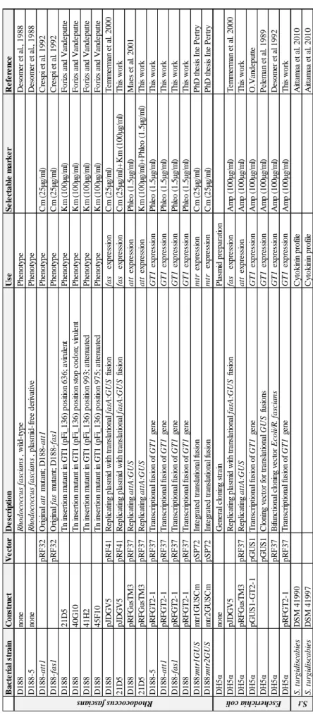
Table 3.1 Characteristics of the bacterial strains used in this work ... 20 Table 4.1 Relative growth of different R. fascians strains on different media based on CFU’s ... 35 Table 4.2 Preliminary data on the cytokinin content of the supernatant of different R. fascians
xv
List of frequently used abbreviations
2-iP 2-isopentenyladenine 2MeS Methylthio-derivatives Amp Ampicillin att Attenuation CKX Cytokinin oxidase/dehydrogenase Cm Chloramphenicol cZ cis-zeatin
DNA Deoxyribonucleic acid dpi Days post-infection fas Fasciation GT Glycosyltransferase GUS β-Glucuronidase His Histidine IM Induction medium Km Kanamycin LB Luria broth
MES 2-(N-morpholino)ethanesulfonic acid MTR Methyltransferase
OD Optical density Phleo Phleomycin Pyr Pyruvate
SAM S-adenosyl methionine TLC Thin layer chromatography tZ trans-zeatin
1
1. Introduction
Organisms living together in ecosystems can interact with each other in different ways. Mutualism is the interaction where both organisms benefit from the relation. Commensalism benefits one organism and does not bring advantage or disadvantage for the other. Pathogenic interactions benefit one of the organisms (the pathogen) and is disadvantageous for the other (the host) (Rédei, 2008). Pathogens can be differentiated according to the mechanism used to obtain nutrients. Necrotrophic pathogens kill the host or the cells of the invaded tissues through the secretion of toxic compounds. Biotrophic organisms extract nutrients directly from living cells (Dyakov et al., 2007).
Pathogens that affect plants are called phytopathogens. Those interactions have important economic implications since diseased plants are unfit for consumption or ornamental purposes (Depuydt et al., 2008). Bacterial interactions with plants can be compatible or incompatible. Compatible interactions happen when the bacteria infect susceptible host plants and are able to cause disease symptoms (Collmer, 1998). Incompatible interactions occur when the bacteria are not able to cause symptoms, because they cannot invade the plant tissues or because the plant secretes compounds that are toxic for the bacterium or that interfere with the pathogen’s virulence mechanism.
A. Rhodococcus fascians, causative agent of the leafy gall syndrome
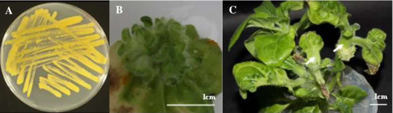
R. fascians is a phytopathogenic Gram-positive, rod-shaped, aerobic, nonmotile, and nonsportulating bacterium that forms orange colonies (Pertry et al., 2010; Stes et al., 2011; Tilford, 1936; Vandeputte et al., 2005) (Figure 1.1.A). The taxonomic position of the bacterium was a matter of debate for several years. Based on phenotypic, biochemical and genetic data the phytopathogen was positioned within the Actinomycetales in the genus Rhodococcus (Goodfellow, 1984). Bacteria from the genus Rhodococcus are known because of their medical, environmental and industrial implications (Gürtler et al., 2004). Despite the advances in the study of the genus and reports referring to plant-association, R. fascians is the only pathogenic member (Stes et al., 2011).
When infecting plants, R. fascians induces the development of leafy galls (Figure 1.1.B), structures consisting of clusters of small often deformed shoots of which further outgrowth
2
is inhibited (Crespi et al., 1992; Vereecke et al., 2000). Generally, plant tissues first grow by cell division and then by cell expansion (Donnelly et al., 1999), but in the formed structures there is continuous growth through cell proliferation (Depuydt et al., 2009a; Vereecke et al., 2002b). Although the important role of cytokinins in the development of those symptoms is established, there is evidence that other hormones are involved as well (Depuydt et al., 2008; Simón-Mateo et al., 2006). Other gall-inducing pathogens, such as bacteria of the genera Agrobacterium, Pantoea, and Pseudomonas, cause the proliferation of undifferentiated tissues (Pitzschke and Hirt, 2010). R. fascians is the only hyperplasia-inducing bacterium that causes differentiated galls (Vereecke et al., 2000), hyperplasia-inducing the formation of shoots from existing and newly formed meristems (de O Manes et al., 2004). The leafy galls represent a niche with the perfect conditions for the survival and maintenance of the pathogen (Bailey et al., 2009; Hayward and Stone, 2005). Other symptoms observed in plants infected with R. fascians are the deformation of the leaves and swelling of the vascular tissues (Stes et al., 2011) (Figure 1.1.C). Symptom development (time to appear and aggressiveness) depends on the infection success, which is influenced by the initial amount of bacteria, the susceptibility of the plant, and the general efficiency of the interaction between the plant and the bacterium.
Figure 1.1 Typical growth of R. fascians on YEB and symptoms on Nicotiana tabacum
infected with the wild-type strain D188 at 28 dpi.
(A) R. fascians grown on YEB; (B) Leafy gall on infected leaf (ISO 100, Diaphragm 16 and closing time 1/100sec); (C) Leaf deformation and swelling of the vascular tissue (arrow) on decapitated plants (ISO 100, Diaphragm 32 and closing time 2,5sec).
With the increase of transport of ornamental plants, this pathogen has become a worldwide threat in horticulture, leading to significant economic losses (Depuydt et al., 2008; Putnam and Miller, 2007). Infections have been recorded throughout the world but are mainly present in temperate regions (CBI, 2006) The reported host range of R. fascians encompasses 164 different plant species belonging to 43 families, mono- and
3
dicotyledonous, but this number is most probably an underestimation of the pathological potential of the species (Depuydt, 2008). Few reports describe plants that are resistant to the morphologic alterations caused by the bacteria. For instance, Dalbergia pervillei synthesizes a compound - perbergin - that is able to inhibit the expression of the virulence genes of the bacteria (Rajaoson et al., 2011).
The bacteria can stay on the plant surface for months before symptoms become visible (Cornelis et al., 2001). Therefore, the propagation of contaminated material is the main transmission way in horticultural practices (Miller et al., 2007). Moreover, the phytohormones that are extensively used in horticulture for plant propagation induce morphological changes that are similar to the ones caused by R. fascians. In that way, infected symptomatic plants can be mistaken for plants deformed by the exogenous hormones. Moreover, other pathogens or herbicides can cause R. fascians-like deformations (Miller et al., 2008; Putnam and Miller, 2006). Altogether, because of these complications, it is likely that the problem with R. fascians in horticulture is underestimated and a good diagnosis combined with strict sanitary measures are imperative to prevent infection spread to other plants (Miller et al., 2007).
Proper diagnosis of the plants is based on the combination of different methods. The traditional method to isolate the bacteria from the plant has proven to be difficult and can lead to false negative results. Additionally, non-pathogenic strains can be obtained as well and, therefore, the pathogenicity of the isolates has to be confirmed by molecular methods which are time consuming, yet extremely sensitive (Lacey, 1961). Pathogenicity can also be established by infecting a plant from the same species as the original host (Putnam, 2014; Vereecke et al., 2000). Finally, serological tests, based on the detection of fluorescent antibodies, are used but those methods are laborious and can raise false results (de Boer et al., 1988). Adding to the complexity, recent work developed at the Oregon State University indicated that the causative agent of the leafy gall syndrome is not a single organism, but two genetically very different bacterial species (Putnam, 2014).
Some plants do not react to the phytohormone-based methods used in in vitro propagation and hence cannot be propagated artificially (Kevers et al., 2004). Since R. fascians triggers vegetative multiplication through the action of phytohormones, this pathology also has a putative positive side and might be useful in different plant research areas. A first line of
4
research is to understand the role of plant hormones in regular plant development. Currently this question is addressed by using plant mutants or exogenous hormones. An alternative approach is to use R. fascians as a source of phytohormones that are delivered directly into the plant tissues in a much more controlled and biologically relevant concentration than exogenous addition (Depuydt et al., 2009a). The direct application of R. fascians in in vitro technologies still lies far ahead because the leafy gall syndrome is the result of a complex interaction between the pathogen and its host, and therefore it is very difficult if not impossible to mimic the shoot formation induced by R. fascians by exogenous hormone addition. Leafy galls induced by R. fascians bacteria on diverse medicinal plants have been reported to generate intact plants without phenotypical abnormalities upon repeated antibiotic treatments (Vereecke et al., 2000). However, it is difficult to convince plant growers to use a phytopathogenic bacterium to propagate their plants. Nevertheless, it is worthwhile to continue exploring the possibilities of implementing R. fascians and/or its morphogens in tissue culture practices since the very broad host range of the pathogen suggests that it might bring solutions for recalcitrant plants (Tarkowski and Vereecke, 2014). Consequently, it is imperative to complete the knowledge on the mechanism of leafy gall development and to get insight into putative novel virulence factors (Depuydt et al., 2008).
B. The molecular basis of the phytopathogenicity of R. fascians strain D188
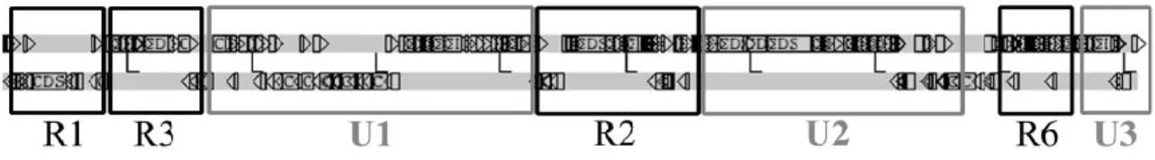
A common strategy in plant-associated microbes is the secretion of phytohormones (Francis et al., 2010). Generally the genes responsible for hormone production are on large circular plasmids (Nester and Kosuge, 1981), but in R. fascians they are uniquely encoded by a linear plasmid (Francis et al., 2007). The wild-type R. fascians strain D188 has a genome consisting of a circular chromosome and two plasmids, one linear, pFiD188, associated with the pathogenicity, and one circular, pD188, related with cadmium resistance (Crespi et al., 1994; Desomer et al., 1988). Two different regions can be considered in the linear plasmid: R regions conserved in other rhodococcal linear plasmids, involved in the plasmid maintenance functions, and U regions unique to R. fascians (Francis et al., 2012) (Figure 1.2).
5
Figure 1.2 Representation of the linear plasmid pFiD188 of R. fascians strain D188.
Conserved (R) and unique (U) regions are indicated (Francis et al., 2012).
Unique region U1 is the largest and contains the loci attenuation (att) and fasciation (fas), described later in this section (Figure 1.3), and hyp, involved in post transcriptional control of virulence-related genes (Crespi et al., 1992; Maes et al., 2001; Rajaoson et al., 2011). Since region U1 contains all genes encoding the known virulence factors and pathogenicity regulators, this region is considered to be the main pathogenicity region on pFiD188. So far, no virulence-associated genes have been identified in the unique regions U2 en U3.
Figure 1.3 Organization and homologies of the fas and att loci located in region U1 of
pFiD188, the linear plasmid of R. fascians strain D188 (Stes et al., 2011).
Infection of a host starts with an epiphytic colonization. R. fascians has several characteristics that are beneficial for a successful establishment of the interaction. For instance, the orange carotenoid pigments (Figure 1.1.A) provide protection against ultraviolet radiation during the establishment of epiphytic colonies on the stems and leaves of the host (Ichiyama et al., 1989; Sundin and Jacobs, 1999). R. fascians produces the hormone indole-3-acetic acid (IAA), an auxin that triggers nutrient release from and suppresses the defense responses mounted by plant cells (Cornelis et al., 2001; Depuydt et al., 2009a; Vandeputte et al., 2005). The formation of a biofilm that is surrounded by a slime layer, probably composed by exopolysaccharides, protects the bacterial cells against environmental conditions and improves the attachment to the plant (Cornelis et al., 2001; Miller et al., 2007; Pertry et al., 2010). All these features are encoded by chromosomal
6
genes because linear plasmid-free bacteria have similar colonization capacities as the wild-type strain (Cornelis et al., 2001).
During the epiphytic colonization the primary metabolism of the host changes. In this process the plant increases the production of carbon and nitrogen sources, which is thought to facilitate the bacterial colonization (Depuydt et al., 2009a). The bacteria sense these metabolic changes of the plant and perceive it as a signal that a compatible host has been found (Maes et al., 2001). The active role of the host in the infection process is very important and complex. However, since the focus of this work is the phytopathogen and not the plant, we will not elaborate on the plant side of the interaction. Information about this topic can be found in recent review articles (Stes et al., 2013; Stes et al., 2011).
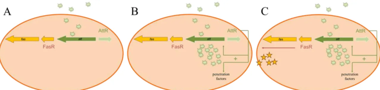
When a specific combination of carbon and nitrogen sources is detected, att gene expression is activated in R. fascians (Figure 1.4.A). This expression is controlled by AttR, a LysR-type transcriptional regulator (Stes et al., 2011). The att operon is essential at the onset of the infection and the first locus to be expressed in this process (Cornelis et al., 2002). An initially low concentration of the att compound is sufficient to activate the expression of the att biosynthetic genes. With this positive feedback mechanism, the production of high amounts of the att compound is established (Figure 1.4.B)(Stes et al., 2013). Such an autoregulatory mechanism resembles the quorum sensing classical concept, a phenomenon that allows the cells to act as a community rather than as single cells. In R. fascians the att autoregulatory mechanism is not considered as genuine quorum sensing since the presence of the plant is required to activate the expression and there is no cell density-dependent production of the regulatory compound (Maes et al., 2001). The att regulatory compound is responsible for the activation of the fas operon (Figure 1.4.C). This operon encodes the genes responsible for the synthesis of the cytokinins and will be described later (Depuydt et al., 2008). The att knockouts require wounding of the plant tissues to infect the plant and therefore it is believed that the regulatory compound also affects, directly or indirectly, the entrance of the bacteria into the plant tissues. Although the exact mechanism is unknown, it is hypothesized that the att compound itself breaks the plant cuticula by phytotoxicity or activates the expression of plant cell wall degrading enzyme genes (Stes et al., 2011). As soon as the bacteria engage in an endophytic stage, att expression is switched off (Cornelis et al., 2002).
7
Figure 1.4 Representation of the molecular processes occurring after the infection of a
host plant by R. fascians.
(A) After contact with a compatible host, the expression of the att genes is increased and the att compound is produced; (B) the accumulation of the att compound triggers the formation of penetration factors and stimulates the production of high amounts of the att compound through a positive feedback; (C) the att compound triggers the expression of the fas genes, responsible for the symptom development (Modified from Stes et al., 2013).
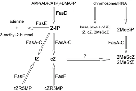
The main pathogenicity strategy of R. fascians is the production of shoot-inducing cytokinins, triggered by the expression of fas operon genes (Stes et al., 2010) (Figure 1.3). fas expression is tightly controlled and involves the AttR regulator and FasR, an AraC-type transcriptional regulator (Temmerman et al., 2000) (Figure 1.4.C). In planta, the fas genes are active both in the epiphytic and the endophytic populations (Cornelis et al., 2002). Through biochemical data and the analysis of cytokinin profiles it was possible to understand that the Fas proteins lead the synthesis of six distinct cytokinins: 2-isopentenyladenine (2-iP), cis-zeatin (cZ), trans-zeatin (tZ) and their methylthio-derivatives – 2MeSiP, 2MeScZ and 2MeStZ (Figure 1.5) (Pertry et al., 2009). In contrast to earlier ideas, the virulence depends more on the cytokinin concentration and ratio of the different compounds that the host is exposed to, than on the production of specialized molecules by the bacterium (Pertry et al., 2009). FasD, an isopentenyltransferase, is the key enzyme in the process. This enzyme synthesizes 2-iP which is the precursor for the other Fas enzymes (Pertry et al., 2009). FasE is a cytokinin oxidase/dehydrogenase (CKX) with a strong affinity for 2-iP-type cytokinins and thought to be required for the optimal functioning of the FasD enzyme. FasA is a putative P450 monooxygenase that hydroxylates 2-iP to form tZ and cZ. This enzyme can also hydroxylate 2MeSiP, allowing the formation of 2MeScZ and 2MeStZ. FasA is absolutely required for the virulence of the phytopathogen (Crespi et al., 1994). FasB and FasC are homologous to the α- and β-subunit of pyruvate decarboxylase, respectively, and are thought to be accessory proteins of FasA providing the required electrons (Crespi et al., 1994; Goethals et al., 2001). The enzymes involved in the direct methylthiolation of 2-iP, tZ, and cZ to produce their 2MeS
8
derivatives remain to be identified. FasF is a phophoribohydrolase and represents a complementary pathway for the formation of Z-type cytokinins. This enzyme directly releases cytokinin bases from their nucleotide precursors and is crucial for symptoms maintenance (Figures 1.3 and 1.5) (Stes et al., 2011).
Figure 1.5 Representation of the cytokinin biosynthetic pathway encoded by the fas
operon of R. fascians D188 (Stes et al., 2011).
The production of a mixture of cytokinins instead of the production of large amounts of a single one has several advantages. In plants, the CKX enzymes are responsible for the degradation of cytokinins and cytokinin homeostasis. Previous work showed that each CKX enzyme has a particular substrate specificity and, consequently, the capacity of different plant tissues and species to degrade specific cytokinins can differ (Galuszka et al., 2007). In Arabidopsis the activation of the CKX enzymes effectively reduces the in planta levels of most of the bacterially produced cytokinins, but they are unable to efficiently degrade cZ and 2MeScZ. The CKX enzymes of Nicotiana tabacum, however, are ineffective against 2-iP (Pertry et al., 2009). Thus, with the strategy of producing a cytokinin mixture, some bacterial cytokinins will always be able to accumulate in the infected plant tissues. The R. fascians cytokinin mixture also modulates the sensitivity of the plant for those morphogens. In Arabidopsis, when infection by the bacteria occurs, the secreted cytokinins are perceived by two of the three cytokinin receptors, the ARABIDOPSIS HISTIDINE KINASES (AHK). At the onset of the interaction, 2-iP
9
strongly accumulates and the upregulation of the AHK3 and especially AHK4 receptor genes occurs. Consequently, even with a relatively low amount of secreted cytokinins, the cytokinin sensitivity of the plant is the highest when symptoms need to be initiated (Pertry et al., 2009; Pertry et al., 2010). Finally, adding to the efficiency of producing a cytokinin mixture, the equimolar mix of the six cytokinins proved to have a stronger effect than an equal final concentration of the individual molecules in different bioassays (Pertry et al., 2009; Stes et al., 2011). R. fascians produces its cytokinins in a very controlled and dynamic way. In the initiation process it secretes mainly high amounts of 2-iP, tZ and cZ and later it produces a maintenance flow consisting of tZ, 2MeStZ, cZ and 2MeScZ (Pertry et al., 2010).
During the endophytic colonization, the bacteria undergo a series of adaptations. In order to adapt to the new environment, the intercellular spaces of plant tissues, their cell wall is modulated (Cornelis et al., 2001) or lost (Lacey, 1961) and their metabolism shifts to the use of C2 compounds (Forizs et al., 2009; Vereecke et al., 2002a; Vereecke et al., 2002b). While the epiphytic bacterial subpopulation triggers the onset of symptoms development, the maintenance of the symptoms depends on the endophytic population (Stes et al., 2011).
C. Glycosyltransferase (GT1), a novel player in the pathology of R. fascians
The molecular identification of the R. fascians virulence determinants started in 1988 with the elimination of both plasmids from strain D188 (Desomer et al., 1988). Recently, a large collection of mutants from this strain has been generated by transposon mutagenesis in the laboratory of Professor Jaziri at the Université Libre de Bruxelles. R. fascians shifts its metabolism during the interaction with plants from the Krebs cycle to the glyoxylate shunt, which allows the bacteria to grow on C2 compounds that are thought to be enriched in infected tissues (Forizs et al., 2009). Due to this link between the glyoxylate shunt and virulence, the obtained mutants were screened for their ability to grow on acetate. Twenty six mutants out of approximately 1400 screened, were unable to grow on acetate, although their growth on rich medium was not seriously affected. From those, strain 21D5 was the only mutant that could not induce malformations on infected plants. The mutation in 21D5 was mapped on pFiD188 (Forizs, 2012).
10
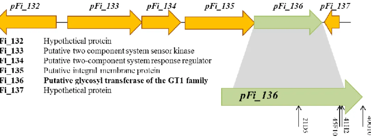
gene of unique region U2 of pFiD188 (Forizs, 2012). Interestingly, this region had not been associated with pathogenicity before. In the mutant library, three additional strains, 45F10, 41H2 and 40G10, were identified with a transposon insertion at different positions in the same gene (Figure 1.6) (Forizs, 2012). The disrupted gene, pFi_136, encodes a putative glycosyltransferase (GT) and was named Rf_GT1. The gene is translationally coupled to the upstream gene, pFi_135, which encodes a putative integral membrane protein. Based on this configuration, it was postulated that these two genes might constitute an operon. Upstream from this putative operon, there are two genes that comprise a two-component regulatory system: pFi_133, a putative sensor histidine kinase and pFi_134, its cognate response regulator that has a LuxR type DNA binding domain. These four genes have the same transcriptional orientation and are flanked by genes with an opposite transcriptional orientation. Altogether this might indicate that they act in the same pathway (Figure 1.6). The current hypothesis is that the sensor kinase (pFi_133) recognizes specific environmental signals and triggers the phosphorylation of the response regulator (pFi_134). This component will then activate de transcription of pFi_135 and Rf_GT1 (Forizs, 2012).
Figure 1.6 Organization and homologies of the GT locus genes in R. fascians.
The arrows in pFi_136 represent the location of the mutations from the different GT1 mutants studied in this work. The mutation in 40G10 is located in the stop codon.
Besides the virulence defect, the colonies formed by 21D5 on rich LB medium had a very different morphology compared to that of the wild-type strain D188. In liquid cultures the mutant cells formed aggregates that precipitated in the medium. An intriguing observation was that the colony morphology and the behavior in liquid cultures of the plasmid-free R. fascians strain D188-5, where Rf_GT1 is also absent, was not similar to that of 21D5. It
11
was hypothesized that other genes located on pFiD188 were involved in the production of the matrix as substrates of Rf_GT1. D188-5 does not make those substrates of Rf_GT1, due to the fact that it does not possess pFiD188, and the pathway cannot be deregulated as in 21D5 (Forizs, 2012).
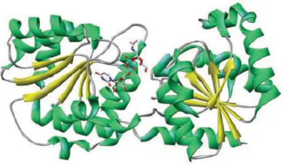
GTs are a large family of enzymes responsible for diverse transglycosylation reactions, involved in the biosynthesis of oligosaccharides, polysaccharides, and glycoconjugates (Berg et al., 2007; Taniguchi et al., 2002). Many of these compounds are cell wall components (Badreddine et al., 2008; Schaeffer et al., 1999; Wagner and Pesnot, 2010) that are critical not only in the maintenance of the structural integrity of cell membranes, but also in the modulation of molecular recognition events (Albesa-Jové et al., 2014). Obtaining a high level of expression, purification and crystallization were the main bottlenecks in the crystal structure determination for GT enzymes, but eventually two structural super-families were identified: GT-A and GT-B. When compared with other protein sequences, Rf-GT1 belongs to the B-fold GTs which consists of two separated Rossmann domains with a connecting linker region and a catalytic site located between these domains (Figure 1.7). Typically, the two Rossmann domains have a high structural homology, but a minimal sequence homology (Hu et al., 2003).
Figure 1.7 The protein structure of Escherichia coli MurG, a typical B-fold GT, in
Introduction
12
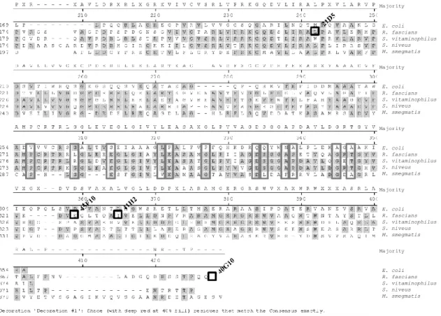
Nevertheless, some of the conserved amino acid residues are thought to be important for catalysis in GT-B glycosyltransferases. When aligning the R. fascians GT1 and related GTs from other organisms, such as Streptomyces vitaminophilus, S. niveus, and Mycobacterium smegmatis, with MurG of E. coli (Guerin et al., 2007), conserved glutamic acid, proline, and glycine residues could be identified in the R. fascians enzyme (Figure 1.8).
Figure 1.8 Sequence alignment of selected GT-B superfamily members.
Conserved residues are boxed in grey and transposon mutations in R. fascians in black. The sequence accession numbers are: P17443 of E. coli, WP_015586190 of R. fascians, WP_018382616.1 of S. vitaminophilus), WP_023538738 of S. niveus), and 2GEJ_A of M. smegmatis.
Another classification system based on amino acid sequence similarities has been proposed for GT function prediction (Campbell et al., 1997; Coutinho et al., 2003). With this sequence-based classification method 92 GT families have been identified. GTs belonging to family 1, to which the protein encoded by pFi_136 of R. fascians belongs, are named GT1 and are typically responsible for the transfer of a saccharide from a glycosyl donor to an acceptor, that can be another saccharide, a protein, a peptide, a lipid or a secondary metabolite (Wagner and Pesnot, 2010).
- - - M S S T L V I T N D F P P R - - - Q G G I E T F V H A L T S R L P P 1 WP _02 3538 738 .1 M G S S H H H H H H S S G L V P R G S H M R I G M V C P Y S F D V P G G V Q S H V L Q L A E V L R D 1 2G EJ X - - Q V V V Y A S A E R G - - - - A A A Y D A G L P F P V V R D R S R T L L P T G R V T R R A V R Ma jor ity 60 70 80 90 10 0 Q G W Q V R W L G T A D R - - - M E A D L V P K H G I E I D F I R I S G L R G K G I K A L 32 P1 744 3.3 E - - S I V V Y T S A E R G - - - - A A A Y D A S L P F L V I R D R S R T L L P T G R V M R R A G R 32 WP _01 5586 190 .1 K - - Q V V V Y A S T T K D T A E - V A R F D A G Q P F P V V R D R S R V L L P S R R V T R R A V R 32 WP _01 8382 616 .1 D - - R V V V Y T S S E P G - - - - A A A Y D A T L P F P V V R D R A H T L L P T A R V T R T A R E 32 WP _02 3538 738 .1 A G H E V S V L A P A S P H V K L P D Y V V S G G K A V P I P Y N G S V A R L R F G P A T H R K V K 51 2G EJ I A R E H G C D X V W F G A A A P L G L M A P A L R X G G X G R I V A T T H G H E A W W A X L P V T Ma jor ity 11 0 12 0 13 0 14 0 15 0 I A A P L R I F N A W R Q A R A I M K A Y K P D V V L G - M G G Y V S G P G G L A A W S L G I P V V 74 P1 744 3.3 I A L E H G C D T A W I A A A A P L G L I A P A L R A C G V K R V V A T T H G H E T W W A K V P L T 76 WP _01 5586 190 .1 L L R E H G C S S V W F G A A A P L G L M A P A L R E A G A R R I V A T T H G H E A G W A A L P V T 79 WP _01 8382 616 .1 T A R S H G C D R V W F G A A A P L A Y M A P E L R N G G I R R M V A T T H G H E I W W A R T P G A 76 WP _02 3538 738 .1 K W I A E G D F D V L H I H E P N A P S L S M L A L Q A A E G P I V A T F H T S T T K S L T L S V F 10 1 2G EJ R Q X L R R I G X X V D V V T A L G E Y T R S R I A P A L G P D A A - - M V R L X P G V D A A X F A Ma jor ity 16 0 17 0 18 0 19 0 20 0 L H E Q N G I A G L T N K W L A K I A T K V M Q A F P G A F P N A E - - - V V G N P - V R T D V L A 12 3 P1 744 3.3 R W G L H T I G R W V D V L T Y I S G H T Y R A I L P A V T P D T A - - L V R L S P G V D V E Q F N 12 6 WP _01 5586 190 .1 R Q L L R R I G D G V D T V T F L G E Y T R S R I A P A L S P R A A G A M V R L T P G V E A E L F H 12 9 WP _01 8382 616 .1 R Q V L R R V G A H V D V V T Y L G E Y T R S R I A P A L G P G A R - - M S R L V P G V D A A M F R 12 6 WP _02 3538 738 .1 Q G I L R P Y H E K I I G R I A V S D L A R R W Q M E A L G S D A V - - - - E I P N G V D V A S F A 15 1 2G EJ P X R - - - X A V L D R X R L X G R X V I V C V S R L V P R K G Q D V L I R A L P X V L A R V P Ma jor ity 21 0 22 0 23 0 24 0 25 0 L P - - - L P Q Q R L A G R E G P V R V L V V G G S Q G A R I L N Q T M P Q V A A K L G 16 9 P1 744 3.3 P V R G S - - - V A G P D P S P D G N S G V I V C T A R L V P R K G Q D S L I R A L P A V L E R H P 17 4 WP _01 5586 190 .1 P G V D R - - - Q A V R D R L R L S T R P V V V C V S R L V P R K G Q D T L I R A W P R V L A R V P 17 9 WP _01 8382 616 .1 P D R A A S C A R D V R D R H G I R G K K I I L C V S R L V T R K G Q D V L I R A L P L I R R S V P 17 4 WP _02 3538 738 .1 D - - - A P L L D G Y P R E G R T V L F L G R Y D E P R K G M A V L L A A L P K L V A R F P 19 7 2G EJ D A V L L V V G X G P D E R R L R E L A R T X A G - - A V R F X G G V P H T E X A S X Y A A A D V F Ma jor ity 26 0 27 0 28 0 29 0 30 0 D S V T I W H Q S G K G S Q Q S V E Q A Y A E A G - - - Q P - Q H K V T E F I D D M A A A Y A W 21 0 P1 744 3.3 D T T L L L V G D G P D R R R L Q R L A R T K G V E N A V T F V G G L P H E L V A Q V L A G A T I F 22 1 WP _01 5586 190 .1 D A V L L V V G G G P G L R R L E R L A E T A G V R E A V R F T G E V P W T E L P A H Y A A G D V F 22 6 WP _01 8382 616 .1 D A V L V V V G Q G P D E R R L R A L A R R H A P - G A V V F A G G R S H T E T A S Y Y A A A D V F 22 4 WP _02 3538 738 .1 D V E I L I V G R G - D E D E L R E Q A G D L A G - - H L R F L G Q V D D A T K A S A M R S A D V Y 24 0 2G EJ A M P C R T R L G G L E V E G L G I V Y L E A S A X G L P V V A G D S G G A P D A V L D G R T G Y V Ma jor ity 31 0 32 0 33 0 34 0 35 0 A D V V V C R S G A L T V S E I A A A G L P A L F V P F Q H K D R Q Q Y W N A L P L E K A G A A K I 25 4 P1 744 3.3 A M P C R T R R L G L E E E G L G I A Y L E A S A M G L P I I A G D S G G A P E T V Q A G R T G Y V 27 1 WP _01 5586 190 .1 A M P C R T R L R G L D V E G L G I V Y L E A S A T G L P V I A G D S G G A P D A V L Q G E T G Y V 27 6 WP _01 8382 616 .1 A M P C R T R K G G L E A E G L G I V F L E A A A S G L P V V V G D S G G A P D A V L D G R T G H V 27 3 WP _02 3538 738 .1 C A P - H - - L G G - - - E S F G I V L V E A M A A G T A V V A S D L D A F R R V L A D G D A G R L 28 7 2G EJ V X G X - - D V D S V A X A L V G L L D D P X L A R A M G E R G R S W V X A X W R W X X X A S R L A Ma jor ity 36 0 37 0 38 0 39 0 40 0 I E Q P Q L S V D A V A N T L A G W S R E T L L T M A E R A R A A S I P D A T E R V A N E V S R V A 30 4 P1 744 3.3 V K - - - - D V E Q L T Q A I V E L L D N P V R A R A M G E R G R S W V A A Q W T W N T A Y S T L L 32 1 WP _01 5586 190 .1 V R G G - - S P A S V A E R V V E L L D D P G L R R R M G E R G R S W V E E S W R W D R L A Q R L A 32 6 WP _01 8382 616 .1 V D G T - - D V P S V A R T L T T L L L A P E R A G A M G E A G R R W V S E E W S W E A S A R R L T 32 3 WP _02 3538 738 .1 V P V D - - D A D G M A A A L I G I L E D D Q L R A G Y V A R A S E R V H R - Y D W S V V S A Q I M 33 1 2G EJ R A L - P - - - N R - - T P - - - - Ma jor ity 41 0 42 0 R A 35 4 P1 744 3.3 T A L F P N V - - - L A D G Q D R S S T P Q Q 36 7 WP _01 5586 190 .1 A L L 37 4 WP _01 8382 616 .1 R L L T P - - - E N T R T T P 37 1 WP _02 3538 738 .1 R V Y E T V S G A G I K V Q V S G A A N R D E T A G E S V 37 8 2G EJ
De cor atio n ' Dec ora tion #1 ': Sha de (wit h d eep re d at 40 % f ill ) re sid ues th at m atc h t he Con sens us exa ctl y.
E. coli R. fascians S. vitaminophilus S. niveus M. smegmatis E. coli R. fascians S. vitaminophilus S. niveus M. smegmatis E. coli R. fascians S. vitaminophilus S. niveus M. smegmatis E. coli R. fascians S. vitaminophilus S. niveus M. smegmatis E. coli R. fascians S. vitaminophilus S. niveus M. smegmatis
13
In pathogenic organisms, the molecules involved in host recognition, adhesion, and colonization, as well as the antigens that target host responses are all located in the cell wall (Albesa-Jové et al., 2014; Netea et al., 2008). The expanding bacterial sequence dataset coupled with developments in mass spectrometry and analytical glycosciences has identified protein glycosylation systems in most bacterial pathogens (Cuccui and Wren, 2013). Those systems have proven to be essential for viability and virulence (Cuccui and Wren, 2013; Dover et al., 2004). As an example, in Streptococcus mutans, a mutant defective in a GT exhibited a modified colony morphology and an impaired aggregation (Kuramitsu, 1976; Taniguchi et al., 2010). In other pathogens as well, including phytopathogens, protein glycosylation systems were shown to be required for biofilm formation and virulence (Cuccui and Wren, 2013). In the pathogen Burkholderia cepacia (Bcc), the inability to produce exopolysaccharide (EPS) was correlated with a deletion in the bceB gene, which encodes a GT. EPS is a putative Bcc virulence factor that is involved in bacterial persistence, interaction with antimicrobial peptides, and biofilm formation (Bartholdson et al., 2008). Xanthomonas campestris causes citrus canker, a leaf-spotting and fruit rind-blemishing disease that can lead to defoliation, shoot dieback and fruit drop when conditions are highly favorable for infection (Gottwald et al., 2002). In this pathogen, silencing of the gpsX gene, that encodes a GT, resulted in a reduction in the amount of EPS and lipopolysaccharides (LPS) in the cell wall, defects in biofilm formation, delayed growth, a higher sensitivity to various stress compounds, and reduced symptoms on the host plant (Chou et al., 1997; Dow et al., 2003; Li and Wang, 2012). Similarly, in Xanthomonas citri, another citrus pathogen causing citrus canker, the knock-out of gpsX, also leads to a decreased biofilm formation and a reduced virulence. Nonetheless, quantitative PCR revealed that the mutation had no effect on the expression of the virulence genes (Li and Wang, 2012).
D. Two methyltransferases, intriguing members of the fas locus
As described above, previous studies on the fas operon identified the functions of all of the Fas proteins in the production of the cytokinin mixture (Pertry, 2009; Pertry et al., 2010). In between fasR, encoding the main regulator of fas gene expression, and fasA, the first gene on the fas operon, two genes are located that are homologous to putative S-adenosyl methionine (SAM)-dependent methyltransferases (MTRs), pFi_075 and pFi_076 (mtr1 and mtr2) (Figure 1.3) (Stes et al., 2011).
14
These genes are conserved in the fas operon of the phytopathogen Streptomyces turgidiscabies (Joshi and Loria, 2007) (Figure 1.9). This organism is the causative agent of potato scab disease, a pathology that leads to significant annual losses to potato growers world-wide. The disease symptoms, characterized by erumpent lesions on the tubers (Figure 1.9-right), is caused by the main phytopathogenicity factor produced by S. turgidiscabies, i.e. the toxin thaxtomin (Loria et al., 2006). Although there are no reports for gall formation caused by this phytopathogen in nature, S. turgidiscabies has a fas operon that strongly resembles and is largely collinear to the one of R. fascians (Figure 1.9-middle). Furthermore, infection of Nicotiana tabacum and Arabidopsis thaliana with a thaxtomin-deficient mutant, lead to the formation of leafy galls similar with those induced by R. fascians (Figure 1.9-left and right). Intriguingly, upstream of the fas operon of S. turgidiscabies there are two mtr genes highly homologous to those of R. fascians (Joshi and Loria, 2007; Kers et al., 2005) (Figure 1.9-middle). Altogether, these observations suggest that the mtr gene products are involved in cytokinin production and/or pathogenicity. Indeed, preliminary data revealed that mutations in the mtr genes in R. fascians cause a complete loss of virulence (Pertry, 2009).
Figure 1.9 Organization of the fas loci of and induced symptoms by R. fascians strain
D188 (left) and S. turgidiscabies (right).
Methyltransferases are a large family of enzymes that catalyze the transfer of a methyl group from a donor to an acceptor. Those enzymes can be divided in several subclasses based on their structural features and used substrates. Most common are the number I methyltransferases, encompassing by SAM-dependent enzymes with a Rossman fold (Katz et al., 2003). SAM-dependent methyl transfers are found in different pathways and mutations is the corresponding genes cause severe or complete impairment of regular behavior of the organism (Struck et al., 2012).
Although it is unlikely to be the case for R. fascians, in other pathogens MTRs are often linked to cell wall formation. In diverse plant pathogens, such as the gram-negative Xanthomonas oryzae, Xanthomonas campestris, Pseudomonas syringae, and Ralstonia
15
solanacearum, MTRs have been associated with the methylation of LPS (Whitfield et al., 1997) which have been shown to be critical virulence determinants and control a number of defense-related responses in plants (Desaki et al., 2006). It has also been suggested that LPS are likely to be involved in the association of bacteria with plant cell walls during the infection process (Boher et al., 1997). In the phytopathogen Xanthomonas albilineans MTRs are important in the biosynthesis of the key pathogenicity factor, i.e. the toxin albicidin (Birch, 2001; Hashimi and Birch, 2010). MTRs in Candida albicans, an opportunistic pathogen, are important for the maintenance of cellular homeostasis and regulation of intra- and interspecies interactions. It was found that those enzymes are also important for virulence-related processes, such as adhesion and membrane transport (Lissina et al., 2014). A gene that encodes a specialized o-methyltransferase is present in all virulent lineages from Francisella, a bacterial human pathogen (Champion, 2011), and therefore is assumed to be related with pathogenicity. Finally, in the gram-positive human pathogen Mycobacterium tuberculosis, MTRs are involved in the biosynthesis of mycolic acids, major components of the cell wall also present in the cell wall of members of the genus Rhodococcus (Nishiuchi et al., 2000). The envelope from M. tuberculosis is partly responsible for its resistance to antibiotics and plays a major role in the virulence and the persistence of the pathogen (Cantaloube et al., 2011; Simeone et al., 2013).
17
2. Aim of this work
Despite extensive mutagenesis screens, the main pathogenicity factors of R. fascians appeared to be linked to unique region U1 of the linear plasmid. Nevertheless, for the majority of genes on pFiD188, the function in the life style of R. fascians remains to be determined. Recently, two loci on pFiD188 were shown to be essential for the pathology of the bacteria: the glycosyltransferase GT1 (pFi_136), situated in unique region U2, and two methyltransferases mtr1 and mtr2 (pFi_075 and pFi_076), located immediately upstream of the fas operon. Here we aim to get insight into the role of GT1 and the two MTRs in the phytopathogenicity of R. fascians.
Preliminary data on GT1 revealed that mutants in the gene have a strong colony phenotype and a severe loss of pathogenicity. Till today, virulence defects of R. fascians are either linked with cytokinin production and the regulation thereof (fas, att, hyp) or with in planta survival (vic) (Stes et al., 2012). GTs are known to glycosylate secondary metabolites. The fas cytokinins can be considered as secondary metabolites of R. fascians and it is currently unknown if their secretion into the environment is active or passive. Moreover, high cytokinin concentrations are toxic for living cells (Pertry et al., 2009). Based on this information we postulated a first working hypothesis that the GT1 mutant 21D5 is non-pathogenic because it is defective in cytokinin production or secretion. The second working hypothesis is based on the knowledge that R. fascians forms an epiphytic biofilm prior to plant tissue penetration (Cornelis et al., 2001) and the finding that, in other pathogens, GTs are important for the survival in host tissues (see Introduction). So, we postulated that the inability of 21D5 to cause disease is the result of a severely impaired capacity to survive on or in the plant tissues. Before testing these hypotheses, we will first confirm the virulence defect by assessing symptom formation upon infection of Nicotiana tabacum cv. Havana and Xanthi (seedlings, decapitated plants and leaves) and Arabidopsis thaliana plants with strain 21D5. We will also evaluate the colony morphology on different defined media. Both phenotypes will be compared to the wild-type strain D188 and its plasmid-free non-pathogenic derivative D188-5; virulence will also be compared to the attenuated att mutant and the non-pathogenic fas mutant. With these experiments, we will get insight into the behavior of the mutant 21D5. The first hypothesis will be tested by measuring the expression of the fas and att genes in the 21D5 mutant, both in vitro and in planta using quantitative assays and histochemical stainings. The secretion of cytokinins
18
will be addressed by determining the cytokinin profile of 21D5 supernatants (in collaboration with Petr Tarkowski, Laboratory of Growth Regulators, Palacký University, Olomouc, Czech Republic). The secretion of the att compound by 21D5 will be verified by assessing the inducing capacity of extracts of plant tissues infected with 21D5. To test the second hypothesis, we will infect tobacco plants with 21D5 and quantify the endophytic and epiphytic populations after different time points. We will also evaluate the behavior of 21D5 on the plant tissues by scanning electron microscopy. Finally, the regulation of GT1 expression will be determined by introducing a transcriptional GT1:GUS fusions into strains D188, D188-5, D188-ΔattR, and D188-ΔfasR and performing in vitro GUS assays with these reporter strains. As a result, we will hopefully have established how the gene is regulated and get some insight on when and where it is expressed during the interaction with a host.
Concerning the MTRs, their location upstream of the fas operon, their conservation in S. turgidiscabies, and the complete loss of phytopathogenicity upon their mutation, strongly indicate that they are involved in the biosynthesis of an essential cytokinin. Based on their homology we postulated that the reaction product of these enzymes is a methylated cytokinin. This hypothesis will be tested by comparing the cytokinin production of R. fascians strains D188, 5, fas1, the two mtr mutants D188-mtr1 and D188-mtr2, and two S. turgidiscabies strains containing a fas operon and the mtr genes (Aittamaa et al., 2010). Therefore, the bacteria will be grown under particular conditions, fed with labeled precursors, and the partially purified supernatants will be evaluated by thin layer chromatography. We will also assess the regulation of the mtr genes both in vitro and in planta by using available translational mtr:GUS reporter strains. As such, we hope to be able to answer the question whether methylated cytokinins are the secret weapon of R. fascians.
Based on the obtained data on GT1 and the mtr’s we anticipate to make a considerable contribution to the understanding of the pathogenicity process of R. fascians and to open new research perspectives that might enable the use of R. fascians or its morphogens in tissue culture practices or help horticulturists in safe-guarding their ornamental crops from R. fascians.
19
3. Materials and methods
3.1 Bacterial strains and growth conditions
An overview of the bacterial strains used in this study is given in Table 3.1.
The R. fascians strains were grown on Yeast Extract Broth (YEB) (5g/L beef extract (Sigma-Aldrich, Mexico), 1g/L yeast extract (Oxoid, England), 5g/L peptone (Duchefa Biochemie, The Netherlands), 5g/L sucrose (C12H22O11, Grand Pont®, Belgium), 500mg/L magnesium sulfate heptahydrate (MgSO4.7H2O, purity >99%, Duchefa Biochemie, The Netherlands) and 15g/L agar (micro agar, Lab associates, The Netherlands) when solid medium was prepared) for two days at 28ºC. Suspension cultures were grown in 5ml liquid YEB and grown for two days at 28ºC under gentle agitation (160 rpm). Bacterial stocks were kept in glycerol (glycerine anhydrous, purity >95%, Duchefa Biochemie, The Netherlands) (25% final volume) at -80ºC.
The S. turgidiscabies strains were grown on Glucose-Yeast-Malt (GYM) medium (4g/L glucose (C6H12O6, purity >99.5%, Duchefa Biochemie, The Netherlands), 4g/L yeast extract and 10g/L malt extract (Duchefa Biochemie, The Netherlands), pH set at 7.2 with potassium hydroxide (KOH, purity 85%, Duchefa Biochemie, The Netherlands) and 2g/L calcium carbonate (CaCO3, purity >99%, UCB, Belgium) and 12g/L agar when solid medium was prepared) for three days at 28ºC. A spore solution stock was prepared to store the S. turgidiscabies. Vegetative mycelium was plated in sporulation medium (for 500mL: 250mL solution with 10g mannitol (purity >98%, Duchefa Biochemie, The Netherlands), 10g agar and 2.5mL of 2M magnesium chloride (MgCl2, UCB, Belgium)added to 250mL solution with 10g soy flower (local bioshop) and grown at 28ºC for seven days. The spore solution was obtained by successive washings of the spores scraped gently with 7mL of sterile water (3mL, 2mL and 2mL). The resulting solution was vortexed, poured over sterile cheesecloth and centrifuged at 14 000rpm for 10min at 4ºC. The obtained pellet was resuspended in 1mL sterile 20% glycerol at 4ºC, divided over 3 eppendorfs in aliquots of 250µL and 500µL and stored at -80ºC.
The E. coli strains were grown on Luria Broth (LB) (1g/L glucose, 10g/L tryptone (Duchefa Biochemie, The Netherlands), 5g/L yeast extract, 10g/L sodium chloride (NaCl, purity ≥95%, Carl Roth) and 15g/L agar when solid medium was prepared) for one day at 37ºC with agitation (160 rpm). Bacterial stocks were kept in glycerol (25% final volume) at -80ºC.