Synthesis, Characterization, and Photocatalytic Properties of Flower-like Mn-doped Ceria
Pei Liab, Wei Zhanga, Xun Zhanga, Zhengde Wanga, Xianpeng Wanga, Songlin Ranb, Yaohui Lva*
Received: March 05, 2018; Revised: May 22, 2018; Accepted: June 08, 2018
Mn-doped CeO2 flower-like microstructures have been synthesized by a facile one-step
composite-hydroxide-mediated method. The structure, morphology, optical and the surface properties of Mn
doped CeO2 have been investigated by X-ray diffraction (XRD), field-emission scanning electron
microscopy (FESEM), UV-Vis absorption spectroscopy and X-ray photoelectron spectroscopy (XPS). The XRD results confirmed the successful incorporation of Mn into the CeO2 lattice through the
formation of face-centered cubic solid solution. The photocatalytic activities of the catalysts were evaluated by measuring the photodegradation efficiency of Rhodamine B (RhB) under ultraviolet light irradiation. With an optimal molar ratio of 1% in Mn/CeO2 the highest rate photodegradation was
achieved under the experimental conditions. The enhanced photocatalytic activity can be attributed to the incorporation of multivalent Mn in CeO2 promoted the separation of photogenerated charges,
inhibited the recombination of photogenerated carriers, and thus prolonged the charges lifetime to participate in the photocatalytic reaction.
Keywords: Cerium oxide, Manganese doped, Photocatalysis.
*e-mail: 396839980@qq.com.
1. Introduction
Semiconductor-based photocatalysis has emerged as one of the most attractive techniques to convert sunlight into chemical energy so as to remove organic pollutants from wastewater for environmental remediation. Among various semiconductor materials, as a high-efficiency, nontoxicity, abundant, photochemical stability and low-cost promising photocatalyst, CeO2 has been an extensively
investigated material for environmental pollution removal and photocatalytic hydrogen evolution1-3. Nevertheless, some
drawbacks limit its practical application in the photocatalyst region, including rapid recombination rate of photogenerated electron-hole pairs, low quantum yield in the reactions, and very poor response to visible light. Therefore, from the viewpoint of photochemistry, numerous methods for suppression of these drawbacks have been attempted to improve CeO2-based photocatalytic activities. To the best of
our knowledge, one of approaches is to develop CeO2 based
heterostructure semiconductor systems, such as TiO2@Pt@
CeO24, Au/CeO 2
5,6, CdS/CeO 2
7, SrTiO 3/CeO2
8, Bi 2O3/CeO2
9,
TiO2/CeO2
10,11 and Cu 2O/CeO2
12. On the other hand, there
have been modifications of CeO2 by doping non-metallic
species13,14 and crystal facet engineering15,16. For instance, Zhu
et al.13 reported the synthesis of N-doped CeO
2 nanoparticles
with controllable doping levels at the nanoscale through a reliable wet chemical approach, showing enhanced visible-light sensitivity and photocatalytic activity. Fuertes et al.14
have doped nitrogen into ceria powder by sintering CeO2
in NH3 flow at very high temperature. Recent reports have shown that CeO2 nanostructures with highly active exposed
crystal planes such as {100} and {110} can significantly enhance their catalytic activity15. Meanwhile, the selective
metal doping of CeO2 to improve its performance started
to appear in the literature. Recently, the metal doping of
CeO2 has also been examined in solar cell devices17, but the
corresponding photocatalytic investigations are relatively few. It has been reported that doping with multivalent transition-metal (TM) cations was considered an effective method to inhibit the recombination of photogenerated carriers in semiconductors18. Theoretical investigation showed
that among the 3d metals, Mn has the greatest potential in permitting significant optical absorption in the visible or even the infrared solar light, through the combined effects of narrowed band gap and the introduction of intermediate bands within the forbidden gap19,20. Recently, there have been
many reports on Mn-doped oxide photo-activity under UV and visible light, such as Mn-ZnO21-24, Mn-TiO
2
25,26, where
Mn exists in the bivalence oxidation state.
Inspired by above-mentioned investigations, flower-like
Mn-doped CeO2 photocatalyst was obtained via a simple
one-step composite-hydroxide-mediated method. In addition, we demonstrated the enhanced photocatalytic performance of the
Mn doped CeO2 flower-like nanostructures by degradation
of Rhodamine B (RhB) solutions, and further investigated the impact of Mn doping concentrations of doped CeO2 on
the resulting photocatalytic activities under ultraviolet light. aSchool of Materials Science and Engineering, Anhui Key Laboratory of Metal Materials and
Processing, Anhui University of Technology, Anhui, Maanshan, 243002, China
bKey Laboratory of Metallurgical Emission Reduction & Resources Recycling, Ministry of Education,
2. Experimental
2.1 Material preparation
Pure and flower-like Mn-doped CeO2 nanostructure were
obtained by a simple one-step composite-hydroxide-mediated method according to our group previously report with tiny modification27. In a typical preparation process, (1) a total
of 20 g of KOH and KI was mixed at a ratio of 70.6:29.4 and placed in a 25mL Teflon vessel. (2) Different molar ratio of MnSO4·H2O and CeO2 was used as the raw material for
reaction, and was placed on the top of the hydroxide in the vessel. (3) The Teflon vessel was put into a furnace preheated to 235 oC. (4) After the hydroxide was totally molten (30
min later), the molten reactants were mixed uniformly by shaking the covered vessel. (5) 24 hours later, the vessel was taken out and cooled to room temperature naturally. The as-prepared samples were removed from the solution, rinsed thoroughly several times with deionized water and ethanol to remove residual salts, and subsequently dried for 12h at vacuum circumstance. The as-prepared samples in the subsequent discussion in this paper are denoted as x%
Mn/CeO2, where x% refers to the Mn/(Mn+Ce) molar ratio.
2.2 Catalyst characterization
The crystal structure of the resultant products was characterized by X-ray powder diffraction (XRD) by using a Bruker AXS D8 advance powder diffractometer with Cu Kα radiation (λ = 0.154056 nm). Field-emission scanning electron microscope (FESEM, S-4800) was employed to characterize the morphologies and size of the synthesized
Mn doped CeO2 samples. UV-vis diffuse reflectance spectra
were obtained for the dry-pressed disk samples by using a Shimadzu UV 2550 recording spectrophotometer, which was equipped with an integrating sphere, and BaSO4 was
used as a reference. X-ray photoelectron spectroscopy (XPS) measurements were performed on a Thermo Fisher Scientific Escalab 250 spectrometer with monochromatized Al Kα excitation, and C1s (284.6 eV) was used to calibrate
the peak positions of the elements. The Raman-scattering experiments were carried out using NEXUS 670 Raman spectrometer at room temperature. The 473 nm line of the solid-state laser was used for excitation.
2.3 Catalyst activity
The photocatalytic performance of the as-prepared samples was characterized by decomposing RhB under ultraviolet light irradiation at room temperature. The photocatalytic experiments were carried out by adding 100 mg photocatalysts into 300 mL of 10 mg L-1 RhB solution in the vessel. A 300
W Hg arc lamp (XPA-1, Nanjing XuJiang Electromechanical Plant) was used as the light source. Prior to the irradiation, the suspensions were magnetically stirred in the dark for 30 min to establish the adsorption/desorption equilibrium.
After different irradiation time, the concentration of the RhB solution was measured on a UV-Vis spectrophotometer (Hitachi UV-3100).
3. Results and Discussion
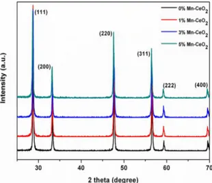
The X-ray diffraction (XRD) patterns of MnxCe1-xO2
photocatalysts are shown in Figure 1 along with pure CeO2.
The diffraction peaks of pure CeO2 can be indexed to the
fluorite cubic phase of CeO2 (JCPDF card: 65-2975) with
lattice constant a =0.5411 nm. The diffraction pattern of
MnxCe1-xO2 photocatalysts was similar to that of pure CeO2.
The XRD pattern does not show any impurity/additional peaks for any Mn doping concentration. This clearly confirms that the Mn ions occupy positions within the fluorite-lattice. Furthermore, the extended X-ray diffraction pattern of MnxCe1-xO2 photocatalysts reveals a slight shift
in the peak positions towards larger angles with increasing Mn-concentration, which may be due to the lattice reduction of CeO2 upon Mn ions doping. Because the ionic radius
of Mnn+ (Mn4+=0.53 Å, Mn3+=0.645 Å, Mn2+=0.83 Å) is
smaller than that of Ce4+ (1.01 Å), when Mnn+ embedded in
CeO2 lattice and takes the place of Ce4+, the contraction and
distortion of the ceria lattice occur, leading to the decrease of the cell parameter28. In present study, the existence of the
Mnn+ oxidation state in MnxCe1-xO2 photocatalysts will be
discussed in the later section.
Figure 2 shows the morphology and size of the source material CeO2 (a) and the flower-like 1% Mn-doped CeO2
sample (b) obtained by typical field-emission scanning electron microscope (FESEM). The anomalous particles morphology of the source material CeO2 can be seen with
the diameter ranging from 2-8 µm (Figure 2 a). From the high-magnification SEM images shown in Figure 2 b, it can be seen that the flower-like micro-/nano-architectures are
Figure 1. XRD patterns of Mn-doped CeO2 from different mole
Figure 3 b. The evaluated band gap values for the pure and the 1% Mn-doped CeO2 sample were observed to be 3.05
eV and 2.88 eV, respectively. It could be seen that the band gap value slightly decreased when the dopant concentration of Mn was increased.
Raman scatting is an effective tool for the investigation of the effects of doping on nanomaterials, as the incorporation of dopants leads to shifts of the lattice Raman vibrational peak positions. Figure 4 displays the Raman spectra of pure
CeO2 as well as Mn-doped CeO2 samples with different Mn
contents. For the pure CeO2 sample, a strong peak at 461 cm -1
can be assigned to the F2g Raman active mode of the cubic fluorite structure of CeO2, which is due to the symmetric
breathing mode of the oxygen atoms around cerium ions. Compared to the pure CeO2, the peak intensity decreased
greatly and became broader and red-shifted for the
Mn-doped CeO2 samples. The red-shift could be attributed to
the changes in lattice parameter with crystallite size, as it built from dozens of flake-like nanopetals with the average
diameter of 1-4 µm.
The effect of Mn substitution on cubic fluorite structure of CeO2 lattice is further confirmed using UV-visible optical
spectroscopy measured in the range of 200-800 nm. Figure 3a shows the room-temperature optical absorption spectra of undoped and flower-like Mn-doped CeO2 photocatalyst.
The absorbance spectra for 1 % Mn-doped CeO2 sample
was found to increase when compared to the undoped CeO2
sample. However, the absorbance intensity for the 3 and 5% Mn-doped CeO2 sample were found to decrease when
compared to the undoped CeO2 sample. This may be due
to the electron, donor atom and electron-hole interactions which increase drastically as doping is increased beyond a critical limit29,30. These interactions
dominate the electron-photon interactions. A plot of variation of (αhν)2versus hν , which is obtained according
to the Kubelka-Munk function transformation, is shown in
Figure 2. FESEM images of (a) pure CeO2 raw materials; (b) 1% Mn/CeO2 sample
Figure 3. (a) UV-Vis absorption spectra of Mn/CeO2 with varying Mn/Ce molar ratio; (b) Plot of (αhν)
2 versus photon energy of pure
Figure 4. Raman spectra of Mn/CeO2 with varying Mn/Ce molar ratio
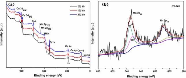
Mn3+ ion couple in the Mn-doped CeO2 samples. Here, the
Mn2P3/2 peak is deconvoluted with the Gaussian-Lorenz model functions, and two peaks at 641.2 eV and 642.3 eV can be assigned to the Mn2+ and Mn3+ ions, respectively, according
to the standard binding energy and previous literature.3 This
results suggest the coexistence of Mn2+ and Mn3+ ions on the
surface of samples. In addition, we can observe that the XPS peaks show some noise, implying that the content of Mn ions at the surface of the sample is low and Mn ions have been doped into the interior of the nanostructure.
The effective incorporation of Mn2+ ions into CeO 2
crystalline lattices can greatly enhance the light photocatalytic performance. The UV-vis spectral changes of RhB solution over 1% Mn doped CeO2 samples during the photodegradation
are shown in Figure 6a, clearly show that the characteristic absorption peaks corresponding to RhB decrease rapidly as the exposure time increases, indicating the decomposition of RhB and the significant reduction in the RhB concentration. Figure 6 b shows the results of RhB photodegradation over Mn
doped CeO2 samples with different Mn doping concentrations.
After 210 min of irradiation, the photodegradation efficiencies of RhB were about 65, 40 and 30% for 1, 3 and 5% Mn
doped CeO2 samples, respectively. It was evident that 1% Mn doped CeO2 samples exhibited excellent photocatalytic
activities for the RhB degradation. However, the photocatalytic performance of Mn doped CeO2 samples decreased with the
increase of Mn doped amount. The results show that there was an optimal Mn doping concentration in CeO2 samples for
the ultraviolet light photocatalysis (1% Mn dopant). When the Mn doping concentration was further increased, the Mn dopant sites could be also act as efficient recombination centers with increased recombination rate due to the reduced average distance between trapped carriers34. The excess Mn
dopant sites could greatly decrease the number of charge carriers and deteriorate the photocatalytic performance of
doped CeO2 samples, as identified from Figure 6b.
was previously explained by phonon confinement model31,32.
Another reason of shifting and broadening may be the increase in oxygen vacancies, which is related to structural defects derived from partially incorporation of manganese
into CeO2 lattice. In the Mn-doped CeO2 samples, the extra
oxygen vacancies were generated by the incorporation of Mn ion into the ceria fluorite lattice to compensate for the valence mismatch between the Mnn+ and Ce4+ ions.
The chemical states of the Mn doping in the as-prepared samples were then investigated by XPS, as presented in Figure 5a, the XPS survey spectra indicated that the
as-prepared Mn doped CeO2 nanoflowers were composed of
Ce, O and Mn elements. Figure 5b shows the XPS spectra of the Mn 2p region. The double peaks with binding energies of ca. 641.0 eV and 653.1 eV correspond to the characteristic of Mn 2p3/2 and Mn 2p1/2 signals, respectively. Since XPS
signals for Mn2+ and Mn3+ are very close to each other ~641.0
eV for the BE of Mn 2p3/2,33 thus the coexistence of Mn2+/
4. Conclusions
In summary, we have synthesized a series of Mn/CeO2
photocatalysts by a facile one-step composite-hydroxide-mediated approach. This work focuses on understanding of the effects of the Mn addition in Mn-doped CeO2 nanoflowers.
Noticeably, there was an optimal Mn doping concentration
in CeO2 samples for the ultraviolet light photocatalysis.
Experiments showed that 1% Mn-doped CeO2 had higher
photocatalytic capability compared with that of the pure CeO2
or 3% and 5% Mn ion-doping catalyst. The enhancement in redox efficiency of CeO2 samples upon Mn doping may
be due to the increase in charge transport rate.
5. Acknowledgements
This research was financially supported by the Natural Science Foundation of AnHui Provincial Education Department (KJ2016A102), Anhui Provincial Natural Science Foundation (1808085ME138), Graduate student Innovation Fund of Anhui University of Technology and National Undergraduate Training Programs for Innovation and Entrepreneurship (201710360024, 201710360026)
6. References
1. Li L, Wang HR, Zou L, Wang X. Controllable synthesis, photocatalytic and electrocatalytic properties of CeO2 nanocrystals.
RSC Advances. 2015;5(52):41506-41512.
2. Lu X, Zhai T, Cui H, Shi J, Xie S, Huang Y, et al. Redox cycles promoting photocatalytic hydrogen evolution of CeO2 nanorods.
Journal of Materials Chemistry. 2011;21(15):5569-5572.
3. Rangaswamy A, Venkataswamy P, Devaiah D, Ramana S, Reddy BM. Structural characteristics and catalytic performance of nanostructured Mn-doped CeO2 solid solutions towards
oxidation of benzylamine by molecular O2. Materials Research
Bulletin. 2017;88:136-147.
4. Li SX, Cai JB, Wu XQ, Liu BW, Chen QY, Li YH, et al. TiO2@Pt@CeO2 nanocomposite as a bifunctional catalyst for enhancing photo-reduction of Cr (VI) and photo-oxidation of benzyl alcohol. Journal of Hazardous Materials. 2018;346:52-61.
5. Li BX, Zhang BS, Nie SB, Shao LZ, Hu LY. Optimization of plasmon-induced photocatalysis in electrospun Au/CeO2 hybrid
nanofibers for selective oxidation of benzyl alcohol. Journal of Catalysis. 2017;348:256-264.
6. Tanaka A, Hashimoto K, Kominami H. Selective photocatalytic oxidation of aromatic alcohols to aldehydes in an aqueous suspension of gold nanoparticles supported on cerium(IV) oxide under irradiation of green light. Chemical Communications. 2011;47(37):10446-10448.
7. Zhang P, Liu Y, Tian BZ, Luo YS, Zhang JL. Synthesis of core-shell structured CdS @CeO2 and CdS @TiO2 composites and
comparison of their photocatalytic activities for the selective oxidation of benzyl alcohol to benzaldehyde. Catalysis Today. 2017;281(Pt 1):181-188.
8. Song S, Xu L, He Z, Ying H, Chen J, Xiao X, et al. Photocatalytic degradation of C.I. Direct Red 23 in aqueous solutions under UV irradiation using SrTiO3/CeO2 composite as the catalyst.
Journal of Hazardous Materials. 2008;152(3):1301-1308.
9. Zou ZJ, Xie CS, Zhang SS, Yu XL, Zou T, Li J. Preparation and photocatalytic activity of TiO2/CeO2/Bi2O3 composite
for Rhodamine B degradation under visible light irradiation. Journal of Alloys and Compounds. 2013;581:385-391.
10. Lu XW, Li XZ, Qian JC, Miao NW, Yao C, Chen ZG. Synthesis and characterization of CeO2/TiO2 nanotube arrays and enhanced
photocatalytic oxidative desulfurization performance. Journal of Alloys and Compounds. 2016;661:363-371.
11. Pavasupree S, Suzuki Y, Pivsa-Art S, Yoshikawa S. Preparation and characterization of mesoporous TiO2-CeO2 nanopowders Figure 6. (a) UV-vis spectral variations of RhB solution over 1% Mn doped CeO2 under ultraviolet light irradiation; (b) Degradation of
respond to visible wavelength. Journal of Solid State Chemistry. 2005;178(1):128-134.
12. Hu S, Zhou F, Wang L, Zhang J. Preparation of Cu2O/CeO2
heterojunction photocatalyst for the degradation of Acid Orange 7 under visible light irradiation. Catalysis Communications. 2011;12(9):794-797.
13. Mao C, Zhao Y, Qiu X, Zhu J, Burda C. Synthesis, characterization and computational study of nitrogen-doped CeO2 nanoparticles with visible-light activity. Physical Chemistry Chemical Physics. 2008;10(36):5633-5638.
14. Belén Jorge A, Fraxedas J, Cantarero A, Williams AJ, Rodgers J, Attfield JP, et al. Nitrogen doping of ceria. Chemistry of Materials. 2008;20(5):1682-1684.
15. Zhou K, Wang X, Sun X, Peng Q, Li Y. Enhanced catalytic activity of ceria nanorods from well-defined reactive crystal planes. Journal of Catalysis. 2005;229(1):206-212.
16. Ren J, Liu X, Gao RH, Dai WL. Morphology and crystal-plane effects of Zr-doped CeO2 nanocrystals on the epoxidation of styrene with tert-butylhydroperoxide as the oxidant. Journal of Energy Chemistry. 2017;26(4):681-687.
17. Corma A, Atienzar P, Garcia H, Chane-Ching JY. Hierarchically mesostructured doped CeO2 with potential for solar-cell use. Nature Materials. 2004;3(6):394-397.
18. Prabaharan DDM, Sadaiyandi K, Mahendran M, Sagadevan S. Investigating the effect of Mn-doped CeO2 nanoparticles by co-precipitation method. Applied Physics A. 2018;124(2):86. 19. Shao G. Electronic structures of manganese-soped rutile TiO2 from first
principles. The Journal of Physical Chemistry C. 2008;112(47):18677-18685.
20. Shao G. Red shift in manganese- and iron-doped TiO2: a DFT+U analysis. The Journal of Physical Chemistry C. 2009;113(16):6800-6808.
21. Yang Y, Li Y, Zhu L, He H, Hu L, Huang J, et al. Shape control of colloidal Mn doped ZnO nanocrystals and their visible light photocatalytic properties. Nanoscale. 2013;5(21):10461-10471. 22. Ullah R, Dutta J. Photocatalytic degradation of organic dyes with
manganese-doped ZnO nanoparticles. Journal of Hazardous Materials. 2008;156(1-3):194-200.
23. Abdollahi Y, Abdullah AH, Gaya UI, Zainal Z, Yusof NA. Enhanced photodegradation of o-cresol in aqueous Mn(1%)-doped ZnO suspensions. Environmental Technology. 2012;33(10-12):1183-1189.
24. Lu Y, Lin Y, Xie T, Shi S, Fan H, Wang D. Enhancement of visible-light-driven photoresponse of Mn/ZnO system: photogenerated charge transfer properties and photocatalytic activity. Nanoscale. 2012;4(20):6393-6400.
25. Chen Z, Li Y, Guo M, Xu F, Wang P, Du Y, et al. One-pot synthesis of Mn-doped TiO2 grown on graphene and the
mechanism for removal of Cr(VI) and Cr(III). Journal of Hazardous Materials. 2016;310:188-198.
26. Deng QR, Xia XH, Guo ML Gao Y, Shao G. Mn-doped TiO2
nanopowders with remarkable visible light photocatalytic activity. Materials Letters. 2011;65(13):2051-2054. 27. Tan J, Zhang W, Lv YH, Xia AL. Facile preparation of
Mn-doped CeO2 submicrorods by
composite-hydroxide-salt-mediated approach and their magnetic property. Materials Research. 2013;16(4):689-694.
28. Murugan B, Ramaswamy AV, Srinivas D, Gopinath CS, Ramaswamy V. Nature of manganese species in Ce1-xMnxO2-d
solid solutions synthesized by the solution combustion route. Chemistry of Materials. 2005;17(15):3983-3993.
29. Van Overstraeten RJ, Mertens RP. Heavy doping effects in silicon. Solid State Electronics. 1987;30(11):1077-1087. 30. Pavan Kumar CHSS, Pandeeswari R, Jeyaprakash BG.
Structural, morphological and optical properties of spay
deposited Mn-doped CeO2 thin films. Journal of Alloys
and Compounds. 2014;602:180-186.
31. Spanier JE, Robinson RD, Zhang F, Chan SW, Herman IP. Size-dependent properties of CeO2-y nanoparticles as studied
by Raman scattering. Physical Review B. 2001;64(24):245407. 32. Fernández-García M, Martínez-Arias A, Hanson JC, Rodriguez
JA. Nanostructured oxides in chemistry: characterization and properties. Chemical Reviews. 2004;104(9):4063-4104. 33. Cong CJ, Liao L, Liu QY, Li JC, Zhang KL. Effects of
temperature on the ferromagnetism of Mn-doped ZnO nanoparticles and Mn-related raman vibration. Nanotechnology. 2006;17(5):1520.