DOI: http://dx.doi.org/10.1590/2446-4740.0380
*e-mail: [email protected]
Received: 07 July 2013 / Accepted: 15 January 2015
Muscle electrical activity during exercises with and without load
executed on dry land and in an aquatic environment
Indira Nayra Paz Santos, Izabela dos Santos Mendes, Mario Oliveira Lima, Alderico Rodrigues de Paula Junior, Andreza Ribeiro Simioni, Paulo Roberto Garcia Lucareli, Fernanda Pupio Silva Lima*
Abstract Introduction: Muscle activity in the aquatic environment was investigated using electromyographic analyses. The physical properties of water and the resistance used may inluence the response of the muscle during exercise. The objective of this study was to evaluate the electrical activity in water and on the loor during lexion and knee extension exercises with and without load and aimed at understanding the muscular response while performing resistance exercises in water. Methods: The sample consisted of 14 volunteers between 18 and 35 years old who were subjected to active exercises involving knee lexion and extension with and without load on the loor and in water. Electromyography was performed during the movement. Results: A signiicant increase was found in the electrical activity of the rectus femoris muscle during exercises on the loor. The biceps femoris muscle showed increased electromyographic activity when resistance was used. A signiicant increase was found in the electrical activity of the rectus femoris muscle compared with exercises with and without load and the moment of rest in immersion. The electrical activity of the rectus and biceps femoris muscles was reduced in exercises with load and without load in a therapy pool compared with on the loor. Conclusion: There was a reduction of the electromyographic activity in the aquatic environment compared with that on the ground, which could be attributed to the effects from hot water. Therefore, it is believed that resistance exercises can be performed early in a therapy pool, which will facilitate the prevention and treatment of musculoskeletal disorders.
Keywords: Electromyography, Hydrotherapy, Kinesiotherapy.
Introduction
Immersion treatment programs have been widely recommended because they provide many therapeutic
beneits (Rahmann et al., 2009; Roper et al., 2013). The aquatic environment favors exercises that often
would be implemented with dificulties on land (Batterham et al., 2011).
The physiological effects from immersion are related
to the physical properties of water (Becker, 2009;
Kisner and Colby, 2000), including the hydrodynamic principles, hydrostatics and thermodynamics, which are
responsible for the differences in the musculoskeletal
system response between exercises on land and in a therapy pool.
The muscular work in immersion is directly inluenced by the thermodynamic property of water. The heat transfer ability of water is twenty-ive times more eficient than that of air, and the heat transfer
begins immediately after the immersion. Thus, a
water temperature between 32°C and 33°C promotes muscle relaxation (Caromano and Nowotny, 2002).
The muscle endurance when performing loor
exercises for therapeutic uses depends on the load and
the action of gravity acting as the liquid medium. The resistance is determined by the hydrostatic pressure, viscosity, buoyancy of the equipment, size and surface
area (Müller et al., 2005) and the speed of movement
(Harrison et al., 1992; Müller et al., 2005). The viscosity and the hydrodynamic forces acting on the displacer require increased muscle torque compared with those
on land (Castillo-Lozano and Cuesta-Vargas, 2013). The assessment of muscle electrical activity can be performed either on the ground or in a liquid medium by means of electromyography, which allows the elucidation of muscle behavior in different
situations in the context of the types of iber and muscle contraction (Müller et al., 2005; Vodusek, 2007). The quantiication of the electromyographic
(EMG) activity in immersion is still considered a challenge due to the dificulty in setting the surface electrodes on the skin (Masumoto et al., 2004). When
the EMG signal is properly carried out in the aquatic
environment, the data integrity is maintained compared
with data acquisition on the land (Castillo-Lozano and Cuesta-Vargas, 2013).
A study by Pinto et al. (2010) showed no differences in the amplitude of the myoelectric signal for maximum strength exercises performed in water and on land. On the other hand, other studies found a reduction of the muscle electrical activity during exercises in the water compared with on the ground
(Cuesta Vargas et al., 2013a; Fujisawa et al., 1998;
Kelly et al., 2000; Masumoto et al., 2004; Pöyhönen et al., 1999). Müller et al. (2005) found that when the move occurred at full speed, there was increased electromyographic activity on land and in water. Cuesta-Vargas et al. (2013b) observed the
most marked increase in the electrical activity of the trunk muscles in the aquatic environment compared
with on land.
There is a need for further studies to quantify the electromyographic signal in the aquatic environment, especially as it concerns the comparison of muscle activity during movement performed with and without resistance from an external force. According to
Escamilla et al. (1998), conducting exercises against a certain resistance has implications for rehabilitation, considering the possible existing mechanisms of injury if not carried out properly.
Therefore, this study aimed to evaluate and
compare the muscle electrical activity during lexion and knee extension, performed on the ground and in
water, with and without load use.
Our hypothesis was that the electromyographic activity during the exercises in the pool with heated water, considered therapeutic, would be lower than on land and that the exercises with resistance would promote increased electromyographic signals in these two environments.
The results of this study may contribute to an improved understanding of the muscle response when performing movements with load in water.
Methods
This was a cross-sectional study, developed in
Sensory Motor Rehabilitation Engineering Laboratory and Sector hydrotherapy at the University of Vale do Paraíba (Univap), using a swimming pool with
a depth of 1.40 m in the shallow end and a depth of 2.10 m in the deeper side. It is important to note that the latter had a mobile platform, which enabled individual adjustment for each individual. This study
was approved by the Research Ethics Committee of the Univap, CAAE: 06708912.8.0000.5503.
The sample consisted of 14 adults (two males and
twelve females), sedentary or who did not practice
regular physical activity (less than three times a week),
aged between 18 and 35 years.
The following exclusion criteria were considered:
• Individuals who had a history of pain, trauma and / or surgery o n the knee joint;
• Clinical evidence of meniscal injury or ligament
instability;
• Musculoskeletal disorders.
After the selection, all of the volunteers were invited to participate in two exercise protocols performed on the ground and in the heated pool with a minimum interval of 24 hours between each protocol. Data collection was performed only with the dominant member of each volunteer. All of the individuals signed the informed consent and were
informed about the measures taken.
Data collection
The acquisition of the muscle electrical signal was performed by an eight channel electromyograph,
EMG830 WF model (EMG System do Brasil Ltda,
São José dos Campos/SP/Brasil), with the following
technical characteristics: analog-to-digital conversion
board (A / D) with 16-bit resolution; bipolar pre-ampliied
electrode with a differential gain of 20 times, totaling
a inal ampliication of 1000 times; impedance > 10 MOhms, signal-to-noise ratio < 3 µVolts RMS;
common mode rejection ratio > 120 dB; analog
band-pass ilter of 20-500 Hz; sampling frequency set to 2 kHz per channel.
Before attaching the electrodes, the removal of impurities was performed at the location with 70% ethanol, and the location was shaved to minimize
the impedance of the electrode-skin interface. To collect the electromyographic signal Ag/AgCl surface electrodes (Double) were used, with a contact diameter
of 10 mm and a distance of 20 mm between centers. The surface electrodes were positioned following surface electromyography protocol recommendations
for the non-invasive assessment of muscles (SENIAM) under the rectus femoris muscle (50% on the line
from the anterior superior iliac spine to the top of
the kneecap) and biceps femoris (50% on the line
between the ischial tuberosity and the tibial lateral epicondyle).
During the test in the therapeutic pool, we used
surface electrodes with preampliiers and a shielded
coaxial extension cable for use in water. We used a
waterproof occlusive tape (Tegaderm - 3M) and plastic
wrap to avoid getting water on the electrodes. The reference electrode, preacher type, made of a metal plate was placed on the styloid process of the ulna.
Experimental procedure
and in heated water, which were classiied as protocol
1 and protocol 2, respectively, as mentioned below:
Protocol 1
- Heating: 5 min walk on a treadmill at a speed
of 2.5 km/h;
- 5 min rest;
- EMG collection at rest in a sitting position for 20 s, during which the knee joint was positioned at 90° lexion.
- Active exercise of knee lexion and extension
of the dominant leg without load in the sitting
position on the ground during the 20 s EMG
collection; - 3 min rest;
- Active exercise of knee lexion and extension
of the dominant leg in a seated position using
a load of 2 kgf positioned at the ankle during 20 s EMG collection;
- Stretching of the muscles of the lower limbs, emphasizing the quadriceps and hamstrings
(3 repetitions of 15 s for each member).
The volunteers were instructed to carry out the
maximum range of knee lexion and extension at a
comfortable and harmonic speed.
Protocol 2
We carried out the same procedure as in Step 1;
however, the pool heated at 34°C, and the heating was
performed on a mat, as described above. The level of immersion to the height of the xiphoid process in the sitting position of each individual was considered. The collection of the electromyographic signal was performed at home and during the exercises with and without load in the water as per the above protocols.
Data analysis
The electromyographic signals were saved in txt
format and processed by EMGworks Analysis (Delsys)
software. After obtaining the signals, we calculated
the mean square value (root mean square - RMS)
of each muscle contraction. Initially, the data were subjected to the D’Agostino normality test. It was
found that all of the parameters satisied the criteria
of normality. The data were analyzed using a paired
t-test for intra-group analysis and ANOVA with a post criterion Tukey test for intergroup analysis (land and therapy pool) using BioEstat software version 5.0 and considering a signiicance level of 5%.
Results
To graphically present the RMS values of the electrical activity of the rectus femoris (Figure 1) and biceps femoris (Figure 2) in the different phases of
Figure 1. Box plots of the mean and two standard errors of the RMS
values of the myoelectric signal of the rectus femoris (a) on the loor and (b) in the therapy pool.
Figure 2. Box plots of the mean and two standard errors of the RMS
the protocol, we used the box plot average and two standard errors.
In this study, there was a signiicant increase
in electrical activity of the rectus femoris muscle performed on the ground with and without load compared with the myoelectric signal collected at
rest. There was a signiicant increase in the electrical
activity of the rectus femoris muscle analyzed with and without load.
Regarding the biceps femoris muscle, a signiicant increase was observed when comparing the EMG
activity at rest with that with loaded exercise; however,
the EMG activity was not signiicant between rest
and movement without load.
During the time in immersion, a signiicant increase
was observed for only the electrical activity of the rectus femoris muscle during movements with and without load for the resting phase. In Table 1, the results obtained after statistical analysis are demonstrated.
There was a signiicant reduction of the electrical
activity of the rectus femoris and biceps femoris at rest with and without load in the therapy pool compared with on land (Table 2).
Discussion
The maintenance and / or improvement of muscle performance are the main objectives set out in the development of exercise programs directed to people
with or without changes in the musculoskeletal system.
In this study, we evaluated the muscle electrical
activity during lexion and knee extension on land and
in the aquatic environment. All of the subjects were instructed to perform the movements at a comfortable and harmonic speed according to procedures adopted in the study by Cardoso and Sousa (2003).
Considering the exercises performed on the ground,
there was an increase in the electrical activity of the biceps femoris only compared with the movements
performed with load for the resting phase. However, the rectus femoris muscle showed a signiicantly
increased myoelectric signal with load compared with the no-load exercises and both movements relative to
the time of rest. This result was justiied considering that the knee extension movement was carried out with
equipment resistance and was held against gravity. According to Devereux et al. (2005), in a terrestrial environment, movements made in favor of gravity required less force production, thus facilitating muscle
contraction. However, the movements performed in the
direction opposite to gravity had a higher resistance. During the movements performed in immersion,
there was a signiicant increase in the electric signal
of the rectus femoris muscle with and without the use of load in comparison with the resting phase. The biceps femoris muscle showed an increased
myoelectric signal; however, it was not signiicant.
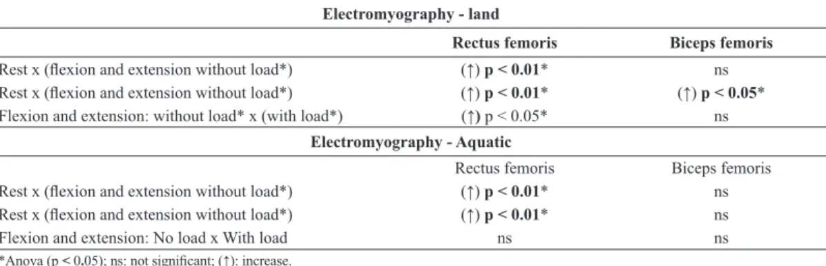
This increase could be attributed to the action of the hydrostatic pressure and the viscosity of the water, in addition to the additional resistance resulting from the load use. In addition, when the member moves in Table 1. Statistical results of electromyography during lexion and knee extension exercises with and without load performed on the loor and in the aquatic environment.
Electromyography - land
Rectus femoris Biceps femoris
Rest x (lexion and extension without load*) (↑) p < 0.01* ns
Rest x (lexion and extension without load*) (↑) p < 0.01* (↑) p < 0.05*
Flexion and extension: without load* x (with load*) (↑) p < 0.05* ns
Electromyography - Aquatic
Rectus femoris Biceps femoris
Rest x (lexion and extension without load*) (↑) p < 0.01* ns
Rest x (lexion and extension without load*) (↑) p < 0.01* ns
Flexion and extension: No load x With load ns ns
*Anova (p < 0.05); ns: not signiicant; (↑): increase.
Table 2. Statistical results of electromyography during lexion and knee extension exercises with and without load comparing that on the
loor with that in the aquatic environment.
Electromyography Rectus femoris Biceps femoris
Land rest x (immersion rest*) (↓) p < 0.001* (↓) p < 0.001*
Flexion and extension: Land without load x (immersion without load*) (↓) p < 0.001* (↓) p < 0.001*
Flexion and extension: Land with load x (immersion with load*) (↓) p < 0.001* (↓) p < 0.001*
the aquatic environment, it is subjected to drag forces
and turbulence (Becker, 2009)
By comparing the data for the movements performed in water and on land, there was a reduction in the electrical activity of the rectus and biceps femoris with and without load in the aquatic environment. These results were consistent with studies conducted by Sugajima et al. (1996) and Devereux et al. (2005), who found that in water, there was less force production compared with the terrestrial environment due to the buoyant force, which acts in the opposite direction to
gravity and reduces the apparent weight (hydrostatic
weight). Pöyhönen et al. (1999) found a signiicant reduction in the amplitude of the electromyographic signal during submaximal and maximal contractions
of the knee extensor muscles in immersion compared
with on the ground. Masumoto et al. (2004) reported
that the individuals walking in the liquid medium
showed a decrease in the muscle electrical activity
compared with walking on the ground. The authors
explained that these results could be attributed to the effect of buoyancy in the water, suggesting that the
individuals minimized their efforts to walk in water,
which would have resulted in reduced muscular endurance.
According to Pöyhönen et al. (1999) and
Kelly et al. (2000), the reduction of muscle electrical activity during immersion exercises can be attributed to the absence of gravity on muscle spindles and proprioceptive receptors in the displacer due to buoyancy.
Another factor that may decrease the electromyographic signal is the water temperature. This study used
immersion at 34°C, which is commonly used in therapeutic pools. It is known that the heated
water temperature contributes to muscle relaxation
(Biasoli and Machado, 2006) and increased blood
supply to the skeletal muscle system by peripheral vasodilation (Becker, 2009; Biasoli and Machado,
2006; Rahmann et al., 2009).
Cuesta-Vargas et al. (2013a) found increased electrical activity of some muscles of the legs on the ground compared with in water; however, when
evaluating the trunk muscles, there was a larger
increase in the electromyographic signal in the aquatic environment. Müller et al. (2005) also found an increase
in the electrical activity of the trunk muscles during
immersion in water compared with on the ground, both at standard speed and full speed.
In contrast with the results obtained in this study,
Pinto et al. (2010) found no signiicant changes in
the electromyographic activity on land or in water during maximal voluntary isometric contractions of
the biceps muscles, triceps brachii, rectus femoris and biceps femoris.
For a study to be comparable on land and in
water, playback is needed the same procedure
in these two environments, as well as the use of adequate protection to prevent water from entering
the electrode during immersion (Castillo-Lozano and Cuesta-Vargas, 2013; Cuesta-Vargas, 2013b). If there is water contact with the electrode during myoelectric evaluation, the myoelectric signal may be decreased
(Carvalho et al., 2010; Pöyhönen et al., 1999). In the research conducted, the same procedures on land and in water were made. Furthermore, to evaluate the electromyographic signal in water, water impermeable occlusive tape was used under the electrodes, as well
as plastic polyvinyl chloride (PVC) in the region of
the body part in question.
Considering the resistance exercises, Pinto et al.
(2014) found that when endurance was used before aerobic exercise, there might be optimization of muscle strength and hypertrophy of the lower limbs. Another
study investigated the effects of a ten-week training
program with progressively increasing resistance in
the aquatic environment and evaluated the lexors and knee extensors of twenty-four healthy women. There was a signiicant increase in the torque, lean
mass and neural activation of the muscles, concluding that training in the aquatic environment could be recommended for neuromuscular conditioning
(Pöyhönen et al., 2002).
In a therapeutic context, authors report that aquatic therapy allows active movement in the early postoperative period without compromising patient
safety (Kelly et al., 2000). According to Bressel et al.
(2012), patients with pain symptoms can more easily
perform muscle exercises irst in an aquatic environment
such that that they could be subsequently transferred to the terrestrial environment. Orselli and Duarte
(2011) observed a signiicant reduction in the joint
torque during marching in water compared with on land, suggesting that the therapeutic pool is a safe environment for the exercise due to the reduction of impact.
The results of this study are in accordance with most of the ongoing research in the literature, although these studies were performed during exercise without load. The heated water may have contributed to the reduction of electromyographic activity. Some studies reported increased electromyographic activity during exercise in immersion compared with on the ground;
however, the analysis was performed on trunk muscles.
Knowing that there is a reduction of electrical
muscle activity while performing movements in the aquatic environment compared with on land, including load use, it is believed that resistance exercises can be performed early in a therapy pool, which will favor
the prevention and treatment of musculoskeletal
disorders, considering that the patient can sooner return to their activities of daily living.
Future studies on exercises using different resistance graduations performed in other positions and velocities and in a pool with water lower temperature may be conducted to improve the results obtained in this
study in order to determine the inluence of all of
these factors on the electrical activity muscle.
References
Batterham SI, Heywood S, Keating JL. Systematic review and meta-analysis comparing land and aquatic exercise for people with hip or knee arthritis on function, mobility and other health outcomes. BMC Musculoskeletal Disorders. 2011; 12(1):123. http://dx.doi.org/10.1186/1471-2474-12-123. PMid:21635746
Becker BE. Aquatic therapy: scientific foundations and clinical rehabilitation applications. PM & R. 2009; 1(9):859-72. http:// dx.doi.org/10.1016/j.pmrj.2009.05.017. PMid:19769921 Biasoli MC, Machado CMC. Hidroterapia: técnicas e aplicabilidades clínicas nas disfunções reumatológicas. Revista Brasileira de Medicina. 2006; 63:79-80.
Bressel E, Dolny DG, Vandenberg C, Cronin JB. Trunk muscle activity during spine stabilization exercises performed in a pool. Physical therapy in sport : official journal of the Association of Chartered Physiotherapists in Sports Medicine. 2012; 13(2):67-72. http://dx.doi.org/10.1016/j. ptsp.2011.06.002. PMid:22498146
Cardoso JR, Sousa LB, Lopes LG. Proposta de manual de aplicação do biofeedback eletromiográfico modelo Pathway MR-20 no músculo tibial anterior de pacientes portadores de sequela de Acidente Vascular Cerebral (AVC). Arquivos de Ciências da Saúde da Unipar. 2003; 7:77-82.
Caromano FA, Nowotny JP. Princípios físicos que fundamentam a hidroterapia. Fisioterapia Brasil. 2002; 3:1-9.
Carvalho RGS, Amorim CF, Perácio LHR, Coelho HF, Vieira AC, Karl Menzel HJ, Szmuchrowski LA. Analysis of various conditions in order to measure electromyography of isometric contractions in water and on air. Journal of electromyography and kinesiology : official journal of the International Society of Electrophysiological Kinesiology. 2010; 20(5):988-93. http://dx.doi.org/10.1016/j.jelekin.2009.12.002. PMid:20385505
Castillo-Lozano R, Cuesta-Vargas AI. A comparison land-water environment of maximal voluntary isometric contraction during manual muscle testing through surface electromyography. BMC Sports Science. Medicine and Rehabilitation. 2013; 5:28.
Cuesta-Vargas AI, Cano-Herrera C, Formosa D, Burkett B. Electromyographic responses during time get up and go test in water (wTUG). SpringerPlus. 2013b; 2(1):217. http://dx.doi.org/10.1186/2193-1801-2-217. PMid:23705108 Cuesta-Vargas AI, Cano-Herrera CL, Heywood S. Analysis of the neuromuscular activity during rising from a chair in water and on dry land. Journal of Electromyography and Kinesiology : Official Journal of The International Society of Electrophysiological Kinesiology. 2013a; 23(6):1446-50. http:// dx.doi.org/10.1016/j.jelekin.2013.06.001. PMid:23834813 Devereux K, Robertson D, Briffa NK. Effects of a water-based program on women 65 years and over: a randomised controlled trial. The Australian Journal of Physiotherapy. 2005; 51(2):102-8. http://dx.doi.org/10.1016/S0004-9514(05)70038-6. PMid:15924512
Escamilla RF, Fleisig GS, Zheng N, Barrentine SW, Wilk KE, Andrews JR. Biomechanics of the knee during closed kinetic chain and open kinetic chain exercises. Medicine and Science in Sports and Exercise. 1998; 30(4):556-69. http://dx.doi.org/10.1097/00005768-199804000-00014. PMid:9565938
Fujisawa H, Suenaga N, Minami A. Electromyographic study during isometric exercise of the shoulder in head-out water immersion. Journal of Shoulder and Elbow Surgery / American Shoulder and Elbow Surgeons ... [et al.]. 1998; 7(5):491-4. http://dx.doi.org/10.1016/S1058-2746(98)90200-2. PMid:9814928
Harrison RA, Hillman M, Bulstrode S. Loading of the lower limb when walking partially immersed: implications for clinical practice. Physiotherapy. 1992; 78(3):164-6. http:// dx.doi.org/10.1016/S0031-9406(10)61377-6.
Kelly BT, Roskin LA, Kirkendall DT, Speer KP. Shoulder muscle activation during aquatic and dry land exercises in nonimpaired subjects. The Journal of Orthopaedic and Sports Physical Therapy. 2000; 30(4):204-10. http://dx.doi. org/10.2519/jospt.2000.30.4.204. PMid:10778797 Kisner C, Colby L. Exercícios terapêuticos: fundamentos e técnicas. São Paulo: Manole; 2000.
Masumoto K, Takasugi S, Hotta N, Fujishima K, Iwamoto Y. Electromyographic analysis of walking in water in healthy humans. Journal of Physiological Anthropology and Applied Human Science. 2004; 23(4):119-27. http:// dx.doi.org/10.2114/jpa.23.119. PMid:15314269
Müller ESM, Black GL, Figueiredo PP, Kruel LFM, Hanisch C, Appell HJ. Comparação eletromiográfica do exercício abdominal dentro e fora da água. Revista Portuguesa de Ciências do Desporto. 2005; 5:255-65.
Orselli MIV, Duarte M. Joint forces and torques when walking in shallow water. Journal of Biomechanics. 2011; 44(6):1170-5. http://dx.doi.org/10.1016/j.jbiomech.2011.01.017. PMid:21334630
Pinto SS, Liedtke GV, Alberton CL, da Silva EM, Cadore EL, Kruel LF. Electromyographic signal and force comparisons during maximal voluntary isometric contraction in water and on dry land. European Journal of Applied Physiology. 2010; 110(5):1075-82. http://dx.doi.org/10.1007/s00421-010-1598-0. PMid:20697905
Pöyhönen T, Keskinen KL, Hautala A, Savolainen J, Mälkiä E. Human isometric force production and electromyogram activity of knee extensor muscles in water and on dry land. European Journal of Applied Physiology and Occupational Physiology. 1999; 80(1):52-6. http://dx.doi.org/10.1007/ s004210050557. PMid:10367723
Pöyhönen T, Sipilä S, Keskinen KL, Hautala A, Savolainen J, Mälkiä E. Effects of aquatic resistance training on neuromuscular performance in healthy women. Medicine and Science in Sports and Exercise. 2002; 34(12):2103-9. http://dx.doi.org/10.1097/00005768-200212000-00036. PMid:12471323
Rahmann AE, Brauer SG, Nitz JC. A specific inpatient aquatic physiotherapy program improves strength after total hip or
knee replacement surgery: a randomized controlled trial. Archives of Physical Medicine and Rehabilitation. 2009; 90(5):745-55. http://dx.doi.org/10.1016/j.apmr.2008.12.011. PMid:19406293
Roper JA, Bressel E, Tillman MD. Acute aquatic treadmill exercise improves gait and pain in people with knee osteoarthritis. Archives of Physical Medicine and Rehabilitation. 2013; 94(3):419-25. http://dx.doi.org/10.1016/j.apmr.2012.10.027. PMid:23131526
Sugajima Y, Mitarai G, Koeda M, Moritani T. Characteristic changes of motor unit activity in hip joint flexor muscles during voluntary isometric contraction during water immersion. Characteristic Changes of Motor Unit Activity in Hip Joint Flexor Muscles During Voluntary Isometric Contraction During Water Immersion. 1996; 6(2):83-95. http://dx.doi. org/10.1016/1050-6411(95)00020-8. PMid:20719666 Vodusek DB. Electromyography. In: Bo K, Berghmans B, Morkved S, Van Kampen M, editors. Evidence based physical therapy for the pelvic floor. Philadelphia: Elsevier; 2007. p. 53-63.
Authors
Indira Nayra Paz Santos1, Izabela dos Santos Mendes1, Mario Oliveira Lima1, Alderico Rodrigues de Paula Junior1, Andreza Ribeiro Simioni2, Paulo Roberto Garcia Lucareli3, Fernanda Pupio Silva Lima1*
1Laboratório de Reabilitação Sensório Motora, Instituto de Pesquisa e Desenvolvimento – IP&D, Universidade do Vale do Paraíba – Univap, Av. Shishima Hifumi, 2911, CEP 12244-000, Urbanova, São José dos Campos, SP, Brazil.
2Laboratório de Síntese Orgânica – IP&D, Universidade do Vale do Paraíba – Univap, São José dos Campos, SP, Brazil.