MANAGEMENT OF TARSAL TUNNEL SYNDROM E
REPORT OF SEVEN CASES
FLAVIOA.R SETTANNI, LINCOLN M. LEANDRO, JOSÉ A.Z. ZULETA, EIDMARA. NERI
SUMMARY - Seven patients with clinical and electroneurographic evidenc e of tarsal tunnel syndrom e wer e managed surgically, after failed attempts for non-surgical treatment. Post-operative results were more satisfactory than the previous response s t o non-surgical therapies . Tarsa l tunne l syndrome appear s t o respond bette r to surgical intervention than to conservative management .
KEY WORDS: tarsal tunnel, posterior tibial nerve, tibial nerve compression, surgical decompression .
Tratamento da síndrome do túnel do tarso: relato de sete casos
RESUMO - Sete pacientes co m evidência clínic a e eletroneurográfica de síndrome do túnel d o tarso fora m tratadas cirurgicamente, após tentativas sem sucesso de tratamento não cirúrgico. Os resultados pós-operatórios foram bem mais satisfatórios que as tentativas prévias com terapêuticas não cirúrgicas. A síndrome do túnel do tarso parece responder melhor à abordagem cirúrgica do que a condutas mais conservadoras.
PALAVRAS-CHAVE: túnel do tarso, nervo tibial posterior, compressão do nervo tibial, descompressão cirúrgica.
Tarsal tunnel syndrome is a well known entity. Studies have described this illness in patients with neurological and electroneurographic evidence of posterior tibial nerve compression3 8 1 0.
From the pertinent literature, there seems to be agreement that this syndrome constitutes a disorder primarily amenable to surgical intervention. The present work was carried out in a series of seven prospectively observed patients, in whom clinical management was unsatisfactory. The latter consisted of systemic administration of non-steroid anti-inflammatory agents, local steroid infiltrations, and a variety of podiatric procedures. Patients eventually underwent surgical decompression of the posterior tibial nerve within the tarsal tunnel.
CASE REPORTS
Case 1 . A 43-year-old woma n presente d wit h a 12-yea r history of progressive burnin g pain ove r the postero-lateral aspec t o f the right leg, irradiated t o the medial an d plantar region s o f the right foot. Thes e symptoms were exacerbated on the upright position, and while deambulating. Relief was achieved during rest. Neurological examination disclosed mild deficit on plantar flexion of the right foot and hallux with hypoesthesia on the medial and dorsal aspects of the right foot. Painful symptom s could be triggered by pressure applied to the medial retromalleolar region. Electroneurographic testing revealed marked slowness in neuroconduction of the dista l segment s o f both posterio r tibia l nerves , mor e pronounced o f the right side. These abnormalitie s
From the Division o f Neurosurgery, Hospita l do Servidor Público Estadual de São Paulo, São Paulo. Aceite: 18-abril-1994 .
suggested bilateral tarsal tunnel syndrome, worse on the right. The patient underwent unilateral exploration of the right tarsal tunnel. At operation a varicose venou s plexus was found, whic h exerte d compressio n o n the posterior tibial nerve and its branches. Nerve decompression was uneventfully carrie d out, and a 2-year follow-up has been asymptomatic.
Case 2. A 45-year-old woma n presente d wit h a 19-mont h history o f progressive burnin g pain on the medial-dorsal aspec t o f th e lef t foot , associate d wit h numbnes s o f th e hallu x an d secon d toe . Neurologica l examination reveale d mil d deficit o n plantar flexion o f the left foo t an d hallux, with associated hypoesthesi a over the dorso-medial aspect of the hallux and second toe. Electroneurographic testing disclosed abnormalitie s consistent with left posterior tibial neuropathy, suggestive of tarsal tunnel syndrome. At operation several varicose venous branche s wer e foun d t o b e compressin g th e lef t posterio r tibia l nerve . Nerv e decompressio n wa s uneventfully carrie d out, and a 2.5-year follow-up has been asymptomatic.
Case 3. A 46-year-old woman presented with a 1-year history of progressive burning pain and numbness over the posterior face of the right leg and heel. Symptoms were exacerbated with deambulation, and relieved during rest. Neurological examination revealed mild deficit o n plantar flexion of the right foot and hallux, with associated hypoesthesia over the entire plantar surface. Painful symptom s could be triggered by pressure over the medial retromalleolar region. Electroneurographic testing showed loss of excitability of plantar nerves, and increased latency on the posterior tibial nerve, suggestive of tarsal tunnel syndrome. At operation, thickening of the retinaculu m o f th e flexo r muscle s wa s found , exertin g compressio n o n the posterior tibial nerve . Nerve decompression was uneventfully carried out, and the patient was asymptomatic for 18 months. She then presented with the same signs and symptoms on the left side, which were also refractory t o clinical management. Surgery was the n carrie d ou t o n th e left foot , wit h th e sam e findings a s o n the right side . Upon discharg e fro m th e hospital she was asymptomatic. The patient was lost for long-term follow-up .
Case 4. A 72-year-old woman presented with a 10-yea r history of progressive burning pain on the left plantar region. Three years after onse t of symptoms, she developed th e same problem on the right side. Pre-morbid history was remarkale for a fracture of the left ankle five years before onset of the left-sided symptoms . Neurological examinatio n revealed mil d defici t o n planta r flexio n o f bot h fee t an d halluces , wit h bilatera l plantar hypoesthesia. Painful symptom s could be triggered by pressure over both medial retromalleolar regions. Electroneurographic testing showed absent sensory conduction on both posterior tibial nerves, with otherwise normal findings. Bilatera l tarsa l tunne l syndrom e wa s therefor e suggested . Th e patien t underwen t bilatera l exploration o f the tarsal tunnel, which revealed thickenin g o f the retinaculum o f th e flexor muscle s o n both sides. Decompression o f both posterior tibial nerves was uneventfully carrie d out, and a 3-year follow-up ha s been essentially asymptomatic, except for slight numbness on the left foot .
Case 5. A 53-year-old woman presented with a 2-year history of progressive numbness of all toes on the right side, associated with burning pain over the plantar and dorsal regions of the foot. Simila r symptoms had subsequently and gradually developed on the left side, over an 8-month period. Neurological examination disclosed hypoesthesia over the dorso-lateral aspect of both feet, without motor deficit. Electroneurographic testing showed sensory potentials on both posterior tibial nerves, in response to medial-plantar stimuli. This was suggestive of bilateral tarsal tunnel syndrome. The patient underwent exploration of the right tarsal tunnel, which disclosed multiple compression of the posterior tibial nerve by large varicose veins. Nerve decompression was uneventfully carried out, with complete relief of painful symptom s and persistent hypoesthesia on the right foot, over a 10-month follow-up period .
Case 6. A 69-year-old woman presented with a 1-year history of progressive burning pain in both plantar regions, without irradiation, exacerbated during deambulation, and relieved during rest. Due to gradual worsening, she lately was unable to walk two blocks. Pre-morbid history was remarkable for a 20-year long diabetes mellitus. Neurological examination was normal. Electroneurographic testing revealed marked decrease of neuroconduction in distal segments of both posterior tibial nerves, suggestive of tarsal tunnel syndrome. The patient underwent bilateral exploration of the tarsal tunnel, which revealed thickening of the retinaculum of the flexor muscles on both sides, and marked venous varicose compression o f the posterior tibial nerve on the left. Upo n discharg e from the hospital she was asymptomatic, with complete relief of the painful symptoms . A long-term follow-u p is pending.
Neurological examinatio n wa s normal . Electroneurographic testin g revealed a predominance o f moderatel y neurogenic motor unit potentials of the abductor hallux and gastrocnemius muscle s on the left, i n contrast to normal motor unit potentials on the right. A complete blockage in neuroconduction o f the left posterio r tibial nerve was also documented. These findings were consistent with tarsal tunnel syndrome on the left. The patient underwent surgical decompression of the left posterior tibial nerve. At operation, pressure was being applied to the posterior tibial nerve by the laciniate ligament, which was satisfactorily relieved . A 3-month follow-up has been asymptomatic .
Anatomical features. Th e tarsal tunnel is an osseofibrous structure, comparable to the carpal tunnel. Its floor is formed by the calcaneal and talar bones, and tendons of the following muscles: posterior tibial, long flexor of the hallux, and long flexor of the small toes. Its roof is formed by the retinaculum of the flexor muscles of the foot, a thin layer of fibrous tissue which extends from the medial malleolus to the heel and proximal border of the abductor muscle of the hallux4.
The posterior tibial nerve runs inferiorly to the medial malleolus and anteriorly to the Achilles tendon, crossing the tarsal tunnel. At the distal end of the tarsal tunnel, the posterior tibial nerve gives off three branches: (a) medial calcaneal branch; (b) medial plantar branch; an (c) lateral plantar branch. Figure 1 illustrates the anatomical landmarks of the tarsal tunnel.
Pathophysiology. An y lesion occupying space inside the tarsal tunnel may result in the tarsal tunnel syndrome, either because of direct compression of the posterior tibial nerve, or because of ischemic neuropathy.
Common etiologic sources in this syndrome are those leading to pathological modification of the spatial configuration of the anatomical elements, secondary to fracture, displacement, or direct pressure as in forced plantar flexion, and acute or chronic eversión of the foot or heel.
Other frequent etiologi c source s ar e lipomas, neuromas , post-traumati c fibrosis , synovia l hypertrophy in reumathoid arthritis, excessive weight, post-traumatic edema, chronic stasis of the posterior tibial veins, thrombophlebitis, and venous ectasis.
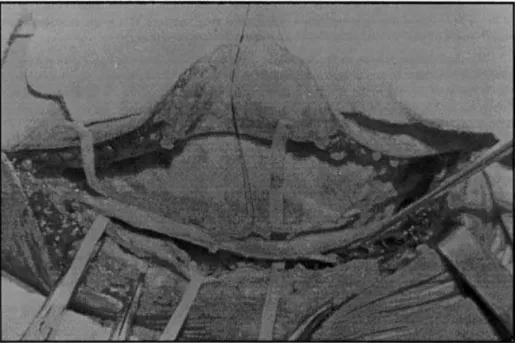
Figure 2. Surgical exposure of the posterior tibial nerve within the tarsal tunnel. The retinaculum of the flexor muscles was sectioned allowing decompression of the nerve.
Anatomopathological findings ar e generally typica l o f a chronic lesion , whic h sometime s leads t o the formation o f neurom a secondar y t o compressio n o f th e posterior tibia l nerv e or its branches. Neuroma formation i s most frequently locate d underneath the retinaculum, behind the malleolus.
Denny-Brow and Brenner2 have demonstrated, experimentally, that pathophysiologic alterations of peripheral nerves occur when mild or moderate nerve compression is applied, mostly during the initial phase of compression.
Differential diagnosis. Befor e definitive confirmatio n of tarsal tunnel syndrome, it is important to rule out other pathologic processes that may mimic this syndrome. Among those are: plantar fasciitis, interdigital neuroma, prolapse of metatarsal head, plantar callus, and reumathoid disease of the foot. In addition to these entities, different source s of sciatic pain, peripheral neuropathy, and peripheral vasculopathy must be excluded as well.
Electroneurographic evaluatio n o f muscle s innervate d b y th e posterio r tibia l nerve , wit h emphasis on the conduction velocity and latency period, have been reported as of diagnostic value5. In fact, the following parameters have been proposed by Distefano and co-workers3 as indicative of nerve compression: a latency period greater than 6.1 m/sec for the medial plantar branch, and greater than 6.7 m/sec for the lateral plantar branch. According to Kaplan and Kernohan6, the lateral plantar branch is more frequently affected than the medial branch.
Treatment. Non-surgical therapeutic measures, such as local infiltration with steroid anti-inflammatory agents, use of elastic stockings, and a variety of podiatric measures, frequently promote just a short-lasting relief os symptoms.
remarkably satisfactor y result s i n response t o surgica l treatment , i t becomes apparen t that early diagnosis and surgical management should be adopted more frequently. This would avoid potential complications associated with long-standing palliative procedures.
Surgical decompression within the tarsal tunnel, by sectioning the retinaculum of the flexor muscles, is indicated. In addition, exploration of the three branches of the posterior tibial nerve1-9 with removal o f fibrou s tissu e involvin g them , a s wel l a s distal exploratio n o f thes e branche s underneath the muscle abductor of the hallux, appear to be important supplementary maneuvers in the surgical approach of the tarsal tunnel syndrome.
Exposure of the retinaculum of the flexor muscles is easily accomplished through a curvilinear incision, starting 2.5 to 3 cm above and behind the medial malleolus, and extending inferiorly and anteriorly to this structure by approximately 4.5 to 5 cm4. Figure 2 illustrates the surgical approach in one of the patients.
Results. The seven female patients above-described presented with immediate post-operative relief of symptoms, which was documented as being long-lasting in four of them. None of these patients had previously experience d a sustained relief o f symptom s b y non-surgica l therapies . Thus, the present results, as well as the pertinent literature, strongly reinforce the notion that the tarsal tunnel syndrome is, up to current days, a primarily surgical entity.
Acknowledgment - Th e author s than k Juli o Cruz , M.D. , fo r criticall y reviewin g an d editin g th e manuscript.
REFERENCES
1. Dellon AL, Mackinnan SE . Tibial nerve branching in the tarsal tunnel. Arch Neurol 1984 , 41: 645-646. 2. Denny-Brown D, Brenner C. Paralysis of nerve induced by direct pressure and by tourniquet. Arch Neurol Psychiat 1944 , 51: 1-26.
3. Distefano V, Sack JT, Whittaker R et al. Tarsal-tunnel syndrome. Clin Orthop 1972 , 88: 76-79.
4. Edwards WG, Lincoln CR , Basset FH et al. The tarsal tunnel syndrome : diagnosis an d treatment. JAM A 1969, 207: 716-720.
5. Johnson EW, Ortiz PR. Electrodiagnosis o f tarsal tunnel syndrome. Arch Phys Med Rehabil 1966 , 47: 776-780.
6. Kaplan PE, Kernahan WT. Tarsal tunnel syndrome: a electrodiagnostic and surgical correlation. J Bone Joint Surg (Am) 1981, 63A: 96-99.
7. Keck C. The tarsal tunnel syndrome. J Bone Joint Surg (Am) 1962, 44A: 180-182. 8. Lam SJS. A tarsal-tunnel syndrome. Lancet 1962 , 2: 1354-1355.
9. Omer G, Spinner M. Management of peripheral neural problems. Philadelphia: Saunders, 1980.