Primary sequence, oxidation-reduction potentials
and tertiary-structure prediction
of
Desulfovibrio
desulfuricans
ATCC
27774 flavodoxin
Jorge CALDEIRA'.', P. Nuno PALMA', Manuela REGALLA*, Jorge LAMPREIA'.*, Juan CALVETE3,4, Wolfram SCHAFER4, Jean LEGALL', Isabel MOURA'.* and JosC J. G. MOURA'
'
Departamento de Quimica, Faculdade de CiCncias e Tecnologia, Universidade Nova de Lisboa, Monte de Caparica, Portugal'
Instituto de Tecnologia Quimica e Biolbgica, Universidade Nova de Lisboa, Oeiras, Portugal Consejo Superior de Investigaciones Cientificas, Madrid, SpainMax-Planck-Institut fur Biochimie, Martinsried, Germany Department of Biochemistry, University of Georgia, Athens, USA (Received December 21, 1993) - EJB 93 1890/3
Flavodoxin was isolated and purified from Desulfovibrio desulfidricans ATCC 27774, a sulfate- reducing organism that can also utilize nitrate as an alternative electron acceptor. Mid-point oxida- tion-reduction potentials of this flavodoxin were determined by ultravioletlvisible and EPR methods coupled to potentiometric measurements and their pH dependence studied in detail. The redox potential E2, for the couple oxidizedsemiquinone forms at pH 6.7 and 25°C is -40 mV, while the value for the semiquinonehydroquinone forms (El), at the same pH, -387 mV. E2 varies linearly with pH, while E, is independent of pH at high values. However, at low pH (<7.0), this value is less negative, compatible with a redox-linked protonation of the flavodoxin hydroquinone. A com- parative study is presented for Desulfovibrio salexigens NCIB 8403 flavodoxin [Moura, I., Moura, J. J. G., Bruschi, M. & LeGall, J. (1980) Biochim. Biophys. Acta 591, 1-81.
The complete primary amino acid sequence was obtained by automated Edman degradation from peptides obtained by chemical and enzymic procedures. The amino acid sequence was confirmed by FAB/MS.
Using the previously determined tridimensional structure of Desulfovibrio vulgaris flavodoxin as a model [similarity, 48,6%; Watenpaugh, K. D., Sieker, L. C., Jensen, L. H., LeGall, J. &
Dubourdieu M. (1 972) Proc. Natl Acad. Sci. USA 69, 3 185 - 3 1881, the tridimensional structure of D. desulfuricans ATCC 27774 flavodoxin was predicted using AMBER force-field calculations.
Flavodoxins are a group of small electron-transfer pro- teins [l-31 (= 15-23 kDa) isolated from different organ- isms, that contain a single FMN group bound non-covalently to the polypeptide chain. They can transfer two electrons at low and differentiated redox potentials.
The way in which a flavodoxin's peptide chain modulates the redox properties of the FMN cofactor has been the major focus of the tridimensional structural studies. Thermodynam- ic data have been discussed in terms of the binding of the cofactor to the polypeptide chain in different redox states of the protein. The pH dependence of the mid-point oxidation- reduction potentials has been extensively explored.
To infer on factors controlling the electron-transfer pro- cess, X-ray diffraction and NMR spectroscopy have been valuable tools to reveal. the environment of the FMN group bound to the protein. Four tertiary structures are available, for Clostridium beijerinckii (former Clostridium CP) [4, 51,
Correspondence to J. J. G. Moura, Departamento de Quimica,
Faculdade de CiCncias e Tecnologia, Universidade Nova de Lisboa, P-2825 Monte de Caparica, Portugal
Fax: +351 12954461.
Note. The novel amino acid sequence data published here have been submitted to the EMBL sequence data bank and are available under the accession number P80312.
D. vulgaris Hildenborough [6- 81, Anacystis nidulans [9] and Chondrus crispus [lo, 111 flavodoxins from X-ray studies ; and structures were proposed from two-dimensional and three-dimensional NMR studies for Megasphaera elsde- nii [12-151 and D. vulgaris [16, 171 flavodoxins. Amino acid sequences are also available for D. desulfuricans ATCC 29577 [18] Desulfovibrio salexigens [ 191 and Desulfovibrio gigas ATCC 29464 and 19364 flavodoxins [20]. Cloning and gene expression on flavodoxins from C. beijerinckii [21], D. vulgaris [3, 22, 231, D. salexigens [19] and D. gigas (two strains) [20] have been reported.
In this study we present the isolation, purification and characterization of D. desulfuricans ATCC 27774 flavo- doxin. Redox potentials were determined by potentiometric titrations coupled with visible and EPR techniques, at dif- ferent pH values. The value of the dissociation constant of the FMN group was estimated by differential spectrophoto- metric titrations of FMN with apo-flavodoxin. The complete amino acid sequence of D. desulfuricans ATCC 27774 flavo- doxin was obtained and its degree of similarity with other Desulfovibrio species flavodoxins is presented. A theoretical model of a tertiary structure obtained using the most similar sequence available (D. vulgaris flavodoxin) is predicted using molecular-modeling tools.
988
MATERIALS AND METHODS Growth of microorganisms
D. desulfuricans ATCC 27774 cells were grown using the medium described by Liu and Peck [24]. Low concentra- tion of iron was a requirement for maximal flavodoxin ex- pression, confirming previous findings indicating the re- placement of flavodoxin by ferredoxin in a rich iron media [25]. Nitrate rather than sulfate was used as a terminal electron acceptor in order to obtain a higher yield of biomass. The cells were harvested by centrifugation and stored at
- 80 "C until use. The cells were thawed and resuspended in
10 mM Tris/HCI, pH 7.6, and broken by means of a French press (6205 kPa). Cells from D. salexigens strain British Gui- ana (NCIB 8403) were grown at 37°C on lactate/sulfate me- dium [26, 271 and treated as previously indicated.
Protein purification
All the purification procedure of D. desulfuricans ATCC 27774 flavodoxin was carried out at 4°C. The crude extract (total volume 1500ml) obtained from 8OOg cells was dia- lyzed and centrifuged at 19000Xg during 30min then at 18OOOOXg for 75 min. The supernatant was applied to a DEAE-52 (Whatman) column ( 5 cmX40 cm) and the bound proteins eluted with a linear gradient from 1 O m M to 500 mM Tris/HCl, pH 7.6 (total volume of 2 1). A yellow fraction eluting at 400 mM was collected, concentrated in a Diaflo apparatus (Amicon) with a YM5 membrane and centrifuged at 4500 g for 30 min. The sample was then fil- tered with a membrane of 0.22 pm, applied to a HPLC with a DEAE-52 TosoHaas column (20 cmX5.5 cm) and eluted with a linear gradient from 1OmM to 500mM Tris/HCl, pH 7.0. Flavodoxin was collected around 380 mM. The same concentrationkentrifugation procedure was then repeated. A final purification step was performed using TSK-GEL G- 3000SW LKB HPLC column (60 cmX2.1 cm; gel filtration), eluted with 0.3 M Tris/HCI, pH 7.0. The flavodoxin was dia- lyzed against 10 mM Tris/HCl, pH 7, concentrated and stored at -20°C. An A,,,,/A,,,purity ratio of 3.7 was obtained. Lio- philization procedure is avoided due to dissociation of the FMN group. Approximately 350 mg pure flavodoxin was ob- tained from 1 kg wet cells under the growth conditions used. D. salexigens flavodoxin was purified as indicated in refer- ence [28].
Oxidationheduction studies
(mid-point oxidation-reduction potentials)
Mid-point oxidation-reduction potentials of D. salexigens and D. desulfuricans ATCC 27774 flavodoxins were deter- mined by ultravioletlvisible potentiometric titrations per- formed under anaerobic conditions in an optical redox cell slightly modified from the design of Dutton et. al. [29, 301. The solution potential was measured with a Crison 2002 po- tentiometer equipped with platinum (P133 2 radiometer) and Ag/AgC1 (K 8040 radiometer) electrodes and quoted relative to the normal hydrogen standard electrode. The following redox mediators were present at the final concentration of 5 pM : 1,4-naphthoquinone, methylene blue, triquat, pheno- safranine, benzylviologen, methylviologen, dichloroindophe- nol, benzoquinone, anthraquinone-2-sulfonic acid, phenazi- namethosulfate, dimethyltriquat, indigo tetrasulfonate, 2-hy- droxy-l,4-naphthoquinone, 5-hydroxy-l,4-naphthoquinone, duroquinone, phenazine and safranine.
Solution redox potentials (in equilibrium) were varied by adding appropriate volumes of deareated dithionite as reduc- tant. Ultravioletlvisible spectra were recorded during titration on a Shimadzu spectrophotometer 265FS. To study the pH dependence of the mid-point redox potentials of flavodoxin, different buffers were used at the following pH values: 0.05/ 0.05 M sodium citrate/sodium phosphate, pH 5.5; 0.1 M, Tris/HCI, pH 6.7-8.0; 0.1 M glycine/NaOH, pH 9.1. All ex- periments were performed under a purified argon atmosphere (passed through an oxygen trap from Chemical Research Supplies). Electrodes were calibrated with quinhydrone at pH 7.0 [31].
EPR redox titrations (at pH 8.0) were also performed for D. desulfuricans ATCC 27774 flavodoxin. The redox poten- tials were varied and measured as described above. The sam- ples (protein at a concentration of 200 pM) were poised at different redox potentials and transferred under purified ar- gon to EPR tubes and frozen in liquid nitrogen for subse- quent quantification. Spectra were recorded at 200 K on a Bruker ESP 300 Spectrometer.
Estimation of dissociation constants for the complex apo-flavodoxin/FMN
Apoprotein was prepared by extracting the FMN from the holoprotein with trichloroacetic acid as described 1321. Apo-flavodoxin was collected by centrifugation and resus- pended after neutralization with Tris/HCI, pH 9. This solu- tion was freshly prepared before conducting the association assay. Commercial FMN (Sigma) or flavodoxin trichloro- acetic acid FMN extracted and repurified by gel filtration (Sephadex G-10) were used. Differential spectrophotometric titration was used to determine the dissociation constant [2, 3, 331. All solutions were buffered at pH 7.0 with 0.1 M Trid HCl. Aliquots of apoprotein were added to one cuvette while the same volume of buffer was added to the reference cu- vette. Temperature was maintained at 25 "C. The titration curve was followed by the difference of optical absorbance (A491 -&).
Sequence of D. desulfuricans ATCC 27774 flavodoxin N-terminal sequence analyses of native flavodoxin and its derived peptides were performed on an Applied Biosys- tem Sequencer model 477A, coupled to an Applied Biosys- tem 120 Analyzer following the manufacturer's instructions. Cleavage of flavodoxin (10 mg/ml in 70%, by vol., for- mic acid) at methionine residues was performed with cyano- gen bromide (100 mg/ml final concentration) for 4 h at room temperature, in the dark, under a nitrogen atmosphere. The reaction mixture was then diluted with water and lyophilized. Proteolytic digestion of flavodoxin ( 5 mg/ml in 100 mM sodium phosphate, pH 7.8) was performed with a-chymo- trypsin (Sigma), endoproteinase Glu-C (Boehringer Mann- heim), endoproteinase Arg-C (Boehringer Mannheim) at an enzyme/substrate ratio of 1 : 50 (by mass) at 37 "C overnight. Alternatively, digestion with endoproteinase Glu-C was car- ried out in 50 mM ammonium bicarbonate, pH 8.0.
Peptides were isolated by reverse-phase HPLC on either a Pep-S (Pharmacia) or Lichrospher RP-100 (Merck) column (25 cmXO.4 cm, C,,, 5-mm particle size) eluting at 1 mllmin with a gradient of 0.1% trifluoroacetic acid in (A) water and (B) acetonitrile, following the absorbance at 220 nm.
Amino acid analyses were performed using a Beckman 6300E analyzer after sample hydrolysis with 6 M HCl at
110°C for 24, 48 and 72 h in sealed evacuated tubes. Values for threonine, serine and tyrosine were corrected after extra- polation to zero-time hydrolysis. Cysteine and methionine were recovered as cysteic acid and methionine sulfone, re- spectively, after performic oxidation [34, 351.
FAB mass spectra of peptides were recorded with a mass spectrometer Mat 900 (Finnigan MaT) equipped with a liquid secondary-ion ionization system. Nominal accuracy was ?0.5Da in the range 500-2500. The molecular mass of na- tive apo-flavodoxin was determined by laser-desorption MS using either a Lasermat (Finnigan Mat) or a Kratos Kompact
MALDI-3 instrument. Equine heart cytochrome c
(12360.1 Da) and soybean trypsin inhibitor (20090.6Da) were used as calibration standards. Accuracy claimed by the manufacturers was -+ 0.1 %.
Prediction of tridimensional structure by similarity modeling
A computational prediction of the tertiary structure of the flavodoxin was performed using a personal Iris-Silicon Graphics workstation. SYBYL 5.5 (Tripos Associates) and AMBER force field [36]. D. vulgaris Hildenborough flavo- doxin was chosen as the template structure due to its high degree of similarity, and its atom coordinates were obtained from the Brookhaven Protein Data Bank [37].
RESULTS AND DISCUSSION
Flavodoxins have been identified and isolated from sev- eral members of the Desulfovibrio genus [18, 26, 331. Most have been characterized in terms of molecular mass, amino acid composition and sequences, as well as visible spectros- copy (oxidized, one-electron-reduced and two-electron-re- duced forms). D. vulgaris flavodoxin structure was eluci- dated by X-ray and two-dimensional 'H NMR [6-8, 16, 171. Many studies have shown structural and physical simi- larities within this family, but differences exist, mainly con- cerning the span of redox potentials and details on the struc- ture around the FMN-binding site [38]. Comparison of amino acid sequences indicate that all Desulfovibrio species flavo- doxins shared a common FMN-binding site with Trp and Tyr flanking residues. Lifetime fluorescence of bound FMN in D. gigas flavodoxin is consistent with a more polar FMN- binding site [39].
D. desulfuricans ATCC 27774 flavodoxin spectral features, redox potentials and Kd values for the apo-flavodoxin/FMN complex
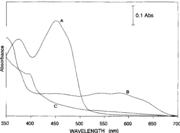
Fig. 1 indicates the spectral characteristics of the three redox forms of D. desulfuricans ATCC 27774 flavodoxin. These visible spectra closely resemble those of other flavo- doxins isolated from different organisms including Desulfovi- brio species [26, 331. The absorption coefficients were calcu- lated to be 37.0 (oxidized, 275 nm), 10.3 (oxidized, 452 nm) and 4.2 mM-' cm-' (semiquinone, 580 nm). The visible spectrum B (Fig. 1) shows a highly stable semiquinone form derived from the full conversion of the FMN moiety. Fig. 2 shows the EPR semiquinone isotropic signal with noticeable hyperfine structure. The linewidth of the radical species ob- served in the semiquinone state narrows when the aqueous solvent is exchanged with a deuterated one (1.9 mT to 1.5 mT), indicating the presence of a neutral radical form
I
/TA 0.1 Abs1
de
P
2 m 350 400 450 500 550 600 650 700 WAVELENGTH (nm)Fig. 1. Ultraviolet and visible spectra of D. desulfuricans ATCC
27774 flavodoxin, (A) native-oxidized, (B) semiquinone and (C)
hydroquinone forms. Spectra were obtained at pH 7.6 in 10 mM
Tris/HCl. The spectra were obtained poising the protein solution at adequate redox potentials (see Materials and Methods) in the pres- ence of dye mediators. The small contribution observed in the fully reduced state (spectrum C) at 400 nm is due to the reduced forms of the mediators present. The semiquinone-form spectrum represents a redox state of the protein where this form attains maximal inten- sity.
t
332 354 336 338 340
MAGNETIC FIELD (mTj
Fig.2. EPR spectrum of the semiquinone form of D. desulfuri-
cans ATCC 27774 flavodoxin. Experimental conditions : temper- ature, 200 K ; microwave power, 2.37 mW; modulation amplitude, 0.1033 mT/point.
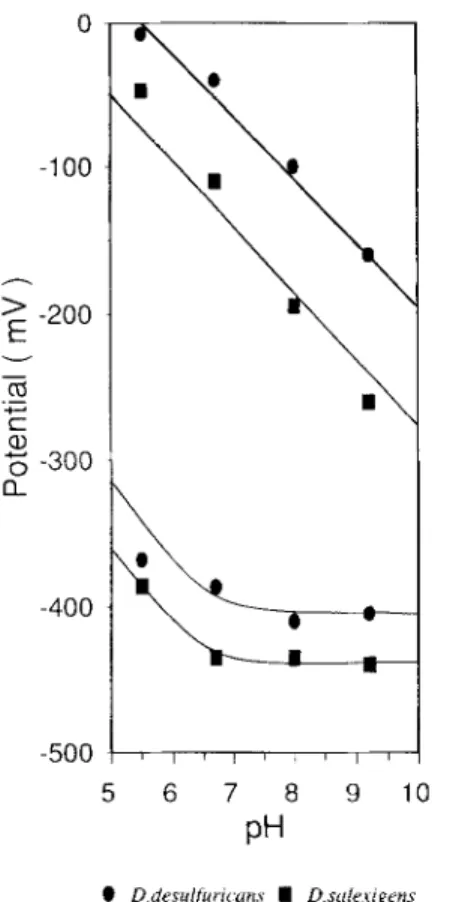
[40]. Redox potentials were determined, at pH 8.0, following the increase and decrease of this signal under non-saturating conditions at 120 K. The fitting of the experimental data (not shown) with two independent one-electron Nernst equations gives El = -100 mV and Ez = -410 mV.
Detailed analysis of the pH dependence of the mid-point redox potentials was made by visible redox titrations per- formed as described in Materials and Methods. Fig. 3 com- piles the measured redox potentials and its pH dependence (including data for D. salexigens flavodoxin). The redox po- tentials of both flavodoxins in the studied pH range show a characteristic behavior, as already reported for other flavo- doxins [l -3, 331. Ez has a linear slope of -59 mV, while E , is pH independent (at high pH) until a pK, value around 6.7 is reached. It then becomes pH dependent, indicating the existence of a pH-linked process.
990 0 -1 00 h
’
-200 E v - cb c al5
-300a
._ -c-. -400 -500 5 6 7 8 9 10PH
e D drsulfur~ uns I D s t i h i g m sFig. 3. pH dependence of the oxidation-reduction potentials of D.
desu@ricans ATCC 27774 ( 0 ) and D. salexigens (B) flavo-
doxins. Experimental details as indicated in Materials and Methods.
The origin of this pH-redox-linked process has been dis- cussed by different authors and its origin has not yet been completely assigned [2, 31. The ratios of Kd values for qui- none, semiquinone and hydroquinone of riboflavin versus those of FMN were used to show that the phosphate group of FMN has an important role on the binding of the flavin to the apoprotein [3]. The large shift of redox potential of the semiquinonehydroquinone couple was proposed to be due to unfavorable electrostatic interactions between the negative charges of the phosphate group of FMN and the negative charge of N1 of the flavin hydroquinone [2,41]. Ludwig and coworkers [2] also supported the existence of charge-charge repulsions but attributed it to the presence of a negative charge on a protein residue only. This conclusion was also reached based on NMR arguments [42-441. However, the problem is not yet completely understood since when Ver- voort and co-workers [41-431 introduced an extra negative charge into the FMN group they observed that the E , value became more negative whilst the removal of the phosphate group produced the opposite effect. Altogether, the presence of a nearby negative charge as well as the existence of the phosphate group, seems to be important in controlling the El values.
The peculiarity of D. desulfuricans ATCC 27774 lies in the fact that, at pH6.7, the E2value is about 70m V more positive than D. salexigens E, and about 130 mV more posi-
tive than D. gigas E, (Moura, I., unpublished results) while, at the pH-independent zone, its El value is 30/35 mV more positive than the typical -440mV [3]. As a consequence, D. desulfuricans ATCC 27774 flavodoxin shows the widest redox-potential span (E,-El) yet known in this family of proteins.
To calculate the dissociation constant of the complex of apo-flavodoxin with FMN, the addition of apo-flavodoxin to FMN was followed spectrophotometrically, as indicated in Materials and Methods. The experimental data were fitted with a theoretical curve based on a simple 1 : 1 binding model (Fig. 4). A Kd value of 0.1 nM was determined, as well as for D. vulgaris flavodoxin, Kd values of 0.24 nM (recombinant protein) and 0.2 nM (wild-type protein) [3].
Dissociation constants for the complexes of apo-flavo- doxin with the semiquinone and hydroquinone forms were calculated, Kdsq (semiquinone) = 24 nM and Kdhq (reduced form) = 4.8 mM, using the E, and E2 values determined, the Kd values for the complex FMN (oxid) and apo-flavodoxin and the values of E , and E2 reported for protein-free FMN [33]. These values indicate that the semiquinone form is more tightly bound to the protein moiety, as observed for other flavodoxins, with a relationship between Kdsq/Kdox = 240, which is one order of magnitude higher than the usually reported value and the consequence of a more positive E2 value. The ratio between Kdsq/Kdhq = 2X lo4 is similar to those previously found [33, 45-471.
D. desuljuricans ATCC 27774 flavodoxin amino acid sequence
The determined primary structure of D. desutfurirans ATCC 27774 flavodoxin is presented in Fig. 5, along with details on peptide analysis. The sequence is compared in Fig. 6 with five other known sequences of flavodoxins iso- lated from Desulfovibrio species (D. vulgaris Hildenborough [33], D. desulfuricans ATCC 29577 [18], D. salexigens [19], D. gigas ATCC 19364 and D. gigas ATCC 29494 [20]). For this purpose two programs were used: MACAW [48] and PC-Genem version 6.7 package [49] with the SWISS-PROT 26 (August 1993) data bank [50]. The multisequential align- ment and the PROSITE program [51] has determined a flavo- doxin signature at positions 6-22 :
ILFGSSTGNTESIAQKL22
which is known from X-ray structural analysis to be the bind- ing region of the FMN phosphate side chain [4-71. The six compared flavodoxins had a consensus length of 149 amino acids, sharing 25.5% identity and 45.6% similarity. Table 1 indicates the degree of identity between each pair of these proteins. The alignment was also used to determine the most significant sequence position of each amino acid on the six flavodoxins, allowing the calculation of the number of iden- tical amino acids in one sequence position. This number (from 6 to 1) is represented in Fig. 6 with different linewidths (thicker line to the most conserved position). It was also used to help visualize the spatial protein regions that had been more or less conserved in the different known Desulfovibrio flavodoxins on the tertiary backbone structure of D. vulgaris (Fig. 7).
Both aromatic residues, Trp60 and Tyr98, that flank the flavin isoalloxazine ring in the crystal structure of D. vul- garis are present in D. desulfuricans 27774 flavodoxin. The FMN-binding site is also preserved in Desulfovibrio sp. fla- vodoxins. However, variability is found in these flanking re- gions when other flavodoxins are considered and the pres- ence of two aromatic residues is not always verified: C. bee- erinckii Met56 and Tyr98 ; M . elsdenii Met56 and Tyr98 and A. nidulans Trp57 and Trp94 (see Table 2).
The molecular mass of apo-flavodoxin determined by mass spectrometry (combined 15420Dak 19) is higher (by
0.2
O.lJ, - 0 . 0 5 5 300 350 400 450 500 550 600 650 700 Wavelength (nm) I I I I I I I I I0
0.2
0.4
0.6
0.8
1
1.2
1.4
1.6
1.8
2
Apo/F M
N
(m
ol/m
o I)
Fig. 4. Determination of the dissociation constant of the FMN group to D. desu&ricans ATCC 27774 apo-flavodoxin. Squares are
experimental points and the line indicates the theoretical fitting. Differential spectrophotornetric titration of 43.8 pM FMN (in 20 rnM Tris/ HCI, pH 7.6, with 417 pM apo-flavodoxin at 25°C. The insert shows equal volumes of FMN introduced into both cuvettes. Difference spectra were recorded after each addition of apo-flavodoxin to the sample cuvette and an equal volume of buffer to the reference cuvette. The fraction of bound FMN was calculated by the change in absorbance at 496 nm caused by the binding of apoprotein. The dissociation constant was calculated from the experimental curve after correction for dilution.
93Da 5 19) than that calculated from its amino acid se- quence (15 327.2Da; the molecular mass of the holoprotein estimated by gel filtration is 16 kDa). This indicates that either the sequence of apo-flavodoxin is longer or that it con- tains a modified residue. Every position within the apo-flavo- doxin sequence was confirmed by FABMS analysis of at least two different peptides, except the polypeptide stretch 56-62. However, this portion was sequenced from a CNBr- obtained peptide providing an unambiguous assignment of all the residues except Cys57, which gave a blank. A cysteine residue at this position is assumed based on its conservation in other known flavodoxin sequences. Recently, we have suc- ceeded in crystallizing D. desulfuricans ATCC 27774 flavo- doxin and the crystals diffract better than 2.2 nm (in collabo- ration with A. Romero and M. J. Romgo). Resolution of the three-dimensional structure indicates the presence of one or two extra amino acid residues in the C-terminus.
Molecular modeling studies
on D. desulfuricans ATCC 27774 flavodoxin
Using D. vulgaris flavodoxin tridimensional structure we have tried to predict a plausible tertiary structure for the D.
desulfuricans ATCC 27774 flavodoxin. A similar tertiary- structure prediction for D. salexigens flavodoxin was re- ported recently following an analogous methodology [52]. These calculations are based on the assumption that if a cer- tain class of proteins shows a high degree of similarity in function, cofactor content and amino acid sequences then a somewhat strong tertiary similarity is also expected [ 5 3 , 541.
Cases are known that even with a very weak similarity in the
amino acids sequences and/or function, the overall tridimen- sional folding pattern can be quite similar. This is the case with the remarkably similar folding pattern of the riboflavin synthetase [55] and N-carbamoyl synthetase [56, 571 and that found in flavodoxins.
Before using this method, the amino acid sequence of both D. vulgaris and D. desulfuricans flavodoxins were aligned. Since no insertions or deletions were required, and due to the high degree of similarity between the two proteins, the alignment was straightforward, and in agreement with the alignment of all other Desulfovibrio flavodoxins.
In the tertiary structure of D. vulgaris, only the side
chains of the different amino acids were changed according to the alignment, conserving all the atoms in the main chain and the a$ carbon bond, to maintain the relative orientation of the side chain to the a carbon.
The resultant computational mutated structure was then released of its energetic tensions using the AMBER force field including all hydrogen atoms.
The strategy of energy minimization was developed as proposed in [58] and consisted of four sequential steps. In the first step a rigidity constraint of the main-chain atoms was defined, which also includes all the atoms of the FMN group. Energy minimization was performed ignoring electro- static interactions, only taking into account the steric repul- sions. A second step was performed with the same con- straints, but considering electrostatic interactions. After these two steps the constraints are removed both from the main- chain and the Fh4N-side-chain atoms. The atoms of the isoa- lloxazine ring were kept rigid and always coplanar, in agreement with X-ray and NMR observations on flavodoxins
992
1 5 10 15 20 25 30 35 40
MSKVLILFGSSTGNTESIAQKLEELVAAGGHEVTLLNAAE
I -K2-1-151.5---~-K3-1565.5 I
I-KK-
]+terminal1-El--1552.4 I -E2-917. -1 -E3-753.4--- I -E4-830.4-- I 1-K1-899. I -K4--143 7. 1- -K7-
I 4 1 - 7 8 8 . P I I-K5-1096.7----
41 45 50 55 60 65 70 75 8 0
ASADNLADGYDAVLMGCSAWGMEDLELQDDFAPLFDEMEN
~-E6-l111 I .- I -Al- I (-A2-1879.-1 IA31 -K6--1381 .-I 1-K10-1593. E I I K11--1066.7 I-E8--1310.0---~ I- I-K9-1312. -1 I -R1- IE7 490.21 (-K12-915.4 81 85 9 0 95 100 105 110 115 120 1-E9-838. E-1 -K7-1780.7 I I K8 425.11MGLKGKKLAAFASGDMEYEHYCGAVPAIEEKAYGLGAEVI
-E10-1867.7 1 I-E13--1720.3 I - I 1 -A4--1445.-1 [-A5 411-1 /-Ell I \ - E 1 2 - 1 1 8 8 . L ~ - E 1 4 - 1 2 4 5 . ~ 4 l - l 1-K13-774.1-~ 121 125 130 135 140 145PEGLKIEGDASSDPDAVSAFAEDVLK
-A5 E14-1 I-K14--1609.2 lK15-546.01 I -.- - - 142-559.21-E15-1438.-1 1-R3-2136.9 I I K16-674.1-1Fig. 5. Amino acid sequence of D. desulfuricans ATCC 27774 flavodoxin. The obtained peptides and their molecular masses are shown.
A, cyanobromogen ; K, a-chymotrypsin; E, endoproteinase Glu-C in sodium phosphate buffer; D, endoproteinase Glu-C in ammonium bicarbonate buffer; R, endoproteinase Arg-C. Molecular masses were determined by FABMS.
D.d.21174 D . d . 2 9 5 7 7 D. salex D.vulgaris D.g. 1 9 3 6 4 D . g . 2 9 4 9 4 " 1 10 19 28 37 46 55 64 73 82 91 100109118 127136 145 1 4 6 148 1 4 6 1 4 8 1 4 6 1 4 7 7 > , I I , c r I , I , I I I ~ ~ i r n r ~ ~ ~ - ~ ~ ~ ~ , - - - q 0 20 40 60 80 100 120 140 160 sequence position
Fig. 6. Comparison of Desulfovibrio species flavodoxin amino acid sequences. The degrees of similarity of amino acid sequences
Table 1. Matrix comparison of amino acid sequence similarity in flavodoxins isolated from sulfate reducers.
Similarity of flavodoxins from
D. gigas 19364 D. gigas 29494 D. vulgaris Hld D. salexigens D. desulfuricans 29577
% D. desulfuricans 27774 44.5 D. desulfuricans 29577 43.8 D. salexigens 54.1 D. vulgaris Hld 56.2 D. gigas 29494 65.8 45.2 43.5 56.9 56.5 48.6 43.8 53.4 53.4 75.3 44.5
Fig. 7. Tridimensional similarity between Desulfovibrio species
flavodoxins based on the D. vulgaris structure. Shadowing refers
to higher or lower degrees of amino acid similarity when comparing the six amino acid sequences (darkest, six conserved amino acids until white, no conserved amino acid in a certain sequence position; see also Fig. 6).
[2, 4-8, 42-44]. The third and fourth steps were performed considering the same type of interactions used in the first and second steps, respectively.
The predicted structure of D. desulfuricans ATCC 27774 flavodoxin is essentially conserved in relation to the original model of D. vulgaris flavodoxin. The maintenance of the aromatic side chains in the same spatial position, coming from non-aligned amino acid in the primary structure was
previously noted [59]. It was also shown that in spite of find- ing a similar general electrostatic potential on the surface of the flavodoxin (with a very acidic character), the a-helices do show a higher mutational frequency in this outer layer, exposed to the solvent, than in its inside.
A similarity of 48.6% between our primary sequence and the template, together with the mutisequential alignment of all six known sequences of Desutfovibrio flavodoxins, gave us confidence in our general assumption, especially near the FMN-binding region.
Analysis of the D. desulfuricans ATCC 27774 flavodoxin
tertiary structure
The tertiary-structure prediction indicates two major mu- tational changes in amino acid position, Serll and Met62.
In all flavodoxins isolated from Desulfovibrio, as well as in many other species, Thrll is conserved, but in D. desul- furicans ATCC 27774 a serine residue is found in this posi- tion. This is the region that binds the FMN ribityl 5’-phos- phate chain. However, the mutation should not affect the binding of the cofactor, since an OH group is present in the side chain. Therefore, the H bond can be conserved as well as all others H bonds around the FMN group, as found in D. vulgaris template structure.
The region of Gly61 -Asp62 is found to be important as the binding region for the semiquinone through an additional H bond. The effect of the flavodoxin polypeptide chain on the redox potentials of the bound FMN was predicted thermodynamically to be caused by an increase of the associ- ation constant value, in the semiquinone state (relative to the oxidized and hydroquinone states), making the oxidized/ semiquinone oxidationheduction potential more positive and the semiquinonehydroquinone more negative, hence produc- ing a stabilization of the semiquinone form. Several X-ray studies [2, 6, 58, 60, 611 have focused on the existence of
Table 2. Comparison of flanking amino acid sequences in flavodoxins. The sequence from A. nidulans was not completed, W98 was
deduced from the X-ray structure [9].
Species FMN upper FMN lower Closest negatively Peptide that H bonded
amino acid amino acid charged group to FMN in the semiquinone
C. beijerinckii Met56 Trp90 Glu59
M. elsdenii Trp56 Tyr98 Glu60
D. vulgaris Trp60 Tyr98 Asp63”
D. desulfuricaus ATCC 27174 Trp60 51-98 G 1 ~ 6 3 ~ A , nidulans Trp57 Trp94 - C. crispus Trp56 Tyr98 - Gly57 - Asp58 - Gly58-Ser59 Asn57 - Thr58 Gly61 -Met62 Gly61- Asp62
994
three redox states in flavodoxins. They indicate significant conformational changes in the oxidized/semiquinone redox step, while only minor alterations in the semiquinonehydro- quinone reduction, in which the control mechanism is consid- ered to be mainly electrostatic [2].
X-ray structures of the semiquinone [5, 6, 581 had re- vealed that the most important conformational changes are in Gly57-Asp58 in C. beijerinckii or in Gly61-Asp62 in
D. vulgaris, both in such way that an additional H bond will
be established between the glycine oxygen (057 or 061) and the FMN group.
The overall control of the redox potentials [12, 13, 581 is achieved by the sum of subtle changes in the protein struc- ture. Our prediction, being only based on the model of oxi- dized D. vulgaris flavodoxin, as well as the employed calcu- lation method, is not accurate enough to explain in what way a different molecular environment affects the FMN binding to the apoprotein in terms of precise distances and angles which dramatically influence H bonds and electrostatic in- teractions.
Mutation of Asp62 in D. vulgaris to Met62 in D. desul-
furicans ATCC 27774 should be noticed, taking into account
the higher stabilization of the semiquinone state and the more positive value of both E , and E2 redox potentials relative to
other flavodoxins with known redox potentials.
The determination of a model structure of D. desulfuri-
cans ATCC 27774 flavodoxin from X-ray crystallography is underway, and a comparison with the present predicted model will be of interest (in collaboration with M. J. RomFio and A. Romero, unpublished results).
This work was supported by Junta Nucional de Investigapfo
CientiJica e Tecnoldgica (PMCT/C/CEN/650/90 and STRDNCI
CEN/538/2). We also thank Eng. Paula Chicau for the excellent technical support and Drs M. J. Romiio and A. Romero for many helpful discussions.
REFERENCES
1. Mayhew, S. G. & Ludwig, M. L. (1975) in The enzymes (Boyer, P. D., ed.) vol. 12 B, pp. 57-109, Academic Press, New York. 2. Ludwig, M. L., Schoper, L. M., Metzger, A. L. & Pattridge, A.
K. (1990) Biochemistry 29, 10364-10375.
3. Curley, G. P., Can; M. C., Mayhew, S. G. & Voordouw, G. (1991) Eur. J. Biochem. 202, 1091-1100.
4. Ludwig, M. L., Burnett, R. M., Daring, G. D., Jordan, S. R.,
Kendal, D. S. & Smith, W. W. (1976) in Flavins andflavopro-
teins (Singer, T. P., ed.) pp. 393-404, Elsevier Scientific Pub-
lishing Co., Amsterdam.
5. Andersen, R. D., Apgar, P. A., Burnett, R. M., Darling, G. D.,
Lequesne, M. E., Mayhew, S. G. & Ludwig, M. L. (1972)
Proc. Nut1 Acad. Sci. USA 69, 3189-3191.
6. Watenpaugh, K. D., Sieker, L. C. & Jensen, L. H. (1976) in
Flavins and flavoproteins (Singer, T. P., ed.) pp. 405-410, Elsevier Scientific Publishing Co., Amsterdam.
7. Watenpaugh, K. D., Sieker, L. C., Jensen, L. H., LeGall, J. &
Dubordieu M. (1972) Proc. Nut1 Acad. Sci. USA 69, 3185-
3188.
8. Watenpaugh, K. D., Sieker, L. C. & Jensen, L. H. (1973) Proc.
Natl Acad. Sci. USA 70, 3857-3860.
9. Smith, W. W., Pattridge, K. A,, Ludwig, M. L., Petsko, G. A., Tsernoglou, D., Tanaka, M. & Yasunobu, K. T. (1983) J. Mol. Biol. 165, 737-755
10. Fukuyama, K., Matusbara, H., Katsube, Y. & Rogers, L. J. (1989) J. Biochem. 105, 348-350
11. Fukuyama, K., Wakabayashi, S., Matusbara, H., Katsube, Y. & Rogers, L. J. (1990) J . Biol. Chem. 105, 348-350.
12. van Mierlo, C. P. M., Lijnzaad, P., Vervoort, J., Muller, F., Be- rendsen, H. J. C. & Vlieg de, J. (1990) Eur: J. Biochem. 194,
185- 198.
13. van Mierlo, C. P. M., Sanden van der, B. P. J., Woensel van, P., Muller, F. & Vervoort, J. (1990) Eur. J. Biochem. 194, 199- 216
14. van Mierlo, C. P. M., Miiller, F. & Vervoort, J. (1990) Eur. J.
Biochem. 189, 589-600.
15. van Mierlo, C. P. M., Vervoort, J., Muller, F. & Bacher, A. (1990) Eur. J. Biochem. 187, 512-541.
16. Knuat, M. A,, Lohr, F., Curley, G. P., O’Farrel, P., Mayhew, S. G. & Ruterjans, H. (1993) Eur: J. Biochem. 213, 167-184.
17. Peelen, J. C. J. & Vervoort, J. (1992) Abstr. XVInt. Con$ M a p .
Reson. Biol. Syst., pp. 165, Jerusalem, Israel.
18. Helms, L. R. & Swenson, R. P. (1991) Biochim. Biophys. Acta 1089,417-419.
19. Helms, L. R., Krey, G. D. & Swenson, R. P. (1990) Biochem.
Biophys. Res. Commun. 168, 809 - 81 7.
20. Helms, L. R. & Swenson, R. P. (1992) Biochim. Biophys. Acta
1131, 325-328.
21. Eren, M. & Swenson, R. P. (1989) J. Biol. Chem. 264, 14874-
14879.
22. Krey, G. D., Vanin, E. F. & Swenson, R. P. (1989) J. Biol. Chem. 263,15 436 - 15 443.
23. Curley, G. P. & Voordouw, G. (1988) FEMS Microbial. Lett. 49, 295-299.
24. Liu, M. C. &Peck, H. D. Jr (1981) J. Biol. Chem. 256, 13159-
13164.
25. Rogers, L. J. (1987) in The Cyanobaceria (Fay, P. & Van Baalen, C., eds) pp. 35-67, Elsevier Science Publishers, Am- sterdam.
26. Czechowski, M., Fauque, G., Galliano, N., Dimon, B., Moura, I., Moura, J. J. G., Xavier, A. V., Barata, B. A. S., Lino, A.
R. & Le Gall, J. (1986) J. Ind. Microbiol. 1, 139-147 27. Starkey, R. L. (1938) Arch. Mikrobiol. 8, 268-304.
28. Moura, I., Moura, J. J. G., Bruschi, M. & LeGall, J. (1980) 29. Dulton, P. L (1971) Biochim. Biophys. Acta 226, 63-80. 30. Moura, I., Teixeira, M., Huynh B. H., Le Gall, J. & Moura, J.
J. G. (1988) Eur. J. Biochem. 176, 365-369.
31. Clark, W. M. (1972) in Oxidation-reduction potentials of or- ganic systems, pp. 365-367, R. E. Krieger Publishing Co., Huntington NY.
32. Wassink, J. M. & Mayhew, S. G. (1975) Anal. Biochem. 86, 609 -616.
33. Dubordieu, M. (1978) PhD thesis, Facult.4 des Sciences de Mar- 34. Moore, S. & Stein, W. H. (1963) Methods Enzymol. 6, 819- 35. Hirs, C. H. W. (1986) Methods Enzymol. 11, 167-199. 36. Weiner, P. K. & Kollman, P. A. (1981) J. Comp. Chem. 2, 287-
303.
37. Abola, E. E., Bernstein, F. C. & Koetzle, T. F. (1988) in Compu-
tational molecular biology sources and methods for sequence analysis (Lesk, A. M., ed.) pp. 69-81, Oxford University
Press, Oxford.
38. Favaudon, V., Le Gall, J. & Lohst, J. M. (1980) in Flavins and
flavoproteins, (Yagi, K. & Yamamoto, T., eds) pp. 373-386, Japan Scientific Societies Press, Tokyo.
39. Leenders, R., Kooijman, M., Hoek, A. V., Veeger, C. & Visser, A. J. W. G. (1993) Eur J. Biochem. 211, 37-45.
40. Palmer, G., Muller, F. & Massey, V. (1971) in Flavins andflavo-
proteins (Kamin, H., ed.) pp. 123-137, University Park Press,
Baltimore .
41. Moonen, C. T. W., Vervoort, J. & Muller, F. (1984) in Flavins
andflavoproteins (Bray, R. C., Engel, P. C. & Mayhew, S. G., eds) pp. 493-496. Walter de Gruyter, Berlin.
42. Vervoort, J., Miiller, F., LeGall, J., Bacher, A. & Sedmaier, H. (1985) Eur: J. Biochem. 151, 49-57.
43. Vervoort, J., van Berkel, W. J. H., Mayhew, S. G., Miiller, F., Bacher, A., Nielsen, P. S. & Le Gall, J. (1986) Eur. J. Bio-
chem. 161, 749-756.
Biochim. Biophys. Acta 591, 1-8.
seille.
44. Franken, H. D., Ruterjans, H. & Muller, F. (1984) Eur. J. Bio-
45. Draper, R . D. & Ingrahan L. L. (1968) Arch. Biochem. Biophys. 46. Rogers, L. J. (1987) in Cyanobacteria (Fay, P. & van Baalen, C., eds) pp. 35-67, Elsevier Science Publishers, Amsterdam. 47. Pueyo, J. J., Gomez-Moreno, C. & Mayhew, S. G. (1991) Eur.
J. Biochent. 202, 1065- 1071.
48. Schulen, G. D., Altschul, S. F. & Lipman, D. J. (1991) Proteins
Struct. Funct. Genet. 9, 180-190.
49. PC-Gene software package, Intelligenetics, Inc. (1992) 700 East El Camino Real, Mountain View CA, USA.
50. Bairoch, A. & Boeckmann, B. (1991) Nucleic Acids Res. 19, 51. Bairoch, A. (1991) Nucleic Acids Res. 19, 2241 -2245. 52. Palma, N. P., Moura, I., Le Gall, J., van Beeurnan, J. J., Wam-
pler, J. E. & Moura, J. J. G., (1994) Biochemistry, in the press.
53. Lesk, A. M. & Chothia, C. H. (1986) Protein Eng. I , 77.
chem. 138,481 -489.
125, 802-808.
2247-2248.
54. Sweet, R. M. (1986) Biopolymers 25, 1565.
55. Ladenstein, R., Scheider, M., Huber, R., Bartnuik, H. D., Wil- son, K., Schott, K. & Bacher, A. (1988) J. Mol. Biol. 203,
1045-1070.
56. Rorngo, M. J., Turk, D., Gomis-Ruth, F. X., Huber, R., Schu- macher, G., Mollering, H. & Lorenz, R. (1992) J. Mol. Biol.
226, 1111-1130.
57. Braden, C. & Tooze, J. (1991) in Introduction to protein struc- 58. Palrna, N. P. (1992) Master thesis, Instituto Superior Tkcnico, 59. Stewart, D. E., Weiner, P. K. & Wampler, J. E. (1987) J. Mol. 60. Smith, W. W., Burnett, R. M., Darling, G. D. & Ludwig, M. L. 61. Watt, W., Tulinsky, A., Swenson, R. P. & Watenpaugh, K. D.
ture, Garland Publishers, New York & London.
Lisboa, Portugal.
Graphics 5, 3.
(1977) J. Mol. Biol. 117, 195-225.