1 Financial support: Foundation for Science and Technology of the State of Pernambuco (FACEPE) and Coordination for the Improvement of Higher Education Personnel (CAPES). This study was approved by the Ethics Committee in Research of Lauro Wanderley University Hospital, Federal University of Paraíba – UFPB (Protocol No. 076/10). Record of research in SISNEP: 322.
EV EVEV EV
EVALALALALUALUUUUAAAAATION OF CTION OF CONTRTION OF CTION OF CTION OF CONTRONTRONTRONTRAST SENSITIVITAST SENSITIVITAST SENSITIVITAST SENSITIVITAST SENSITIVITY AMONGY AMONGY AMONGY AMONGY AMONG P
PP P
PAAAATIENTATIENTTIENTTIENTTIENTS S S WITH MIGRS S WITH MIGRWITH MIGRWITH MIGRWITH MIGRAINEAINEAINEAINEAINE11111
Liana Chaves Mendes Melyssa Kellyane Cavalcanti Galdino Jákina Guimarães Vieira Maria Lúcia de Bustamante Simas Natanael Antonio dos Santos
Abstr Abstr Abstr Abstr
Key KeyKey Key
Keywwwwwororororords:ds:ds:ds: visual perception; contrast sensitivity; sinusoidal angular grid; migraine.ds:
Migraine is a common neurological disorder characterized by moderate to severe headache, unilateral or bilateral, throbbing, lasting on average 4-72 hours, often accompanied by photophobia, phonophobia and nausea (Wolthausen, Sternberg, Gerloff & May, 2008). There is a close connection between this condition and the visual system, for some vi-sual stimulation triggers migraine and neuro-ophthalmic changes are frequent during and between episodes (Friedman, 2008; Shepherd, 2000). These changes may be caused by dysfunctions in the subcortical (Drummond & Anderson, 1992; McKendrick, Vingrys, Badcock & Heywood, 2000) and cortical processing (Friedman, 2004; McKendrick & Badcock, 2003; Wolthausen et al., 2008).
Patients with migraine have shown losses in adaptation and masking techniques (McColl & Wilkinson, 2000; Shepherd, 2000) and changes in visual processing of motion (Granziera, Smith, Snyder, David & Hadjikhan, 2006, McKendrick & Badcock, 2004), visual field (McKendrick & Badcock, 2003; McKendrick & Sampson, 2009), discrimination of visual orientation (Tibber, Guedes & Shepherd, 2006) and contrast sensitivity (McKendrick & Sampson, 2009; Shepherd, 2000 ).
The Contrast Sensitivity Function (CSF) is a technique used for the study of visual perception and represents the smallest amount of contrast that the visual system needs to detect each frequency in the visible spectrum (Cornsweet, 1970). Spatial frequency is the number of cycles per degree of visual angle or the number of cycles per unit of space. For example, a frequency of 4 cycles per degree of visual angle (cpd) has four light and four dark stripes on a given space (Schwartz, 2004).
The CSF is also an useful tool for assessing the development of the visual system (Adams & Courage, 2002; Benedek, Benedek, Keri & Janáky, 2003, Santos & French, 2008) and the visual impairment caused by various diseases such as glaucoma (Silva & Rodrigues, 2002), cataract (Elliott & Situ, 1998; Maraini et al., 1994), Down syndrome (Courage, Adams & Hall, 1997) and schizophrenia (Keri, Antal, Szekeres, Benedek & Janka, 2000; Slaghuis, 1998).
McKendrick and Badcock (2003) using the technique of constant and pulsed pedestal rated M and P pathways of people with migraine, who also had visual field loss, and people without the pathology. These authors demonstrated the impairment of contrast sensitivity of subjects with migraine, particularly towards M. McKendrick and Sampson (2009) obtained similar results with the same technique, voluntary groups and with visual stimuli of vertical sine-wave gratings with spatial frequencies of 0.25, 0.5, 1, 2 and 4 cpd.
Benedek, Tajti, Janáky, Vecses and Benedek (2002) measured contrast sensitivity with static and dynamic visual stimuli of sinusoidal vertical gratings with spatial frequencies of 0.5, 1.2, 1.9, 2.9, 3.6, 4, 8, 5.7, 7.2 and 14.3 cpd in conditions of photopic and scotopic luminance and found a decrease in the sensitivity of patients with migraine, especially in lower frequencies in the scotopic condition.
Shepherd (2000) used a supra-scale contrast and found that participants with migraine had on average lower values to see contrast, and a loss in the detection of spatial frequency of 4 cpd. Yenice et al. (2007) evaluated the contrast sensitivity cards through the spatial frequencies of 1.5, 3, 6, 12, 18 cpd and showed that the group with migraine had lower contrast sensitivity at all frequencies tested.
Shibata, Yamane, Iwata and Ohkawa (2005) used high levels of contrast, medium and low visual stimuli with spatial frequencies of 0.5, 1 and 4 cpd in patterns through the checkerboard visual evoked potential. These researchers found impaired visual processing in people with migraine with and without aura in the frequency of 4 cpd.
In summary, the data from these studies show that migraine contrast alters the perception of visual stimuli in a variety of methods, there is a greater involvement of the M pathway and the low spatial frequencies. Until today, no studies relating the effects of migraine using CSF and the stimulation of sinusoidal angular grating were found.
This stimulus is defined in terms of amplitude modulation of contrast and spatial frequency. The angular grating is a sinusoidal pattern whose luminance varies in angular direction, according to the sine or cosine in a polar coordinate system. This stimulus is a dimensionless, integer and its frequency is independent of distance, because the frequency is defined as the number of whole cycles per 360°. More information about angular grating stimuli, originally proposed by Simas (1985), can be found in the literature (Santos & Simas, 2001; Simas & Santos, 2002).
Method
Participants
Participated these study 12 adult females, aged from 20 to 37 years. Six volunteers with migraine, mean age 29 ± 5.4 (two with visual aura and four without such symptoms) and six free of disease, mean age 27.8 ± 4. All had normal or corrected visual acuity, confirmed by chart of optotypes “E” Rasquin. Participants who presented eye diseases or any disease that affects visual functions were excluded.
An investigation of the history of the disease and its episodes was conducted through a questionnaire and showed that the average time that participants had migraine was 17 years. All had accompanying factors and common triggers such as nausea, photophobia, phonophobia, stress, sleep deprivation and exposure to the computer screen. Five volunteers presented a family history of migraine and headache. Bilateral pain predominated in the frontal, temporal and occipital region radiating to the back of the neck. For all participants with the disease, the search for analgesia was through rest and medication, such as paracetamol and sodium dipyrone.
During of 15 days, participants recorded their migraine episodes in the Journal of Pain, an assessment tool of the crises, available in the website of Brazilian Headache Society (2009). The characterization of these episodes shows that there was an average of four crises in this period. The qualifications of pain showed a predominance of moderate to severe, constant, or throbbing, lasting over two hours.
Measures of contrast sensitivity occurred between seizures and with an interval of at least 48 hours. Therefore, the volunteers were subjected to visual tests without the use of medication.
Participation in the survey was by signing a term of informed consent, according to Resolution 196/96 of the National Health Council’s work recorded by the Ethics Committee in Research of the Center for Health Sciences at this university.
Equipment and Visual Stimuli
16,384 gray scale levels. This equipment allowed running experiments with high contrast resolution in a common computer. A computer mouse to the participants answer to visual stimuli presented and a ColorCal photometer (Cambridge Research Systems) to measure the average luminance of the monitor screen, which was 41 cd / m², and perform gamma correction were also used.
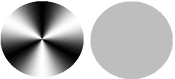
The visual stimuli used in the experiments were: a neutral stimulus with uniform luminance and angular sine-wave standards with spatial frequencies of 2, 3, 4, 24 and 64 ° cycles/360 (Figure 1). These were static, generated grayscale, circularly symmetric, with a diameter of about 7 degrees of visual angle at 150 cm (standard distance used between the monitor and the volunteer).
Figure 1. Example of angular grating stimulus of spatial frequencies of 3 cycles/ 360º and neutral stimulus, respectively. The stimuli were originally calibrated to be seen at a distance of 150 cm.
Procedure
We used the time forced-choice psychophysical method (Wetherill & Levitt, 1965) between two alternatives, in which the volunteer, with binocular vision, had to choose between two stimuli: a test stimulus and a neutral stimulus, which contained the spatial frequency. There were two experimental sessions on different days for each frequency. During the experimental sessions, participants were subjected to successive simple discrimination of pairs of stimuli displayed on the monitor screen. The order of stimulus presentation was random. Each stimulus was presented for two seconds, with a one second interval between them.
volunteers were instructed to press the left mouse button (or number 1) when they felt the test stimulus was presented first, and the right mouse button (or number 2) when they felt the test stimulus was presented second.
Each session began with the contrast of the test stimulus supra-level. The criterion was: three consecutive hits to lower contrast and a unit of an error to increase contrast in a unit (20%). The number of presentations needed to determine the threshold contrast varied according observer arrangements to provide a total of ten reversals (five maximum and minimum values of five contrast), required for the automatic end of the session.
Results
The contrast values obtained for each spatial frequency were gathered into spreadsheets according to each group (participants with migraine and participants without migraine). The grand average was used as an estimate of contrast sensitivity
Figure 2. Contrast sensitivity curves for angular sine wave grating stimuli of spatial frequencies of 2, 3, 4, 24, 64 cycles/360º of volunteers with migraine and volunteers without migraine. The error bars represent standard error of the mean for each frequency.
The analysis of variance for repeated measures (ANOVA) showed main effect of group (F11, 228 = 6.66, p <0.001), the main effect of frequency (F4, 912 = 1152.86, p <0.001) and interaction effect of group versus frequency (F44, 912 = 10.60, p <0.001).
The post hoc analysis with Newman Keuls test showed significant differences between the group with migraine and the group without migraine for spatial frequencies of 2, 4, 24 and 64° cycles/360º (p <0.0001).
Discussion
The initial proposal of this research was to investigate the effects of migraine on visual perception of contrast in adult female, using the CSF of angular sine-wave gratings measured with forced-choice psychophy-sical method. The hypothesis was to assess whether migraine changes the visual processing of the angular sine-wave gratings in photopic luminance conditions.
because at photopic luminance levels, the range of maximum sensitivity occurs at about 24 degrees to cycles/360º angular stimuli (Simas, 1985; Simas & Santos, 2002). Moreover, according to Figure 2, the contrast sen-sitivity curves of both groups show a similar profile to the general pattern characteristic found in the literature for adults in this age group (Simas, Walnut, and Santos, 2005; Simas & Santos, 2002 ). In other words, these curves show a peak in the middle frequencies, with reduced sensitivity to values below or above this range.
Statistical analysis showed that the participants without migraine are more sensitive to the spatial frequencies of 2, 4 and 64 cycles/360° compared to participants with migraine. The group with migraine was more sensitive than the group without migraine frequency of 24 cycles/ 360º. These results support the hypothesis that migraine changes the visual processing of angular sine-wave gratings with photopic contrast levels. The result with angular sine-ware gratings reinforces some studies in the literature, which showed losses in visual perception of contrast for vertical sine-wave grating patterns (Benedek et al. 2002; McKendrick & Badcock, 2003; McKendrick & Sampson, 2009; Yenice et al ., 2007), even some of these studies only reported damage, for example, in the spatial frequency 4 cpd (Shepherd, 2000, Shibata et al., 2005).
On the other hand, the results of this study were different from the study of Benedek et al. (2002). The researchers found no damege in the perception of scotopic vertical sine-wave gratings of patients with migraine. They found differences only for scotopic low vertical spatial frequencies. However, these studies can not be compared directly, since they have different experimental and methodological conditions. For example, studies so far have been vertical sine-wave gratings but the present study was conducted with angular sine-wave gratings.
However, the decreased sensitivity in the lower frequencies (2, 3 and 4 cycles/360º) may be an indication of impairment of the M visual pathway in patients with migraine, confirming some studies (McKendrick & Sampson, 2009; McKendrick, Vingrys, Badcock, & Heywood, 2001; Shepherd, 2000). The low frequencies correspond to the general shape of the object (Simas & Santos, 2001). On the other hand, the commitment of the highest spatial frequency tested (64 cycles/360°) may be related to changes in P pathway. It is not possible to say yet that migraine interacts differently with different levels of luminance or P and M pathways, further research must be conducted to investigate this question.
verti-cal and angular sine-wave gratings are processed in different visual areas, while the vertical sine-wave grating is processed in the primary visual area (V1), the angular sine-wave grating is processed in extra visual areas, V4 and inferior temporal visual cortex – IT (De Valois & De Valois, 1990; Gallant, Connor, Rakshit, Lewis & van Essen, 1996; Merigan, 1996; Wilson & Wilkinson, 1998). These findings are also reinforced with functional imaging technique in humans (Wilkinson et al., 2000).
Tibber et al. (2006) confirmed the involvement of visual areas V4 and IT in subjects with migraine, and McKendrick, Badcock, and Gurgone (2006), specifically in V4. The literature also reports changes in the processing of visual in the cortical areas primary and secondary (Puca, Tommaso, Savarese, Genco & Prudenzano, 1992; Schoenen, 1996), auditory cortex (Schoenen, 1996) and in the frontal cortex (Kropp & Gerber, 1993; Schoenen, 1992) related to migraine.
In this study, the sensitivity was evaluated only in female participants, this choice was made because of the higher prevalence of migraine in this population (Dahlem & Müller, 2003; WHO, 2005). Studies of psycho-physical techniques generally use a few subjects and multiple measurements of the frequencies. This fact is also seen in the classic papers (Blakemore & Campbell, 1969; Campbell & Robson, 1968).
Studies of this nature have to consider some variables of the sample that may confound or interact with migraine, such as drug use, frequency and duration of crisis etc. (Shepherd, 2000). Several studies described here did not mention the control of these variables, but they can directly influence the results. In this study, the subjects answered a structured questionnaire with questions about the history of migraine, and the Journal of Pain, a tool for assessing the crisis. The average time the participants had the disease was 17 years, with an average of four attacks for 15 days and pain intensity from moderate to strong. The experimental sessions to measure visual perception occurred between crisis, with an interval of at least 48 hours, and participation has not happened under the influence of pain medication.
Only two participants had the symptom of visual aura, but changes in visual functions of people with migraine do not seem to be the effect of the aura, as these losses were also reported in participants with migraine who did not have visual aura (McKendrick & Badcock, 2003; McKendrick & Sampson, 2009). The visual aura is a neurological symptom that prece-des, accompanies, or rarely succeeds migraine, characterized by scintillating scotomas, photopsia, hemianopsia unilateral or bilateral, or flashes of light colored lines (Queiroz et al., 1997).
A AA A
Avvvvvaliação da sensibilidade ao caliação da sensibilidade ao caliação da sensibilidade ao contraliação da sensibilidade ao caliação da sensibilidade ao controntrontrontrastastaste em pacientastaste em paciente em paciente em pacientes ce em pacientes ces ces ces com migrâneaom migrâneaom migrâneaom migrâneaom migrânea
Resumo: Resumo:Resumo: Resumo:
Resumo: O objetivo foi medir a Função de Sensibilidade ao Contraste (FSC) de pa-cientes com migrânea e de voluntários saudáveis sem a patologia. Participaram dos testes 12 voluntários do sexo feminino, seis com migrânea e seis sem migrânea na faixa etária de 20 a 37 anos. As medidas de FSC foram realizadas utilizando estímulos visuais estáticos de grades senoidais angulares com frequências espaciais de 2, 3, 4, 24 e 64 ciclos/360º Foi utilizado o método psicofísico da escolha forçada entre duas alternativas temporais, condições de luminância fotópica (luminância média da tela de 41 cd/m²) e visão binocular com pupila natural. Os resultados demonstram que a percepção visual de contraste dos voluntários com migrânea foi menor nas frequên-cias de 2, 3, 4 e 64 ciclos/360º. Esses achados preliminares sugerem alterações na FSC relacionadas a essa patologia.
P PP P
Palaalaalaalaalavrvrvras-chavrvras-chaas-chaas-chavvvvve: as-cha e: e: e: Percepção visual. Sensibilidade ao contraste. Grade senoidal angu-e: lar. Enxaqueca.
E EE
EEvvvvvaluación de la sensibilidad al caluación de la sensibilidad al caluación de la sensibilidad al contraluación de la sensibilidad al caluación de la sensibilidad al controntrontrontrastastaste en pacientastaste en paciente en paciente en paciente en pacientes ces ces ces ces cononononon
Résumé: Résumé:Résumé: Résumé:
Résumé: Dans ce travail, l’objectif était celui de mesurer la Fonction de Sensibilité au Contraste (FSC) chez des patients souffrant de migraine et des volontaires sains sans cette pathologie. Ont participé des tests 12 volontaires du sexe féminin, âgées de 20-37 ans, six avec de la migraine et six sans migraine. Les mesures de FSC ont été réalisées avec des stimuli visuels statiques des grilles d’ondes sinusoïdales angulaires, avec des fréquences spatiales de 2, 3, 4, 24 et 64 cycles/360º. La méthode utilisée a été la psychophysique, avec choix forcé entre deux alternatives temporelles, les conditions de luminosité photopique (luminance moyenne par écran de 41 cd/m²) et la vision binoculaire avec des pupilles naturelles. Les résultats démontrent que la perception visuelle de contraste par les volontaires souffrant de migraine a été plus faible dans les fréquences de 2, 3, 4 et 64 cycles/360°. Ces résultats préliminaires suggèrent des changements dans le FSC liés à cette pathologie.
M MM M
Év Év Év Év
Évaluation de sensibilité au caluation de sensibilité au caluation de sensibilité au caluation de sensibilité au caluation de sensibilité au controntrontrontrastontraste chez des patients aastastaste chez des patients ae chez des patients ae chez des patients ae chez des patients avvvvvec deec deec deec deec de
Resumen: Resumen: Resumen: Resumen:
Resumen: En este trabajo, el objetivo era medir la función de sensibilidad al contras-te (CSF) en paciencontras-tes con y voluntarios sanos sin esta patología. Los sujetos de los ensayos fueron 12 mujeres voluntarias, con edades entre 20-37 años – seis de ellos con la migraña, y seis otros sin migraña. Mediciones LCR se realizaron mediante estí-mulos visuales estáticos de rejilla angular de onda sinusoidal, con frecuencias espaciales de 2, 3, 4, 24 y 64º cycles/360º. El método utilizado fue el psicofísico con elección forzada entre dos alternativas temporales, las condiciones de luminosidad fotópica (luminancia de pantalla media de 41 cd / m²) y la visión binocular con pupi-la natural. Los resultados demuestran que pupi-la percepción visual de contraste por los voluntarios con migraña fue menor en las frecuencias de 2, 3, 4 y 64 cycles/360°. Estos hallazgos preliminares sugieren cambios en el LCR en relación aesta patología.
P PP P
Ref RefRef Ref
Refererererencerencencencenceseseseses
Adams, R. J., & Courage, M. L. (2002). Using a single test to measure human contrast sensitivity from early childhood to maturity. Vision Research, 42, 1205-1210.
Benedek, G., Benedek, K., Kéri, S., & Janáky, M. (2003). The scotopic low-frequency spatial contrast sensitivity develops in children between the ages of 5 and 14 years. Neuroscience, 345, 161-164.
Benedek, K., Tajti, J., Janáky, M., Vécsei, L., & Benedek, G. (2002). Spatial contrast sensitivity of migraine patients without aura. Cephalalgia, 22, 142-145.
Blakemore, C., & Campbell, F. W. (1969). On the existence of neurones in the human visual system selectively sensitive to the orientation and size of retinal images. Journal of Physiology, 203, 237-260
Campbell, E. W., & Maffei, L. (1974). Contrast and spatial frequency. Scientific American, 231, 106-114
Campbell, E. W., & Robson, F. G. (1968). Application of the Fourier analysis to the visibility of gratings. Journal of Physiology, 197, 551-566
Cornsweet, T. N. (1970). Vision perception. New York: Academy Press.
Courage, M. L., Adams, R. J., & Hall, E. J. (1997). Contrast sensitivity in infants and children with Down syndrome. Vision Research, 37(11), 1545-1555.
Dahlem, M. A., & Müller, S. C. (2003). Migraine aura dynamics after reverse retinotopic mapping of weak excitation waves in the primary visual cortex.Biological Cybernetics, 88, 419-429.
De Valois, R. L., & De Valois, K. K. (1990). Spatial vision (2ª ed.). New York: Oxford University Press.
Drummond, P. D., & Anderson, M. (1992). Visual field loss after attacks of migraine with aura. Cephalalgia, 12, 349-352.
Elliott, D. B., & Situ, P. (1998). Visual acuity versus letter contrast sensitivity in early cataract. Vision Research, 38, 2047-2052.
Friedman, D. (2004). The eye and headache. Ophthalmology Clinics of North America, 17(3), 357-369.
Gallant, J. L., Connor, C. E., Rakshit, S., Lewis, J. W., & van Essen, D. C. (1996). Neural responses to polar, hyperbolic, and cartesian gratings in area V4 of the macaque monkey. Journal of Neurophysiology, 76, 2718-2739.
Granziera, C., Silva, A. F. M., Snyder, J., David, S., T., & Hadjikhan, N. (2006). Anatomical alterations of the visual motion processing network in migraine with and without aura. PLoS MEDICINE, 3(10), 1915-1921.
Hubel, D. H., & Livingstone, M. S. (1990). Color and contrast sensitivity in the late-ral geniculate body and primary visual cortex of the macaque monkey. The Journal of Neuroscience, 10(7), 2223-2237.
Kéri, S., Antal, A., Szekeres, G., Benedek, G. & Janka, Z. (2000). Visual information processing in patients with schizophrenia: Evidence for the impairment of central mechanisms. Neuroscience Letters, 293, 69-71.
Kropp, P., & Gerber, W. D. (1993). Is increased amplitude of contingent negative variation in migraine due to cortical hyperactivity or to reduced habituation? Cephalalgia, 13, 37-41.
Maraini, G., Rosmini, F., Graziosi, P., Tomba, M. C., Bonacini, M., Cotichini, R. et al. (1994). Influence of type and severity of pure forms of age-related cataract on visual acuity and contrast sensitivity. Investigative Ophthalmology & Vi-sual Science, 35(1), 262-267.
McColl, S. L., & Wilkinson, F. (2000). Visual contrast gain control in migraine: Measures of visual cortical excitability and inhibition. Cephalalgia, 20, 74-84.
McKendrick, A. M., Badcock, D. R. (2003). Contrast-processing dysfunction in both magnocellular and parvocellular pathways in migraineurs with or without aura. Investigative Ophthalmology & Visual Science, 44, 442-448.
McKendrick, A. M., & Badcock, D. R. (2004). Motion processing deficits in migraine. Cephalalgia. 24, 363-372.
McKendrick, A. M., Badcock, D. R., & Gurgone, M. (2006). Vernier acuity is normal in migraine, whereas global form and global motion perception are not? Investigative Ophthalmology & Visual Science, 47(7), 3213-3219.
McKendrick, A. M., & Sampson, G. P. (2009). Low spatial frequency contrast sensiti-vity deficits in migraine are not visual pathway selective.Cephalalgia, 29, 539-549.
McKendrick, A. M., Vingrys, A. J., Badcock, D. R., & Heywood, J. T. (2001). Visual dysfunction between migraine events. Investigative Ophthalmology & Visual Science, 42(3), 626-633.
Merigan, W. H. (1996). Basic visual capabilities and shape discrimination after lesions of extrastriate area V4 in macaques. Visual Neuroscience, 13, 51-60.
Puca, F. M., de Tommaso, M., Savarese, M. A., Genco, S., & Prudenzano, A. (1992). Topographic analysis of steady-state visual evoked potentials (SVEPs) in the medium frequency range in migraine with and without aura. Cephalalgia, 12, 244-249.
Queiroz, L. P., Rapoport, A. M., Weeks, R. E., Sheftell, F. D., Siegel, S. E., & Baskin, S. M. (1997). Characteristics of migraine visual aura. Headache, 37(3), 137-141.
Santos, N. A., & França, V. C. R. M. (2008). Sensibilidade ao contraste a grades senoidais de freqüências espaciais baixas em crianças. Estudos de Psicolo-gia, 25, 177-184.
Santos, N. A., & Simas, M. L. B. (2001). Função de sensibilidade ao contraste: Indica-dor da percepção visual e da resolução espacial. Psicologia:Reflexão & Críti-ca, 14(3), 589-597.
Schoenen, J. (1992). Clinical neurophysiology studies in headache: A review of data and pathophysiological hints. Functional Neurology, 7, 191-204.
Schoenen, J. (1996). Abnormal cortical information processing between migraine attacks. In M. Sandler, M. Ferrari & S. Harnett (Eds.), Migraine: Pharmacology and Genetics (pp. 233-246). London: Chapman & Hall.
Schwartz, S. H. (2004). Visual perception: A clinical orientation (3ª ed.). New York: McGraw Hill.
Shepherd, A. J. (2000). Visual contrast processing in migraine. Cephalalgia, 20, 865-880.
Shibata, K., Yamane, K., Iwata, M., Ohkawa, S. (2005). Evaluating the effects of spatial frequency on migraines by using pattern-reversal visual evoked potentials. Clinical Neurophysiology, 116, 2220-2227.
Silva, A. C., & Rodrigues, M. de L. V. (2002). Importância do estudo da função da sensibilidade ao contraste no glaucoma. Revista Medicina, 35, 497-504.
Simas, M. L. B., Nogueira, R. M. T. B. L., & Santos, N. A. (2005). Radial frequency stimuli and sine-wave gratings seem to be processed by distinct contrast brain mechanisms. Brazilian Journal of Medical and Biological Research, 38, 419-430.
Simas, M. L. B., & Santos, N. A. (2002). Narrow-band 1, 2, 3, 4, 8, 16 and 24 cycles/360 angular frequency filters. Brazilian Journal of Medical and Biological Research, 35, 243-253.
Simas, M. L. B., Santos, N. A., & Thiers, F. A. (1997). Contrast sensitivity to angular frequency stimuli is higher than that for sinewave gratings in respective middle range. Brazilian Journal of Medical and Biological Research, 30, 633-636.
Slaghuis, W. L. (1998). Contrast sensitivity for stationary and drifting spatial frequency gratings in positive and negative-symptom schizophrenia.Journal of Abnormal Psychology, 107(1), 49-62.
Sociedade Brasileira de Cefaléia. (2009, Março). Diário da Cefaléia. Recuperado em 20 jul. 2009: http://www.sbce.med.br/index.php?option=com_ content&view=article &id= 45:diario-da-cefaleia&catid=25:technology &Itemid=70
Tibber, M. S., Guedes, A., & Shepherd, A. J. (2006). Orientation discrimination and contrast detection thresholds in migraine for cardinal and oblique angles. Investigative Ophthalmology & Visual Science, 47, 5599-5604.
Wetherill, G. B., & Levitt, H. (1965). Sequential estimation of points on a psychometric function. The British Journal of Mathematical and Statistical Psychology, 48, 1-10.
Wilkinson, F., James, T. W., Wilson, H. R., Gati, J. S., Menon, R. S., & Goodale, M. A. (2000). An fMRI study of the selective activation of human extrastriate form vision areas by radial and concentric gratings., Current Biology, 10,1455-1458.
Wilson, H. R., & Wilkinson, F. (1998). Detection of global structure in Glass patterns: implication for form vision. Vision Research, 38, 2933-2947.
Wolthausen, J., Sternberg, S., Gerloff, C., & May, A. (2008). Are cortical spreading depression and headache in migraine causally linked? Cephalalgia, 29, 244-249.
ş
Liana Cha Liana Cha Liana Cha Liana Cha
Liana Chavvvvves Mes Mes Mes Mendeses Mendesendesendesendes, PhD Student, Neuropsychiatry and Behavioral Sciences Program. Visual Perception Laboratory (LabVis). Federal University of Pernambuco (UFPE). Mailing address: Juvenal Mario da Silva St., 377, Apt. 501, Ed Maria Luiza Manaíra District, Zip code: 58038-510, João Pessoa-PB. Email: [email protected]
M M M M
Melyssa Kellyane Celyssa Kellyane Celyssa Kellyane Celyssa Kellyane Caaaaavvvvvalcanti Gelyssa Kellyane C alcanti Galcanti Galcanti Galcanti Galdinoaldinoaldinoaldinoaldino, PhD student, Neuropsychiatry and Behavioral Sciences Program. Visual Perception Laboratory (LabVis). Federal University of Pernambuco (UFPE). Mailing address: Vigolvino Florentino da Costa St., 581, Apto. 303, Ed. Lucídia Tavares, Bairro Manaíra. Zip code: 58038-580, João Pessoa-PB. E-mail: [email protected]
Ják Ják Ják Ják
Jákina Guimarães ina Guimarães ina Guimarães ina Guimarães Vina Guimarães VVVVieirieirieirieirieiraaaaa, Specialist in Morphology, Federal University of Pernambuco (UFPE). Masters student, Neuropsychiatry and Behavioral Sciences Program. Visual Perception Laboratory (LabVis). Federal University of Pernambuco (UFPE). Mailing address: Wilson Flávio Moreira Coutinho St., 161, Apto. 202, Ed. Monte Sinai, Bairro Jardim Cidade Universitária, Zip code: 58052-510, João Pessoa-PB. E-mail: [email protected]
M M M M
Maria Lúcia de Bustamantaria Lúcia de Bustamantaria Lúcia de Bustamantaria Lúcia de Bustamante Simasaria Lúcia de Bustamante Simase Simase Simase Simas, Professor, Department of Psychology, Federal University of Pernambuco. Visual Perception Laboratory (LabVis). PhD in Psychology, Queen’s University, Kingston, Canada Mailing address: Gomes de Matos Junior St., 91, Apto. 502, Bairro Encruzilhada, Zip code: 52050-420, Recife-PE. E-mail: [email protected]
Nathaniel Ant Nathaniel Ant Nathaniel Ant Nathaniel Ant
Nathaniel Antonio dos Santonio dos Santonio dos Santonio dos Santonio dos Santososososos,,,,, Associate Professor, Department of Psychology at the Federal University of Paraíba (UFPB). Perception, Neuroscience and Behavior Laboratory (LPNeC). PhD in Neuroscience and Behavior at the Federal University of São Paulo (USP). Address: Federal University of Paraiba, Center of Humanities, Arts and Letters, Department of Psychology, Joint Castelo Branco, s / n, Bairro Ci-dade Universitaria. Zip Code: 58059-900, João Pessoa-PB. E-mail: [email protected]