Original article
Prevalence of low bone mineral density in children and
adolescents with celiac disease under treatment
Prevalência de baixa densidade mineral óssea em crianças e
adolescentes com doença celíaca em tratamento
Maria Eugênia Farias Almeida Motta
I, Maria Eduarda Nóbrega de Faria
II, Gisélia Alves Pontes da Silva
IIIDivision of Pediatric Gastroenterology, Instituto Materno Infantil Professor Fernando Figueira (IMIP), Recife, Pernambuco, Brazil
IMD, PhD. Adjunct professor, Division of Pediatric Gastroenterology, Universidade Federal de Pernambuco (UFPE), Recife, Pernambuco, Brazil. IIMD, MSc. Physician, Division of Pediatric Gastroenterology, Instituto Materno Infantil Professor Fernando Figueira (IMIP), Recife, Pernambuco, Brazil. IIIMD, PhD. Associate professor, Division of Pediatric Gastroenterology, Universidade Federal de Pernambuco (UFPE), Recife, Pernambuco, Brazil.
ABSTRACT
CONTEXT AND OBJECTIVE: Low bone mineral density may be a inding among children and adolescents with celiac disease, including those undergoing treatment with a gluten-free diet, but the data are contradictory. The aim of this study was to determine the frequency of bone mineral density abnormalities in patients on a gluten-free diet, considering age at diagnosis and duration of dietary treatment.
DESIGN AND SETTING: Cross-sectional prevalence study at the Pediatric Gastroenterology Outpatient Clinic of Instituto Materno Infantil Professor Fernando Figueira.
METHODS: Thirty-one patients over ive years of age with celiac disease and on a gluten-free diet were enrolled. Bone mineral density (in g/cm2) was
measured in the lumbar spine and whole body using bone densitometry and categorized using the criteria of the International Society for Clinical Densitometry, i.e. low bone mineral density for chronological age ≤ -2.0 Z-scores. Age at diagnosis and duration of dietary treatment were conirmed according to the date of starting the gluten-free diet.
RESULTS: Low bone density for chronological age was present in 3/31 patients in the lumbar spine and 1/31 in the whole body (also with lumbar spine abnormality). At diagnosis, three patients with low bone mineral density for the chronological age were more than 7.6 years old. These patients had been on a gluten-free diet for six and seven months and 3.4 years.
CONCLUSION: Pediatric patients with celiac disease on long-term treatment are at risk of low bone mineral density. Early diagnosis and long periods of gluten-free diet are directly implicated in bone density normalization.
RESUMO
CONTEXTO E OBJETIVO: Baixa densidade mineral óssea pode ser encontrada em crianças e adolescentes com doença celíaca, incluindo aqueles em tratamento com dieta sem glúten, mas dados são contraditórios. O objetivo deste estudo foi determinar a frequência de alteração da densidade mineral óssea nos pacientes em dieta sem glúten, conforme a idade ao diagnóstico e o tempo de tratamento dietético.
TIPO DE ESTUDO E LOCAL: Foi realizado estudo transversal de prevalência no Ambulatório de Gastroenterologia Pediátrica do Instituto Materno Infantil Professor Fernando Figueira (IMIP).
MÉTODOS: Trinta e um pacientes maiores de cinco anos com doença celíaca que estavam em dieta sem gluten foram avaliados. A densidade mineral óssea (em g/cm2) foi medida na coluna lombar e no corpo inteiro utilizando densitometria óssea, categorizando-a pelo critério da Sociedade Internacional
para Densitometria Clínica (baixa densidade mineral óssea para a idade cronológica ≤ -2.0 escores Z). A idade ao diagnóstico e o tempo de tratamento foram conirmados pela data de início da dieta sem glúten.
RESULTADOS: Baixa densidade mineral óssea para a idade cronológica foi detectada em 3/31 pacientes na coluna lombar e 1/31 no corpo inteiro (também apresentava alteração da coluna lombar). Ao diagnóstico, três pacientes com baixa densidade mineral óssea para a idade cronológica estavam com mais de 7,6 anos. Esses pacientes estavam em dieta sem glúten por seis e sete meses e 3,4 anos.
CONCLUSÃO: Pacientes pediátricos com doença celíaca em tratamento a longo prazo são de risco para baixa densidade mineral óssea. Diagnóstico precoce e longo período de dieta sem glúten são diretamente implicados na normalização da densidade óssea.
KEY WORDS: Celiac disease. Bone mineral density. Glutens.
Diet therapy. Child. Therapeutics.
PALAVRAS-CHAVE: Doença celíaca. Densidade mineral óssea. Glutens.
INTRODUCTION
Celiac disease is a chronic inlammatory enteropathy induced by ex-posure to gluten. It may be associated with decreased bone mineraliza-tion secondary to chronic inlammamineraliza-tion of the intestinal mucosa. his causes poor absorption of calcium and vitamin D, which reduces serum calcium and stimulates the release of parathormone, thereby exacerbat-ing bone reabsorption from the mobilization of bone calcium. It may also be associated with systemic inlammation, with increased concen-trations of interleukin 1 and 6.1-7
Several studies have demonstrated that, independent of the clini-cal presentation, patients with untreated celiac disease present low bone mineral density (BMD).1-3,5,7,8 Osteopenia is a common public health problem in adults, but can be diagnosed and prevented in childhood. Considering that most of the bone mass is acquired during the irst two decades of life, early diagnosis of celiac disease and adherence to a glu-ten-free diet are of fundamental importance for ensuring adequate bone metabolism in such cases.9,10 An early start to treatment for pediatric pa-tients with celiac disease ensures signiicantly higher bone metabolism rates, since the treatment reverses the inlammatory process and pre-vents impairment of bone mass acquisition during the most important period for its acquisition.3,5,11 On the other hand, failure to adhere to a gluten-free diet reduces bone metabolism. here is controversy regard-ing the time required for a gluten-free diet to normalize bone mineral-ization. Some authors have detected this recovery over a short duration of adequate follow-up (up to one year), while others have stated that it can only be veriied over a longer term.1,3,7,11-13
OBJECTIVE
Since early diagnosis of celiac disease may avoid repercussions re-garding bone metabolism, the present study was conducted with the aims of determining the frequencies of low BMD among children and adolescents with celiac disease under treatment and evaluating the pres-ence of these abnormalities, considering the patient’s age at diagnosis, duration of treatment, failure to adhere to a gluten-free diet and indices of weight and height for age.
METHODS
Type of study
his was a cross-sectional prevalence study.
Setting
he study was conducted at the Pediatric Gastroenterology Outpa-tient Clinic of a tertiary-level hospital, Instituto Materno Infantil Pro-fessor Fernando Figueira (IMIP), in Recife, Pernambuco, Brazil.
Patients
he initial study population consisted of 37 patients aged 5 to 18 years who were undergoing treatment for celiac disease. Six patients aged less than ive years were excluded because their bone densitometry could be evaluated with the pediatric software used in this study. he
re-maining 31 patients were evaluated for the presence of bone mineraliza-tion abnormalities. None of the patients was receiving calcium, vitamin supplements or drugs that could have altered their bone metabolism. his study was performed in accordance with the principles of the Dec-laration of Helsinki and it was approved by the Ethics Committee of IMIP. Informed consent was obtained from all of the patients’ parents.
Procedures
he diagnosis of celiac disease had been made in accordance with the following criteria: clinical condition and irst biopsy on the small in-testine that were compatible with celiac disease, and/or clinical remis-sion on withdrawal of gluten.14 he biopsy on the small intestine was performed by means of upper digestive tract endoscopy. he sample was analyzed and classiied using the criteria of Marsh.15
he dual energy x-ray absorptiometry (DXA)method was used to perform bone densitometry on the lumbar spine and whole body except for the head, by using a DPX-L scanner (Lunar Corp, Madison, Wis-consin, United States). he patients were evaluated wearing trousers and a cotton shirt. All the measurements were made using the same appara-tus, equipped with pediatric software (version 1.35), and were analyzed by a single experienced examiner. he bone mineral density (in g/cm2) of the lumbar vertebrae L2-L4 and whole skeleton was measured and categorized in accordance with the criteria of the International Society for Clinical Densitometry. Low bone density for chronological age was thus deined as less than or equal to -2.0 Z-scores.16
A questionnaire asking about signs and symptoms prior to the di-agnosis and after starting the gluten-free diet was applied to the par-ents. One question asked whether there had been any failures to adhere to the diet over the preceding three months. Patients were deemed to be non-compliant with the gluten-free diet if they had had intake of gluten once a week or more frequently. A test for anti-human tissue transglutaminase (anti-tTG) antibody [ImmuLisa anti-human tissue transglutaminase antibody immunoglobulin A (IgA) Enzyme Linked ImmunoSorbent Assay (Elisa), IMMCO Diagnostics, United States] was performed on 23 patients in order to verify compliance with the gluten-free diet.
Weight and height measurements were made in accordance with the technique standardized by Gibson.17 Briely, the patients were weighed while wearing underwear and t-shirt on a calibrated balance with a maximum capacity of 150 kg and sensitivity of 0.1 kg
(
Filizola, Brazil). Height was measured with the patients standing upright and keeping their eyes ixed on the horizon, using a stadiometer (profes-sional model, Gofeka, Brazil), with an accuracy of 0.1 cm. he mea-surement of height was performed twice and the average meamea-surement was recorded. he anthropometric indices of weight for age, height for age and body mass index (weight (kg)/height (m2)) were expressed as Z-scores, using the curves and graphs of the Centers for Disease Con-trol and Prevention (CDC, Atlanta, United States) as the reference.18 he cutof point for low weight and linear growth deicit was taken to be -2 Z-scores.18catego-ries of gluten-free diet periods (two, three and four years). Statistical sig-niicance was taken as P ≤ 0.05.
RESULTS
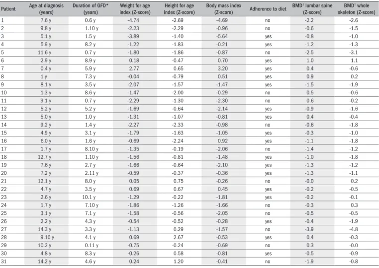
he median age of the 31 patients (41.9% males) at the time of agnosing celiac disease was 10.3 years (range: 5.2 to 18.7 years). his di-agnosis was based on histopathological analysis in 25/31 cases (80.6%) and on clinical remission after starting a gluten-free diet in 6/31 cases (19.4%). At the time when the DXA was performed, the patients had been on a gluten-free diet for 0.6 to 10.1 years. he clinical manifesta-tions at the time of diagnosis were diarrhea in 23/31 (74.2%) and ab-dominal distension and pain in 27/31 (87.1%), separately or in combi-nation. At the time of the interview, all the patients were asymptomatic. Information about the patients’ age at diagnosis, beginning of the glu-ten-free diet, nutritional assessment, adherence to treatment and BMD are shown in Table 1.
Analysis of the lumbar spine using DXA showed low bone density in 3/31 patients (9.7%), while analysis of the whole skeleton identiied low bone density in 1/31 patient (3.2%). his patient had both lumbar spine and whole body abnormalities. he patients with low bone den-sity were diagnosed at the ages of 7.6, 11.6 and 14.3 years. With regard to the duration of the gluten-free diet, two patients presenting low bone
Table 1. Clinical information and bone mineral density results for patients with celiac disease undergoing treatment
Patient Age at diagnosis (years)
Duration of GFD* (years)
Weight for age index (Z-score)
Height for age index (Z-score)
Body mass index
(Z-score) Adherence to diet
BMD† lumbar spine
(Z-score)
BMD† whole
skeleton (Z-score)
1 7.6 y 0.6 y -4.74 -2.69 -4.69 no -2.2 -2.6
2 9.8 y 1.10 y -2.23 -2.29 -0.96 no -0.6 -1.5
3 5.1 y 1.5 y -3.89 -1.40 -5.64 yes -0.8 -1.0
4 5.9 y 8.2 y -1.22 -1.83 -0.21 yes -1.2 -1.3
5 11.6 y 0.7 y -1.80 -1.86 -0.87 no -2.5 -3.1
6 2.9 y 8.9 y 0.18 -0.47 0.70 yes 1.0 1.1
7 0.4 y 5.9 y 2.77 0.65 3.20 yes 0.4 -0.6
8 1 y 7.3 y -0.04 -0.79 0.51 yes 0.9 0.2
9 8.1 y 3.5 y -2.07 -1.57 -1.47 yes -1.5 -1.9
10 1.3 y 8.6 y -1.47 -2.00 -0.29 no 0.5 -0.6
11 9.1 y 0.7 y -2.29 -1.30 -2.30 no 0.6 -0.2
12 5.2 y 5.2 y -1.69 -0.64 -2.14 yes -0.9 -1.6
13 5.0 y 1.0 y -1.31 -1.07 -0.81 yes 0.4 -0.4
14 9.2 y 1.4 y -2.27 -2.33 -0.98 no -0.6 -1.8
15 4.9 y 3.1 y -1.79 -1.63 -1.05 yes -0.3 -1.0
16 6.0 y 1.6 y -0.69 -2.24 0.92 yes -1.1 -1.8
17 1.7 y 8.10 y -1.35 -0.19 -2.06 no -1.4 -1.2
18 12.7 y 1.10 y -1.56 -0.81 -1.48 yes -1.0 -1.8
19 7.6 y 2.7 y -1.66 -0.64 -2.10 yes -1.3 -1.2
20 7.2 y 2.11 y -0.59 -0.37 -0.36 yes -1.3 -1.1
21 12.1 y 8.0 y 0.05 0.75 -0.26 no -0.0 0.2
22 4.7 y 3.5 y 0.69 0.67 0.45 yes -0.2 -0.5
23 2.6 y 10.1 y -1.29 -0.22 -1.81 yes -0.2 -0.1
24 1.7 y 7.10 y -1.86 -1.26 -1.66 no -0.3 0.3
25 3.1 y 7.1 y -1.58 -0.56 -2.05 no -0.5 -0.5
26 2.2 y 4.3 y -0.54 -0.52 -0.28 yes -0.4 -1.9
27 14.3 y 3.3 y -1.13 0.29 -1.57 no -3.9 -4.8
28 9.10 y 4.1 y 0.69 2.67 -0.53 yes 0.4 -0.3
29 10.2 y 0.11 y -0.75 -0.24 -0.69 no 0.3 -0.0
30 4.8 y 8.3 y -0.26 0.58 -0.81 yes -0.5 -0.9
31 14.2 y 4.6 y 0.24 1.20 -0.41 no -1.9 -0.8
*GFD = gluten-free diet; †BMD = bone mineral density.
density had been on a gluten-free-diet for six and seven months and one of them for 3.3 years. Failure to adhere to the gluten-free diet over the preceding three months was conirmed by three patients with low bone density. here were positive indings of anti-tTG antibodies in 47.1% (8/23) of the patients who underwent this test, and one of these pre-sented low bone density.
he weight/age index was low in 6/31 patients (19.4%). he height/ age index was low in 5/31 patients (16%), among whom one presented low bone density and had been on a gluten-free diet for seven months. his patient was positive for anti-tTG.
Analysis of the duration of the gluten-free diet showed that there was no statistical diference (P > 0.05) in the median BMD in the lum-bar spine and whole skeleton for two and three years of gluten-free diet (data not shown). However, there was a trend towards signiicance with four years of gluten-free diet (Table 2).
DISCUSSION
were low. he sample in this study presented suicient statistical pow-er (95%): 31 patients wpow-ere needed for this study, according to Statcalc (Epi-Info, version 6.04 b), with 25% of the patients presenting BMD lower than -2.5 Z-scores.1 Among our patients, 9.7% presented low BMD and thus this “worst result” was acceptable.
Diminished BMD is frequently found prior to diagnosing celiac disease.1-5,8,19,20 Just like in our study, other authors also found that when the gluten-free diet was started late, because of delays in diagnosing the disease, there was a relationship with lower BMD. Achieving normal bone mass was dependent on early diagnosis of the disease.1,3-5,12,13,21-24
here is currently a discussion in the literature about the duration of gluten-free diet that is needed to reverse the changes in bone metab-olism relating to celiac disease. Recovery from bone abnormalities has been found to be correlated with the duration of the gluten-free diet. 1-3,11,13,19,25 Increases in or stabilization of BMD have particularly been
observed during the irst year of the diet and are maintained over the long term (more than four years) in most patients.1-5,11,13,19,20,24 However, great variability in BMD during treatments for celiac disease have been described. his is related to the severity of atrophy of the villi at the time of diagnosis, the patient’s nutritional status and whether the diet has been followed strictly.1 Such variability may explain why some pa-tients continue to present bone mineralization abnormalities, even over the long term, as detected in the present study.1,21,26 For this reason, it is still unclear whether low BMD due to celiac disease can be completely eliminated.1,27
he clinical and histological recovery is related to the extent of at-rophy of the villi. Patients with severe histological lesions may achieve reductions in the severity of their lesions, to attain lower grades or even full recovery of the integrity of the mucosa after more than two years on a gluten-free diet. On the other hand, patients with mild lesions may achieve incomplete histological recovery, or may take years to recov-er, thus continuing to present persistent residual abnormalities.25,28,29 For this reason, it is questionable whether patients without symptoms after following a gluten-free diet are truly free from bone disease and have fully recovered their bone mass losses, since there is a tendency towards lower BMD indices in patients with almost total atrophy of the villi than in those with partial atrophy.4,12,30 A proportion of the patients who are asymptomatic after following a gluten-free diet con-tinue to present reduced bone mineralization.12 Failure to adhere to the diet, celiac disease of greater severity due to late diagnosis, genet-ic factors and recurrent inlammatory stimuli on the intestinal mu-cosa from other causes may contribute towards delays in histologi-cal recovery and thus towards changes in bone metabolism.28 Most of the patients in the present study had severe histological lesions before starting on a gluten-free diet. It is possible to speculate that the pa-tients who presented low bone density would be the ones who had not achieved full recovery from their histological lesions, thus continuing to present inlammation and consequently, BMD abnormalities. Al-though only eight cases were positive for anti-tTG, Tursi et al.29 did not observe a good correlation between anti-tTG and histological le-sions. hus, even the patients whose serological tests did not indicate any failure to adhere to the diet may have continued to have some de-gree of histological lesion.
It has been observed that patients who fail to adhere to the diet pres-ent low BMD, and this was also seen among our patipres-ents.20 In general, patients who follow a gluten-free diet without straying from it recover their BMD more rapidly, but simply complying with the diet does not ensure normalization, even after years.2,5,8,19-21,25,30-33 As is seen among chronic diseases in which dietary treatment is required, there is no guar-antee that a gluten-free diet will be followed strictly over the long term, and failure to adhere to the diet was conirmed in 13 of our patients.30 Serological tests for celiac disease have been recommended as an indi-rect measurement for monitoring the compliance with the diet. Once the gluten has been eliminated and the symptoms have disappeared, its reintroduction causes no apparent symptoms.34 Anti-tTG antibod-ies form complexes with the gluten present in the diet and, after a short time on a gluten-free diet, the test for these antibodies become nega-tive.20 his may elucidate why these antibodies were not detected in two of our patients with low BMD who said they had not been adhering to the diet, considering that such failures tend to be occasional rather than continuous and there is no single laboratory method to detect small di-etary transgressions.34
he quantitative method most used for investigating bone mass in pediatrics is DXA. It is a method in which the measurements are based on two-dimensional projection of the three-dimensional bone structure, and this may induce measurement errors that are dependent on the size of the bone under evaluation.11,21,35 hus, BMD abnormalities evaluated using DXA may only relect the growth stage, such that the BMD of shorter individuals will be underestimated and the BMD of taller indi-viduals will be overestimated.11,19,25 here needs to be caution in evalu-ating the presence of BMD abnormalities in patients with celiac disease who are undergoing treatment. Since low BMD is a chronic condition in which nutrient absorption and growth are abnormally low, it may be due to an inadequately treated underlying disease or it may just relect inappropriate skeletal growth resulting from the length of time with the disease prior to starting on a gluten-free diet. hus, catch-up growth may be impeded even when treatment is administered (an efect from chronic malnutrition).11-13,36 In our study, ive patients presented abnor-mal weight-for-age indices, among whom one had low BMD. his may relect nutrition that has been afected by the length of time with the disease, but it may also relect the persistence of histological abnormali-ties due to inadequate adherence to the diet. On the other hand, since most of the patients of normal height did not present bone abnormali-ties, it is possible that in those with low BMD, this may have been the result of the celiac disease itself (late diagnosis, with insuicient length of treatment for histological recovery). he American Gastroenterology Association guidelines suggest that DXA scans are unnecessary in chil-dren with newly diagnosed uncomplicated celiac disease.4 Nonetheless,
Table 2. Bone mineral density in lumbar spine and whole skeleton according to duration of gluten-free diet
Duration of gluten-free diet
≤ 4 years > 4 years P
Bone mineral density (g/cm2)
Lumbar spine 0.625 (0.585-0.655) 0.721 (0.586-0.890) 0.11
bone densitometry should be assessed in relation to each patient indi-vidually, and clinical evolution, histological indings and growth pattern should also be considered.
here were some limitations to our study. Firstly, not all of the pa-tients were diagnosed by means of jejunal biopsies. However, the diag-nostic criteria of the European Society of Paediatric Gastroenterology and Nutrition14 were followed, and these criteria are often used.7 here-fore, we believe that all of the patients included in the study had celiac disease. Secondly, the cross-sectional design may have led to diiculty in understanding the contributions of late treatment, good adherence to gluten-free diet and duration of treatment towards bone mass.
CONCLUSION
his study detected that some patients with celiac disease who were on a gluten-free diet might present low bone density for chronological age, which could have resulted from older age at the time of diagnosis and short duration of gluten-free diet or inadequate compliance.
REFERENCES
1. Kalayci AG, Kansu A, Girgin N, Kucuk O, Aras G. Bone mineral density and importance of a gluten-free diet in patients with celiac disease in childhood. Pediatrics. 2001;108(5):E89. 2. Mora S, Barera G, Beccio S, et al. A prospective, longitudinal study of the long-term effect of treatment on bone density in children with celiac disease. J Pediatr. 2001;139(4):516-21. 3. Kavak US, Yüce A, Koçak N, et al. Bone mineral density in children with untreated and
trea-ted celiac disease. J Pediatr Gastroenterol Nutr. 2003;37(4):434-6.
4. Bernstein CN, Leslie WD, Leboff MS. AGA technical review on osteoporosis in gastrointestinal diseases. Gastroenterology. 2003;124(3):795-841.
5. Mora S. Celiac disease: a bone perspective. J Pediatr Gastroenterol Nutr. 2003;37(4):409-11. 6. Fornari MC, Pedreira S, Niveloni S, et al. Pre- and post-treatment serum levels of cytokines IL-1beta, IL-6, and IL-1 receptor antagonist in celiac disease. Are they related to the asso-ciated osteopenia? Am J Gastroenterol. 1998;93(3):413-8.
7. Mora S. Celiac disease in children: impact on bone health. Rev Endocr Metab Disord. 2008;9(2):123-30.
8. Barera G, Beccio S, Proverbio MC, Mora S. Longitudinal changes in bone metabolism and bone mineral content in children with celiac disease during consumption of a gluten-free diet. Am J Clin Nutr. 2004;79(1):148-54.
9. Chesnut CH 3rd. Is osteoporosis a pediatric disease? Peak bone mass attainment in the adolescent female. Public Health Rep. 1989;104 Suppl:50-4.
10. Lu PW, Briody JN, Ogle GD, et al. Bone mineral density of total body, spine, and femoral neck in children and young adults: a cross-sectional and longitudinal study. J Bone Miner Res. 1994;9(9):1451-8.
11. Mora S, Barera G, Beccio S, et al. Bone density and bone metabolism are normal after long-term gluten-free diet in young celiac patients. Am J Gastroenterol. 1999;94(2):398-403. 12. Szathmári M, Tulassay T, Arató A, Bodánszky H, Szabó A, Tulassay Z. Bone mineral content
and density in asymptomatic children with coeliac disease on a gluten-free diet. Eur J Gastroenterol Hepatol. 2001;13(4):419-24.
13. Pludowski P, Karczmarewicz E, Socha J, Matusik H, Syczewska M, Lorenc RS. Skeletal and muscular status in juveniles with GFD treated clinical and newly diagnosed atypical celiac disease--preliminary data. J Clin Densitom. 2007;10(1):76-85.
14. Revised criteria for diagnosis of coeliac disease. Report of Working Group of European Society of Paediatric Gastroenterology and Nutrition. Arch Dis Child. 1990;65(8):909-11. 15. Marsh MN. Gluten, major histocompatibility complex, and the small intestine. A molecular
and immunobiologic approach to the spectrum of gluten sensitivity (‘celiac sprue’). Gastro-enterology. 1992;102(1):330-54.
16. ISCD. The International Society for Clinical Densitometry. 2007 Pediatric Oficial Positions of the International Society for Clinical Densitometry. Available from: www. http://www.iscd. org/visitors/pdfs/ISCD2007OficialPositions-Pediatric.pdf. Accessed in 2009 (Oct 7).
17. Gibson RS.. Anthropometric assessment of body composition. In: Gibson RS, editor. Princi-ples of nutritional assessment. New York: Oxford University; 1990. p. 187-208. 18. Centers for Disease Control and Prevention. 2000 CDC growth charts. Available from: www.
cdc.gov/growthcharts. Accessed in 2009 (Oct 7).
19. Mora S, Weber G, Barera G, et al. Effect of gluten-free diet on bone mineral content in growing patients with celiac disease. Am J Clin Nutr. 1993;57(2):224-8.
20. Hartman C, Hino B, Lerner A, et al. Bone quantitative ultrasound and bone mineral density in children with celiac disease. J Pediatr Gastroenterol Nutr. 2004;39(5):504-10. 21. Mora S, Barera G, Ricotti A, Weber G, Bianchi C, Chiumello G. Reversal of low bone density
with a gluten-free diet in children and adolescents with celiac disease. Am J Clin Nutr. 1998;67(3):477-81.
22. Scotta MS, Salvatore S, Salvatoni A, et al. Bone mineralization and body composition in young patients with celiac disease. Am J Gastroenterol. 1997;92(8):1331-4.
23. Walters JR, Banks LM, Butcher GP, Fowler CR. Detection of low bone mineral density by dual energy x ray absorptiometry in unsuspected suboptimally treated coeliac disease. Gut. 1995;37(2):220-4.
24. Carvalho CN, Sdepanian VL, de Morais MB, Fagundes-Neto U. Doença celíaca em tratamen-to: avaliação da densidade mineral óssea [Celiac disease under treatment: evaluation of bone mineral density]. J Pediatr (Rio J). 2003;79(4):303-8.
25. Kemppainen T, Kröger H, Janatuinen E, et al. Bone recovery after a gluten-free diet: a 5-year follow-up study. Bone. 1999;25(3):355-60.
26. De Lorenzo A, Di Campli C, Andreoli A, Sasso GF, Bonamico M, Gasbarrini A. Assessment of body composition by bioelectrical impedance in adolescent patients with celiac disease. Am J Gastroenterol. 1999;94(10):2951-5.
27. González D, Mazure R, Mautalen C, Vazquez H, Bai J. Body composition and bone mineral density in untreated and treated patients with celiac disease. Bone. 1995;16(2):231-4. 28. Yachha SK, Srivastava A, Mohindra S, Krishnani N, Aggarwal R, Saxena A. Effect of a
gluten-free diet on growth and small-bowel histology in children with celiac disease in India. J Gastroenterol Hepatol. 2007;22(8):1300-5.
29. Tursi A, Brandimarte G, Giorgetti GM. Lack of usefulness of anti-transglutaminase antibodies in assessing histologic recovery after gluten-free diet in celiac disease. J Clin Gastroenterol. 2003;37(5):387-91.
30. Matysiak-Budnik T, Malamut G, de Serre NP, et al. Long-term follow-up of 61 coeliac pa-tients diagnosed in childhood: evolution toward latency is possible on a normal diet. Gut. 2007;56(10):1379-86.
31. Barera G, Mora S, Brambilla P, et al. Body composition in children with celiac disease and the effects of a gluten-free diet: a prospective case-control study. Am J Clin Nutr. 2000;72(1):71-5.
32. Bai JC, Gonzalez D, Mautalen C, et al. Long-term effect of gluten restriction on bone mineral density of patients with coeliac disease. Aliment Pharmacol Ther. 1997;11(1):157-64. 33. Smecuol E, Gonzalez D, Mautalen C, et al. Longitudinal study on the effect of treatment
on body composition and anthropometry of celiac disease patients. Am J Gastroenterol. 1997;92(4):639-43.
34. Jadresin O, Misak Z, Sanja K, Sonicki Z, Zizić V. Compliance with gluten-free diet in children with coeliac disease. J Pediatr Gastroenterol Nutr. 2008;47(3):344-8.
35. Prentice A, Parsons TJ, Cole TJ. Uncritical use of bone mineral density in absorptiometry may lead to size-related artifacts in the identiication of bone mineral determinants. Am J Clin Nutr. 1994;60(6):837-42.
36. Pooni PA, Chhina RS, Jaina BK, Singh D, Gautam A. Clinical and anthropometric proile of children with celiac disease in Punjab (North India). J Trop Pediatr. 2005;52(1):30-3.
Sources of funding: None
Conlict of interest: The authors conirm that there is no conlict of interest
Date of irst submission: September 3, 2008
Last received: October 8, 2009
Accepted: October 9, 2009
Address for correspondence:
Maria Eugênia Farias Almeida Motta Rua Amaraji, 80/1.001 Casa Forte — Recife (PE) — Brasil CEP 52060-440