e-mail: [email protected]
Received: 27 July 2015 / Accepted: 15 March 2016
System for open-chest, multidirectional electrical deibrillation
Marcelo Almeida Viana*, Rosana Almada Bassani, Orlando Petrucci, Denilson Antônio Marques, José Wilson Magalhães Bassani
Abstract Introduction: Cardiomyocytes are more sensitive to stimulatory electrical ields when the latter are applied
longitudinally to the cell major axis. In the whole heart, cells have different spatial orientations, which may limit the effectiveness of conventional electrical deibrillation (i.e., shock delivery in a single direction). This article describes the constructive aspects of a portable system for rapidly-switching, multidirectional stimulus delivery, composed of an electrical deibrillator and multielectrode-bearing paddles for direct cardiac deibrillation.
Methods: The deibrillator delivers monophasic, truncated monoexponential waveforms with energy up
to 7.3 J. Upon selection of the deibrillation modality (unidirectional or multidirectional), shock delivery is triggered through 1 or 3 outputs. In the latter case, triggering is sequentially switched to the outputs, without interval or temporal overlap. Each paddle contains 3 electrodes that deine shock pathways spaced by 60°. The system was tested in vivo for reversal of experimentally-induced ventricular ibrillation in healthy swine,
using 30- and 20-ms long shocks (N= 4 in each group). Results: The deibrillator delivers identical stimulus
waveforms through all outputs in both stimulation modalities. In all animals, successful deibrillation required lower shock energy when 20 ms-long stimuli were applied in 3 directions, compared to a single direction. However, performance was poorer with multidirectional deibrillation for 30 ms-long shocks. Conclusion: The
delivery of identical shock waveforms allowed conirmation that multidirectional deibrillation can promote restoration of sinus rhythm with lower shock energy, which may reduce myocardial electrical damage during deibrillation. Nevertheless, increase in shock duration greatly impairs the effectiveness of this deibrillation
modality.
Keywords: Cardiac arrhythmia, Ventricular ibrillation, Cardiac deibrillator, Shock duration.
Introduction
Cardiac arrest caused by ventricular ibrillation (VF) is a leading cause of sudden death, which is estimated to account for 15-20% of all deaths (Hayashi et al., 2015). The chance of survival drops by ~5% for every minute of untreated VF (Larsen et al., 1993). Electrical deibrillation, which consists of the application of a brief, high-intensity electrical shock to the heart, is the most effective therapy in the early phase of VF (Koster et al., 2006; Patil et al., 2015).
Nonetheless, exposure to high-intensity electrical ields may cause harmful effects on the heart muscle, attributed to electrical cell membrane damage and consequent Ca2+ overload (Fedorov et al., 2008;
Krauthamer and Jones, 1997; Oliveira et al., 2008; Yabe et al., 1990). Although the minimum electrical ield required for successful deibrillation is estimated as 3-9 V/cm (Malmivuo and Plonsey, 1995), during deibrillation the ield may reach ~100 V/cm near the electrodes (Kroll and Swerdlow, 2007; Yabe et al., 1990), which is higher than the threshold values for membrane electroporation (> 25 V/cm, Cheek and
Fast, 2004) and cell death (> 50 V/cm; Goulart et al., 2012; Oliveira et al., 2008).
Optimizing the direction of stimulation might decrease the ield intensity required for effective stimulation, since, for ield application parallel to the cell major axis, the threshold intensity is only 50% of that when the ield is applied in the transversal direction (Bassani et al., 2006; Oliveira et al., 2008). A problem of the practical application of this approach to deibrillation is that myocytes are disposed in several directions in the whole heart (Smerup et al., 2009). Nevertheless, using randomly-oriented isolated ventricular myocytes as an experimental model for heterogeneous cell spatial orientation, Fonseca et al. (2013) showed that rapidly switching stimuli among 3 directions was able to more than double the percentage of cells excited by near-threshold stimulus amplitude.
The effectiveness of the in vivo multidirectional
1986; Jones et al., 1988; Kerber et al., 1994; Pagan-Carlo et al., 1998; Zheng et al., 2002). However, in these studies, different deibrillators were used for shock delivery through different pathways. This may impair the comparison of the effectiveness of unidirectional and multidirectional deibrillation because the applied peak voltage and current for a given level of shock energy may markedly vary among deibrillators. Conventional deibrillators deliver shocks via a single pair of electrodes, thus in a single direction, and, to our knowledge, deibrillators with multiple outputs are not commercially available, even for experimental purposes.
In a recent study, we reported improved in vivo
deibrillation by applying stimuli through 3 pathways using a single deibrillator for both unidirectional and multidirectional stimulation (Viana et al., 2014). In the present article, we describe, from the constructive point of view, the system used in the previous study, composed of an electrical deibrillator and electrode-bearing paddles for rapid shock delivery on the epicardial surface in up to 3 directions, with a special focus on the inluence of shock duration on the effectiveness of deibrillation.
Methods
Instrumentation
The multidirectional deibrillator
The present instrument, developed at the Center for Biomedical Engineering of the University of Campinas (CEB/UNICAMP), was based on the combination of a switching circuit that allows the output to be changed among different pairs of electrodes (Fonseca et al.,
2013), and a capacitive pulse generator that delivers monophasic, truncated exponential voltage pulses (American National Standards Institute…, 1996; Associação Brasileira de Normas Técnicas…, 2005). In its present version, the instrument can deliver sequential shocks up to 3 outputs (pairs of electrodes).
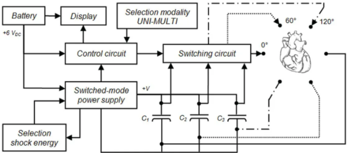
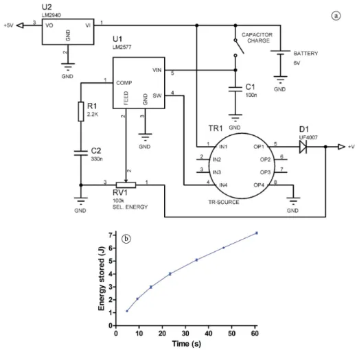
The block diagram of the multidirectional deibrillator is shown in Figure 1. A rechargeable sealed lead-acid battery (UP-645, Unipower, Extrema, MG, Brazil; 6 V) powers the switched-mode power supply (SMPS) circuit (Figure 2a), which operates in the lyback mode. This circuit contains an adjustable switch regulator (LM2577-ADJ, National Semiconductor Co., Santa Clara, CA, USA) that switches the electrical current at high frequency (52 kHz) to a step-up voltage transformer (TR 70415, Ralp Industrial, Alvorada, RS, Brazil; 1:10), as to stabilize the DC output voltage that charges three 100 µF electrolytic capacitors, one for each output. The desired charging voltage level (up to 382 V, corresponding to a maximum of 7.3 J shock energy) can be adjusted by means of a rotary linear potentiometer (100 kΩ) on the front panel of the equipment. The relationship between the time required for fully charging the capacitors and the shock energy level is shown in Figure 2b, where it can be seen that capacitor charging can take as long as 1 min for the maximum shock energy available.
The SMPS circuit energizes the microcontroller-based control circuit (PIC16F818, Microchip Technology, Inc., Chandler, AZ, USA) and the liquid crystal display (LCD-016M002B, Vishay Intertechnol., Shelton, CT, USA) on the control panel via a linear low dropout voltage regulator (LM2940CT-5.0, National Semiconductor Co., Santa Clara, CA, USA; 5 V).
Figure 1. Summarized block diagram of the multidirectional deibrillator. The battery powers the display, control circuit and the
switched-mode power supply (which provides controlled voltage to charge the capacitors C1-C3). The switching circuit controls shock delivery through
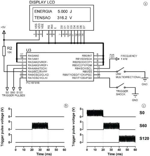
The modality of shock delivery (unidirectional or multidirectional, i.e., through 1 or 3 electrodes pairs, respectively) can also be preset on the front panel (Figure 3). These settings conigure the operating mode control circuit (Figure 4a), which commands the output switching. The microcontroller functions were programmed in assembly language, and the program was written and loaded in the microcontroller using the MPLAB IDE recorder software (Microchip Technol. Inc., Chandler, AZ, USA).
When shock delivery is triggered, a command pulse chopped at 1 kHz is generated by the microcontroller, as an interval-modulated train of rectangular voltage pulses. The train duration (set at either 20 or 30 ms) deines the duration of the each deibrillatory shock. If the unidirectional delivery modality is selected (Figure 4b), a single pulse train voltage (S60) is generated, in this case to trigger stimulus delivery through the central electrodes in the paddles (see next). If the multidirectional modality is set, triggering pulse trains are generated sequentially (S0, S60 and S120,
Figure 4c), without interval or temporal overlap, to trigger successive shock delivery though each of the 3 pairs of electrodes, thus allowing switching the deibrillator outputs. Current backlow from the fully charged capacitors is prevented by six blocking diodes (6A10, Rectron Semiconductor Inc., Chino, CA, USA), which ensures the establishment of a loating potential coniguration. Pulse transformers (TP-1:1/4T, Thornton, Vinhedo, SP, Brazil) were used with chopped pulses to ensure that switching was triggered, and to isolate the high-voltage switching circuit from the low-voltage control circuit. The discharge switching circuit (one for each output, Figure 5a) was implemented by using bidirectional thyristors (triodes) for alternating current (TRIAC 226M, Power Innov., Inc., Lindon, UT, USA) suficiently fast for delivery of up to 3 shocks within 60 ms (Figure 5b, c), i.e., in a shorter period than the duration of the absolute refractory period of action potential in the swine ventricle (Roscher et al., 2001). This ensures that a
Figure 2. (a) Switched-mode power supply circuit; (b) Time required for charging the three 100 µF electrolytic capacitors by the SMPS
cardiac myocyte excited by one of the shocks will not be reexcited by the subsequent stimulus.
Internal deibrillation paddles and electrodes
A pair of deibrillation paddles was designed to contain 3 electrodes each, as to allow direct multidirectional electrical stimulation of the ventricular surface (Figure 3). The paddle handle is a 32 mm diameter, 140 mm long, cylindrical nylon tube with labeled pushbuttons on the top, for enabling capacitor voltage charging after the shock energy level is selected (green), and for triggering shock delivery (red). A hollow support shaft in the handle lengthens the paddles, facilitating shock application and minimizing the possibility of contact of the operator with the electrode conductive area. The cables for electrical connection of the deibrillator outputs and the electrodes were inserted into the shaft central cavity. A semicircular electrode support, made of polycarbonate/acrylnitrile-butadiene-styrene by rapid
Figure 3. The multidirectional deibrillator and the paddles. From
the left to the right in the instrument´s control panel: (1) display that shows the charged voltage and energy level; (2) switch for selection of shock delivery modality; (3) rotary potentiometer for adjusting shock energy; (4) LED that indicates stimulus delivery; (5) outputs for connection to the electrode cables; and (6) on/off switch. Each paddle bears 3 electrodes, which allow shock delivery in 3 directions spaced by 60° (see text for details). Labeled pushbuttons on the top of each handle enable capacitor charging (7) or discharge triggering (8).
Figure 4. (a) Control circuit; (b) Upon triggering shock delivery, pulse trains are generated by the control circuit to control shock discharge
prototyping at the Institute of Biofabrication of the Faculty of Chemical Engineering of the University of Campinas (BIOFABRIS/UNICAMP), was ixed at the inferior extremity of the paddle. The electrodes, shaped as concave discs, were made of polished surgical stainless steel (25 mm radius, 1 mm thickness, 2 mm depth) and ixed to the polycarbonate support. Lateral electrodes were positioned on both sides at 60° with respect to the central electrode, as to conigure the 3 directions of shock delivery (0°, 60° and 120°). Thus, the electrode support pieces of both paddles encircle the heart in a way that prevents contact between electrodes, which, on the other hand, can make full contact with the ventricular epicardial surface. After electrode ixation, electrostatic paint was applied to the support surface for electrical insulation.
Bench testing of the deibrillator
In the initial tests, monophasic, truncated exponential waveforms were obtained by the rapid discharge of the capacitors through a 50 Ω resistive test load
connected to the electrode output (assuming that cardiac impedance is equivalent to 50 Ω; American National Standards Institute…, 1996; Associação Brasileira de Normas Técnicas…, 2005). The output waveform was recorded with a DSO3062A oscilloscope (Agilent Technologies Inc., Santa Clara, CA, USA) for conirmation of the pulse peak voltage, current and duration.
In vivo test of the deibrillator
These experiments were conducted at the Laboratory of Surgical Techniques of the Nucleus of Medicine and Experimental Surgery, School of Medical Sciences of the University of Campinas (NMCE/FCM/UNICAMP).
Healthy Landrace Large White pigs (female, 8 week-old; N = 8) of conventional sanitary standard were maintained at the NMCE/FCM/UNICAMP swine vivarium area, receiving iltered water and industrial chow ad libitum. The animal care and experimental protocols were approved by the Committee of Ethics in
Figure 5. (a) Switching circuit. One of these circuits was used for each output; (b) and (c) Monophasic, truncated exponential shocks (5
Animal Use of the Institute of Biology of UNICAMP (protocol number 2251-1).
The in vivo preparation was similar to that
described by Petrucci et al. (2003) and Viana et al. (2014). Briely, the animals, sedated with ketamine (10 mg.kg–1, i.m.) and anesthetized with fentanyl
hydrochloride and sodium thiopental (12.5 μg.kg–1
and 25 mg.kg–1, i.v., respectively), were artiicially
ventilated via an orotracheal cannula (10 ml.min–1.kg–1
body weight, 50% O2), under electrocardiographic
monitoring.
Experiments lasted no longer than 4 h. Within 15 s after VF induction by epicardial, low energy DC stimulation (Euler et al., 1999; Viana et al., 2014), either unidirectional or multidirectional deibrillation was attempted. If the arrhythmia could be reversed, a shock of the same modality, but lower energy, was applied 5-15 min later. If the attempt was not successful, VF was terminated using a conventional deibrillator (Cardiomax, Instramed, Porto Alegre, RS, Brazil), and a higher energy shock of the same modality was used in the next trial. Termination of VF was considered to be successful only if sinus rhythm was completely restored without spontaneous arrhythmia recurrence. After a pair of shock energy values (i.e., effective and ineffective at reverting VF) was obtained, the protocol was repeated for the other deibrillation modality. The modalities were alternated until the animals developed cardiovascular instability, spontaneous arrhythmia and/or unresponsiveness to electrical deibrillation, in which case thiopental anesthesia was deepened and intraventricular injection of 3 M KCl solution was used for euthanasia.
Two groups of animals (each with N = 4) were deined based on the shock duration, which was 20 or 30 ms. Overall, shock energy varied from 0.3 to 7 J, although the energy range used in each experiment depended on the sensitivity of the heart to electrical deibrillation (i.e., the average effective energy level was lower for more sensitive hearts).
Data Analysis
Data are presented as means accompanied by the standard error of the mean (SEM).
The relationship between the probability of deibrillation and shock energy was determined by survival analysis (Altman, 1991) from the pairs of shock energy values obtained in succession. A sigmoid function was itted to the points resulting from the survival analysis (R2 > 0.94) for estimation of the energy
value associated with a probability of deibrillation equal to 0.5 (SE0.5), which was compared between
deibrillation modalities in each animal with the F test. In addition, the deibrillation failure index (DFI), i.e.,
the ratio of the number of shocks with energy ≥ 6 J that failed at deibrillation and the total number of applied shocks of this energy level, was determined in each animal for 20 and 30 ms-long shocks, independently of the number of shock directions used for stimulation. The DFI values for the different shock durations were compared with Mann-Whitney test. Statistical signiicance was considered to occur for p < 0.05.
Results
The deibrillator is portable (300 mm width, 260 mm depth, 65 mm height; 2 kg) and of simple operation. The paddle design allowed complete contact between the ventricular epicardial and electrode surfaces. The bench tests showed that, for different resistive loads (9, 50, 115, 229, and 560 Ω), the pulse waveforms had similar peak voltage for both unidirectional and multidirectional stimulus delivery within the whole energy range, except for the lowest tested resistance (9 Ω), at which the pulse peak voltage was decreased (Figure 6). As expected, the exponential voltage decay time course was markedly inluenced by the resistive load. Nevertheless, except for 9 Ω resistance, for which the stimulus completely decayed in less than 10 ms, stimuli presented similar durations. It is also important to stress that, at a given energy level, identical shock waveforms (peak voltage and time course) were delivered at the 3 pairs of electrodes for multidirectional stimulation, as well as via the pair of central electrodes in the unidirectional modality (Figure 5b, c).
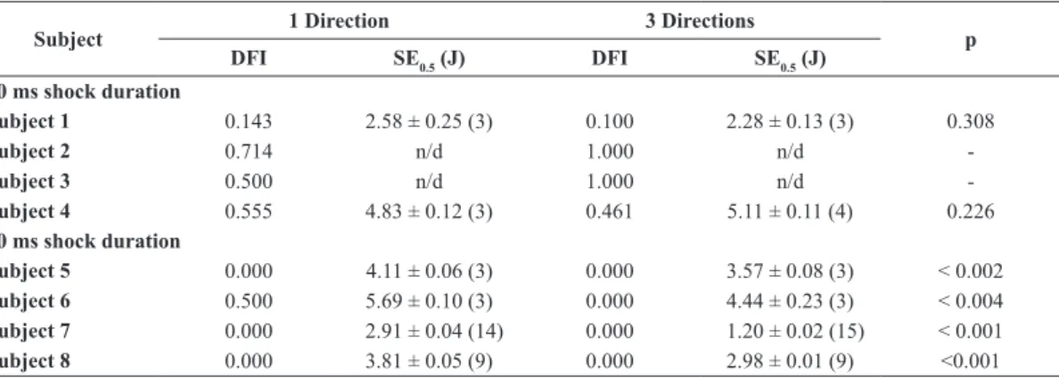
Table 1 shows data of individual experiments using shocks of either 20 or 30 ms duration, applied in one or three directions. Failure at deibrillation at the high shock energy range was more common for the longer shocks (DFI median values were 0.634 and 0.000 for 30 and 20 ms duration, respectively; p < 0.001, Mann-Whitney test). As it can be seen in Table 1, it was not possible to determine the deibrillation curves for 2 of the 4 animals to which 30 ms-long shocks were applied, because none of attempts with the multidirectional modality was able to reverse the arrhythmia (DFI = 1). In contrast, when multidirectional 20 ms-long shocks were used, successful deibrillation was achieved in all cases (DFI = 0).
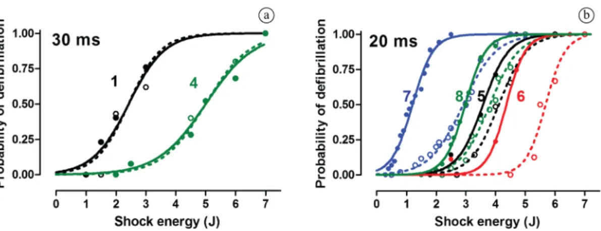
multidirectional deibrillation. Each color represents the data from a different animal, which is identiied by a number in Figure 7 and Table 1. As it can be seen in the igure, sensitivity to shocks was highly variable among the animals. However, it is possible to observe that, in all experiments using 20 ms-long shocks (Figure 7b), the curve obtained with multidirectional deibrillation was shifted to the left with respect to that determined with unidirectional deibrillation in the same animal. Nevertheless, this did not happen when shocks lasted 30 ms (Figure 7a). Accordingly, the SE0.5 values estimated from the curves (Table 1) were not statistically different between the unidirectional and multidirectional modalities in the few animals that responded to 30 ms-long shocks, whereas the a statistically signiicant decrease in the SE0.5 values
(p < 0.01) occurred for the multidirectional modality in
all tested animals receiving shocks with 20 ms duration. These results indicate that, provided that the stimulus was suficiently short, application of deibrillatory shocks in 3 directions decreased the stimulus energy required for deibrillation, compared to the use of the conventional, unidirectional approach. For further comparison in a larger experimental sample with 20 ms-long shocks, see Viana et al. (2014).
Discussion
Although several authors have reported greater eficiency of biphasic shocks (Shelton et al., 2011; Tanabe et al., 2012), the deibrillatory monophasic truncated exponential waveform was chosen in present study to better isolate the inluence of stimulation direction by removing possible interference of waveform-dependent effects on myocardial polarization (Trayanova and Bray, 1997). The delivered discharges in the two deibrillation modalities, and at the 3 deibrillator outputs during multidirectional stimulation, reached the same peak voltage and had similar time course at a given energy level, which eliminated a possible bias caused by variation in the pattern of stimulation in different directions and/or modalities introduced by the use of different equipments for each pathway. The cardiac impedance estimated during application of the deibrillatory pulses (61 ± 11 Ω; Viana et al., 2014) was in agreement with the standard 50 Ω resistive load assumed for development of cardiac stimulation devices (American National Standards Institute…, 1996; Associação Brasileira de Normas Técnicas…, 2005) and in the project of the present deibrillator.
In this instrument, the duration of the trigger pulse train at the TRIAC gate deines the duration of the
Figure 6. Monophasic truncated exponential waveforms (5 J peak
energy) for different resistive test loads (9, 50, 115, 229, 560 Ω).
Table 1. Individual data from electrical deibrillation experiments in swine using shocks applied in one or 3 directions. DFI: deibrillation failure index, i.e., the ratio of the number of shocks with energy ≥ 6 J that failed to restore sinus rhythm and the total number of applied shocks in this energy range. SE0.5: estimated shock energy associated with a probability of successful deibrillation equal to 0.5 (mean ±
standard error), obtained from the deibrillation curves shown in Figure 7. The number of degrees of freedom of the non-linear regression is shown in parentheses. The p values obtained with the F test for comparison of the SE0.5 values between the deibrillation modalities are
also shown. n/d: not determined.
Subject 1 Direction 3 Directions p
DFI SE0.5 (J) DFI SE0.5 (J)
30 ms shock duration
Subject 1 0.143 2.58 ± 0.25 (3) 0.100 2.28 ± 0.13 (3) 0.308
Subject 2 0.714 n/d 1.000 n/d
-Subject 3 0.500 n/d 1.000 n/d
-Subject 4 0.555 4.83 ± 0.12 (3) 0.461 5.11 ± 0.11 (4) 0.226
20 ms shock duration
Subject 5 0.000 4.11 ± 0.06 (3) 0.000 3.57 ± 0.08 (3) < 0.002
Subject 6 0.500 5.69 ± 0.10 (3) 0.000 4.44 ± 0.23 (3) < 0.004
Subject 7 0.000 2.91 ± 0.04 (14) 0.000 1.20 ± 0.02 (15) < 0.001
deibrillatory shock. This electronic type of switch was chosen to avoid prolonging of the total discharge duration, which is observed when relays are used for switching among outputs (Fonseca et al., 2013). The results of the bench and in vivo tests showed that this option resulted in reproducible discharge duration, in agreement with the preset value.
The present study shows that the superior effectiveness of multidirectional deibrillation over the conventional approach is highly dependent on the duration of the deibrillatory pulse. For 30 ms-long stimuli, multidirectional stimulation generally presented a poorer performance than unidirectional deibrillation, as it completely failed at restoring sinus rhythm in half of the tested subjects, even at high shock energy levels. Moreover, in the animals that responded to this modality, deibrillation eficiency (evaluated by the SE0.5 values) was not statistically different from
that of the conventional, unidirectional stimulation. However, the sole decrease of shock duration to 20 ms not only did enable hearts to respond to multidirectional stimulation, but also revealed the greater eficiency of this deibrillation modality.
Even though the time elapsed for delivery of 3 pulses with 30 ms duration would be shorter that the reported action potential duration recorded in vitro in swine
ventricle at physiological temperatures (> 100 ms; Roscher et al., 2001), it is possible that the in vivo
conditions, such as transient myocardial ischemia and increased catecholamine release, resulting from the interruption of cardiac pumping during VF, may have resulted in action potential shortening (Christé et al., 2006; Hoeker et al., 2014). In this case, it is likely that deibrillatory pulses would reach some cells during the relative refractory period (vulnerable period), which would favor arrhythmia reinitiation (Corbisiero et al., 1999), thus masking or even reverting
the beneicial effect of multidirectional deibrillation. These observations point out the importance of using short shocks for eficient multidirectional deibrillation.
Despite the choice of 100 µF electrolytic capacitors, aimed at minimizing potential damaging effects of the application of high peak voltage and current to the myocardium, the time for attainment of the maximal capacitor charge exceeded the recommended period of 15 s (American National Standards Institute…, 1996; Associação Brasileira de Normas Técnicas…, 2005). In our experimental setup, this has not posed a problem because, in the case of deibrillation failure with the developed instrument, a conventional deibrillator was ready for use. Nevertheless, this limitation needs to be addressed during the development of a prototype for clinical application. Additionally, because 3 electrodes must be accommodated in each paddle support, it was necessary to reduce the contact area of each electrode to less than the minimal area recommended for pediatric internal use (American National Standards Institute…, 1996; Associação Brasileira de Normas Técnicas…, 2005), as to keep them suficiently apart as to avoid their contact, but still close enough to allow them to fully contact the epicardium,. Again, redesigning the electrode disposition in the paddles is necessary prior to their use in humans. Nevertheless, in the experimental scenario, the smaller contact area did not result in macroscopic changes indicative of burn or other kind of damage in the ventricular epicardium after repeated shock delivery (up to 36 shocks).
It is known that electric ields applied to the heart may produce excitatory or deleterious effects, depending on the intensity. The sensitivity of cardiac cells to both kinds of effect markedly depends on the direction of the ield application with respect to the cell or iber bundle major axis: lower ield intensities are required
Figure 7. Probability of successful deibrillation as a function of the shock energy estimated from in vivo experiments in swine. Shocks
to produce both excitation and cell injury when the ield direction is parallel to the major axis. This seems to be due to the direction-dependent ability of the ield to cause a variation in transmembrane electrical potential suficient for attainment of the excitation threshold or massive electroporation (Bassani et al., 2006; Goulart et al., 2012; Knisley and Baynham, 1997; Oliveira et al., 2008;). As in the whole heart the muscle ibers are arranged in different directions (Smerup et al., 2009), a shock applied between a pair of electrodes in a given orientation will have different impact on cells with different spatial orientations. Thus, multidirectional stimulation should be able to promote electrical recruitment of cells that might remain unexcited by unidirectional stimulation, thus allowing effective stimulation with lower and safer ield intensities. This proposal was conirmed for both near-excitation threshold and high-intensity ield stimulation of isolated cardiac myocytes (Fonseca et al., 2013) and of the whole heart in vivo (Viana et al.,
2014; present study).
In the present coniguration, multidirectional deibrillation consisted of the sequential delivery of shocks in 3 directions, in a 60° angle from each other, which resulted in an average decrease in shock energy of 30%, compared to a single direction, for a probability of deibrillation success of 0.5. Although other authors have found an even greater reduction of shock energy requirement using more than one stimulation direction (Chang et al., 1986; Jones et al., 1988; Kerber et al., 1994; Pagan-Carlo et al., 1998), one must consider that, in addition to the possible bias introduced by the use of different deibrillators for shock delivery in different pathways, some authors used temporally overlapping multidirectional shocks (Kerber et al., 1994; Pagan-Carlo et al., 1998), which was avoided in our study. Even though temporal superposition should further enhance excitatory recruitment, it should also increase the damaging potential of high-intensity ield stimulation, which may result in failure of deibrillation due to arrhythmia reinduction and/or myocardial injury.
Fonseca et al. (2013) identiied two mechanisms involved in the enhanced electrical recruitment of cardiac myocytes during rapidly-switching, sequential application of electrical ield in different directions. One of them is direction-dependent, probably due to the induction of threshold depolarization in a greater number of cells, as explained earlier. The other mechanism is time-dependent, and probably involves temporal summation of the depolarization responses evoked by the stimuli delivered in rapid succession, as excitatory recruitment was facilitated also during unidirectional stimulation with pulses applied at very short intervals. The effects of multidirectional
and sequential stimulus presentation were additive (Fonseca et al., 2013), which might explain the increased eficiency of our present protocol of rapidly-switching multidirectional deibrillation. It is also important to observe that, despite the large inter-individual variability in sensitivity to deibrillatory shocks (e.g., compare subjects 6 and 7, in which the difference in SE0.5 was at least of 100%, Table 1,
Figure 7), multidirectional deibrillation with 20 ms shocks resulted in a consistent, statistically signiicant decrease in effective shock energy.
Overall, the developed deibrillation system, unique in its design that allows rapidly-switching multidirectional stimulation with identical waveforms, allowed the conirmation of decrease in shock energy requirement for in vivo direct deibrillation by using
shocks sequentially delivered in 3 directions within a short period (60 ms), in comparison with the conventional approach with a single pair of electrodes. The present data also show that the beneicial effects of multidirectional deibrillation are dependent on shock duration, and abolished with longer stimuli.
Acknowledgements
We are indebted to Mr. Mauro Martinazo, Mr. Renato S. Moura and Mr. Carlos A. Silva (CEB/UNICAMP), and to Ms. Ana Cristina de Moraes and Mr. William A. Silva (NMCE/FCM/UNICAMP) for technical support to the development of the deibrillator and project documentation, and to the in vivo experiments,
respectively. We are also grateful to Mr. André Jardini and Mr. Luis F. Bernardes (BIOFABRIS/UNICAMP) for the contribution to the development of the paddles. This study was supported by the Conselho Nacional de Pesquisa e Desenvolvimento (CNPq), grant nº 302996/2011-7 (JWM Bassani) and scholarship nº 135204/2008-9 (MA Viana).
References
Altman DG. Practical statistics for medical research. London:
Chapman and Hall; 1991.
American National Standards Institute. Association for the Advancement of Medical Instrumentation. DF2: cardiac defibrillator devices. New York: ANSI; 1996.
Associação Brasileira de Normas Técnicas. NBR IEC 60601-2-4: equipamento eletromédico - Parte 60601-2-4: prescrições particulares para segurança de desfibriladores cardíacos. Rio de Janeiro: ABNT; 2005.
Bassani RA, Lima KA, Gomes PAP, Oliveira PX, Bassani
Chang M, Inoue H, Kallok M, Zipes DP. Double and triple sequential shocks reduce ventricular defibrillation threshold in dogs with and without myocardial infarction. Journal of the American College of Cardiology. 1986; 8(6):1393-405. http://dx.doi.org/10.1016/S0735-1097(86)80313-8.
PMid:3782643.
Cheek ER, Fast VG. Nonlinear changes of transmembrane potential during electrical shocks: role of membrane electroporation. Circulation Research. 2004; 94(2):208-14. http://dx.doi.org/10.1161/01.RES.0000111526.69133.DE.
PMid:14670844.
Christé G, Hadour G, Ovize M, Ferrera R. Brain death does
not change epicardial action potentials and their response
to ischemia-reperfusion in open-chest pigs. The Journal of Heart and Lung Transplantation. 2006; 25(7):847-53. http://
dx.doi.org/10.1016/j.healun.2006.03.018. PMid:16818129.
Corbisiero R, Kabell G, Cook JR, Fitzgerald TF, Kirchhoffer JB. Effects of adenosine on local stimulus-response latency and induction of atrial fibrillation by premature stimuli. Pacing and Clinical Electrophysiology. 1999; 22(9):1378-85. http://dx.doi.org/10.1111/j.1540-8159.1999.tb00632.x. PMid:10527020.
Euler DE, Whitman TA, Roberts PR, Kallok MJ. Low
voltage direct current delivered through unipolar transvenous
leads: an alternate method for the induction of ventricular fibrillation. Pacing and Clinical Electrophysiology. 1999; 22(6):908-14. http://dx.doi.org/10.1111/j.1540-8159.1999. tb06815.x. PMid:10392389.
Fedorov VV, Nikolski VP, Efimov IR. Effect of electroporation
on cardiac electrophysiology. Methods in Molecular
Biology (Clifton, N.J.). 2008; 423:433-48. http://dx.doi.
org/10.1007/978-1-59745-194-9_34. PMid:18370220.
Fonseca ASV, Bassani RA, Oliveira PX, Bassani JWM. Greater cardiac cell excitation efficiency with rapidly switching multidirectional electrical stimulation. IEEE Transactions on Biomedical Engineering. 2013; 60(1):28-34. http://
dx.doi.org/10.1109/TBME.2012.2220766. PMid:23033428.
Goulart JT, Oliveira PX, Bassani JWM, Bassani RA. The influence of cell dimensions on the vulnerability of ventricular
myocytes to lethal injury by high-intensity electrical
fields. Revista Brasileira de Engenharia Biomédica. 2012; 28(4):337-45. http://dx.doi.org/10.4322/rbeb.2012.040.
Hayashi M, Shimizu W, Albert CM. The spectrum of
epidemiology underlying sudden cardiac death. Circulation
Research. 2015; 116(12):1887-906. http://dx.doi.org/10.1161/
CIRCRESAHA.116.304521. PMid:26044246.
Hoeker GS, Hood AR, Katra RP, Poelzing S, Pogwizd SM. Sex differences in β-adrenergic responsiveness of action
potentials and intracellular calcium handling in isolated
rabbit hearts. PLoS One. 2014; 9(10):e111411. http://dx.doi. org/10.1371/journal.pone.0111411.
Jones DL, Klein JG, Rattes MF, Sohla A, Sharma AD. Internal cardiac defibrillation: single and sequential pulses and a variety of lead orientations. Pacing and Clinical Electrophysiology. 1988; 11(5):583-91. http://dx.doi.
org/10.1111/j.1540-8159.1988.tb04554.x. PMid:2456537.
Kerber RE, Spencer KT, Kallok MJ, Birkett C, Smith R, Yoerger D, Kieso RA. Overlapping sequential pulses: a new waveform for transthoracic defibrillation. Circulation. 1994; 89(5):2369-79. http://dx.doi.org/10.1161/01.CIR.89.5.2369.
PMid:8181163.
Knisley SB, Baynham TC. Line stimulation parallel to myofibers enhances regional uniformity of transmembrane
voltage changes in rabbit hearts. Circulation Research. 1997;
81(2):229-41. http://dx.doi.org/10.1161/01.RES.81.2.229.
PMid:9242184.
Koster RW, Walker RG, Van Alem AP. Definition of successful defibrillation. Critical Care Medicine. 2006; 34(12 Suppl):423-6. http://dx.doi.org/10.1097/01.CCM.0000246008.95156.78.
PMid:17114971.
Krauthamer V, Jones JL. Calcium dynamics in cultured
heart cells exposed to defibrillator-type electric shocks. Life Sciences. 1997; 60(22):1977-85. http://dx.doi.org/10.1016/
S0024-3205(97)00162-8. PMid:9180351.
Kroll MW, Swerdlow CD. Optimizing defibrillation waveforms for ICDs. Journal of Interventional Cardiac Electrophysiology. 2007; 18(3):247-63. http://dx.doi.
org/10.1007/s10840-007-9095-z. PMid:17541815.
Larsen MP, Eisenberg MS, Cummins RO, Hallstrom AP. Predicting survival from out-of-hospital cardiac arrest: a graphic model. Annals of Emergency Medicine. 1993; 22(11):1652-8. http://dx.doi.org/10.1016/S0196-0644(05)81302-2. PMid:8214853.
Malmivuo J, Plonsey R. Bioelectromagnetism: principles
and applications of bioelectric and biomagnetic fields. New York: Oxford University Press; 1995.
Oliveira PX, Bassani RA, Bassani JWM. Lethal effect of electric fields on isolated ventricular myocytes. IEEE Transactions on Biomedical Engineering. 2008; 55(11):2635-42. http://
dx.doi.org/10.1109/TBME.2008.2001135. PMid:18990634.
Pagan-Carlo LA, Allan JJ, Spencer KT, Birkett CL, Myers R, Kerber RE. Encircling overlapping multipulse shock waveforms for transthoracic defibrillation. Journal of the American College of Cardiology. 1998; 32(7):2065-71. http://
dx.doi.org/10.1016/S0735-1097(98)00486-0. PMid:9857894.
Patil KD, Halperin HR, Becker LB. Cardiac arrest: resuscitation and reperfusion. Circulation Research. 2015;
116(12):2041-9. http://dx.doi.org/10.1161/CIRCRESAHA.116.304495.
PMid:26044255.
Petrucci O Jr, Oliveira PP, Carmo MR, Vieira RW, Braile
DM. Standardization of an isolated pig heart preparation with parabiotic circulation: methodological considerations. Brazilian Journal of Medical and Biological Research. 2003; 36(5):649-59. PMid:12715085.
Roscher R, Arlock P, Sjöberg T, Steen S. Effects of dopamine
on porcine myocardial action potentials and contractions
at 37 °C and 32 °C. Acta Anaesthesiologica Scandinavica. 2001; 45(4):421-6. http://dx.doi.org/10.1034/j.1399-6576.2001.045004421.x. PMid:11300379.
International Journal of Cardiology. 2011; 147(3):405-8. http://
dx.doi.org/10.1016/j.ijcard.2009.09.545. PMid:19861229.
Smerup M, Nielsen E, Agger P, Frandsen J, Vestergaard-Poulsen P, Andersen J, Nyengaard J, Pedersen M, Ringgaard S, Hjortdal V, Lunkenheimer PP, Anderson RH. The three-dimensional arrangement of the myocytes aggregated together within the mammalian ventricular myocardium. The Anatomical Record. 2009; 292(1):1-11. http://dx.doi.
org/10.1002/ar.20798. PMid:19051244.
Tanabe S, Yasunaga H, Ogawa T, Koike S, Akahane M, Horiguchi H, Hatanaka T, Yokota H, Imamura T. Comparison of outcomes after use of biphasic or monophasic defibrillators among out-of-hospital cardiac arrest patients: a nationwide
population-based observational study. Circulation:
Cardiovascular Quality and Outcomes. 2012; 5(5):689-96. http://dx.doi.org/10.1161/CIRCOUTCOMES.112.965319.
PMid:22967787.
Trayanova N, Bray MA. Membrane refractoriness and excitation induced in cardiac fibers by monophasic and biphasic
shocks. Journal of Cardiovascular Electrophysiology. 1997; 8(7):745-57. http://dx.doi.org/10.1111/j.1540-8167.1997. tb00833.x. PMid:9255682.
Viana MA, Bassani RA, Petrucci O, Marques DA, Bassani
JW. Rapidly switching multidirectional defibrillation: reversal of ventricular fibrillation with lower energy shocks. The Journal of Thoracic and Cardiovascular Surgery. 2014; 148(6):3213-8. http://dx.doi.org/10.1016/j.jtcvs.2014.07.035. PMid:25173125.
Yabe S, Smith WM, Daubert JP, Wolf PD, Rollins DL, Ideker
RE. Conduction disturbances caused by high current density
electric fields. Circulation Research. 1990; 66(5):1190-203. http://dx.doi.org/10.1161/01.RES.66.5.1190. PMid:2335021.
Zheng X, Benser ME, Walcott GP, Smith WM, Ideker RE. Reduction of the internal atrial defibrillation threshold with balanced orthogonal sequential shocks. Journal of Cardiovascular Electrophysiology. 2002; 13(9):904-9. http://dx.doi.org/10.1046/j.1540-8167.2002.00904.x.
PMid:12380930.
Authors
Marcelo Almeida Viana1*, Rosana Almada Bassani2, Orlando Petrucci3, Denilson Antônio Marques1, José Wilson Magalhães Bassani1,2
1 Department of Biomedical Engineering, School of Electrical and Computer Engineering, Universidade Estadual de
Campinas – UNICAMP, Av. Albert Einstein, 400, Sala 221, CEP 13083-852, Campinas, SP, Brazil.
2 Center for Biomedical Engineering, Universidade Estadual de Campinas – UNICAMP, Campinas, SP, Brazil. 3 Department of Surgery, School of Medical Sciences, Universidade Estadual de Campinas – UNICAMP, Campinas, SP,