DOI: http://dx.doi.org/10.1590/2446-4740.00316
*e-mail: [email protected]
Received: 16 January 2016 / Accepted: 28 February 2016
A digital approach for design and fabrication by rapid prototyping
of orthosis for developmental dysplasia of the hip
Rodrigo Munhoz, Cícero André da Costa Moraes, Harki Tanaka, Maria Elizete Kunkel*
Abstract Introduction: Immobilization in a hip spica cast is required in surgical and nonsurgical treatments for children aged three months to four years diagnosed with developmental dysplasia of the hip. Skin complications are associated with the use of the spica cast in 30% of the cases. This research explores the use of photogrammetry and rapid prototyping for the production of a lighter, shower friendly and hygienic hip orthosis that could replace the hip spica cast. Methods: Digitalized data of a plastic dool was used for design and fabrication of a customised hip orthosis following four steps: 1) Digitalization of the external anatomical structure by photogrammetry using a smartphone and open source software; 2) Idealization and 3D modeling of the hip orthosis; 3) Rapid prototyping of a low cost orthosis in polymer polylact acid; 4) Evaluation tests. Results: Photogrammetry provided a good 3D reconstruction of the dool’s hip and legs. The manufacture method to produce the hip
orthosis was accurate in itting the hip orthosis to the contours of the doll. The orthosis could be easily placed
on the doll ensuring mechanical strength to immobilize the region of the hip. Conclusion: A new approach and the feasibility of both techniques for hip orthosis fabrication were described. It represents an exciting advance for the development of hip orthosis that could be used in orthopedics. To test the effectiveness of this orthosis for developmental dysplasia of the hip treatment in newborns, material and mechanical tests, design optimization and physical tests with patients should be carried.
Keywords: Developmental dysplasia of the hip, Orthosis, Rapid prototyping.
Introduction
Developmental dysplasia of the hip (DDH) is a spectrum of anatomical abnormalities in the immature hip joint in which the femoral head has an abnormal relationship with the acetabulum (Storer and Skaggs, 2006). DDH occurs in newborn infants and it varies from milder acetabular dysplasia to complete dislocation of the femoral head (Yang and Quanjun, 2012) (Figure 1). DDH is usually asymptomatic during infancy - Guarniero (2010) demonstrated an incidence in Brazil of 5 per 1,000 cases. The etiology of DDH is multi factorial including ligament laxity, breech presentation, postnatal positioning and primary acetabular dysplasia (Schwend, 2014). Uncorrected DDH is associated with long-term morbidity such as gait abnormalities, chronic pain and degenerative arthritis (Noordin et al., 2010; Schwend et al., 1999).
The aim of the DDH treatment is to achieve and maintain concentric reduction of the femoral head
into the acetabulum. If diagnosed during the irst six
months of life, DDH can be corrected with nonsurgical methods that includes the use of orthotic devices for hip repositioning and to keep the femoral head in the correct position in the acetabulum. Pavlik harness,
a brace straps and fasteners, is used to it around a
child’s chest, shoulders and legs being the standard
treatment for DDH. This is a dynamic positioning device that avoids complete immobilization of the joint, prevents hip extension and adduction but allows
lexion and abduction (Storer and Skaggs, 2006; Wahlen and Zambelli, 2015). If a dislocated hip is not reduced within three weeks, the Pavlik harness should be discontinued and an alternative treatment selected. The next option involves closed reduction under anesthesia with the use of a hip spica casting, a plaster cast used to keep the affected hip joint of the newborn in the best position for normal growth (Rampal et al., 2008; Storer and Skaggs, 2006; Vitale and Skaggs, 2001). The position of the femoral head in a spica cast of a child with DDH needs to
be conirmed accurately and monitored routinely
by imaging methods (Teng et al., 2012). The shape of the hip spica cast varies and can be extend from the mid-chest down to the ankle; sometimes a bar between the legs strengthens the cast. An opening in the genital area allows normal urine and bowel elimination. Hip spica casts are usually worn for two up to three months and cannot be removed before this period of time (Steps, 2010).
Caring for a child in a hip spica cast is extremely challenging. The spica must be kept dry, if water is
absorbed into the spica the plaster will become weak and may crack. Daily tasks such as changing nappies and bathing of a child in a hip spica takes extra time and practice. It is not possible to bath a child using a hip spica and thorough wash with a damp cloth at least once a day is required. The lack of bathing and the pressure of the cast on the spine cause skin complications in 30% of the cases (Difazio et al., 2011; Halanski and Noonan, 2008). In general, children in casts only sleep for short periods and often become restless and distressed. Disturbed nights can be caused due to the occurrence of cramp, itching and the inability to turn over. It is necessary to change the child’s position every 2-4 hours (The Royal…, 2015; Steps, 2010).
Surgical methods are an option of treatment for children in whom nonoperative treatment with hip spica has failed or in children diagnosed after six months of age. The treatment includes open reduction of the hip with femoral or acetabular osteotomy also followed by the use of hip spica cast (Mulpuri et al., 2015). Families, especially mothers, experience problems providing home care after their child’s surgery, the most frequently related problems are hygiene/care after toileting, cast and skin care, and personal hygiene (Demir et al., 2015).
The literature has reported the use of some orthotic devices in treatment of the DDH, such as rigid and semirigid abduction braces, to replace the use of Pavlik harness and the spica cast (Dyskin and Ferrick, 2015; Hedequist et al., 2003; Ibrahim et al., 2013; Uras et al., 2014; Wahlen and Zambelli, 2015; Wilkinson et al., 2002). The use of an abduction brace is easier for the patient, parents and the physician. However, current literature provides controversial results of treatment of DDH with these plastic abduction orthoses and since the results depends on severity of hip dysplasia more research is needed.
Because every patient with DDH has an unique anatomy (Yang and Quanjun, 2012), custom made orthoses remain the ‘gold standard’ once the orthotic geometry is tailored to each patient. Improved clinical effectiveness has been linked to customised orthesis over massproduced ones (Brncick, 2000). However,
the manual fabrication of a custom-it orthosis is a
laborious, time-consuming and imprecise process performed by skilled orthotists in order to create comfortable and functional devices. Due to the reduced cost, in some contexts there has been an increasing preference for prefabricated orthoses (Majumdar et al., 2012). There is a great need for customised products
in the ield of orthotics since not all patients can be
treated with standard sized orthesis.
In recent years, some attempts to use new technologies
in the manufacture of patient speciic orthotics have
shown good results. Computerized techniques for
manufacture of patient-speciic orthotic devices have
the potential to provide excellent comfort. Otherwise it allows changes in the standard design to meet
the speciic needs of each patient (Mavroidis et al., 2011). The main phases of this process includes: a) 3D Scanning of an anatomical structure and 3D digital model reconstruction, b) Orthosis design and c) Production of the orthosis by a manufacture process.
Laser scanners, structured light scanners, scanners with video cameras and photogrammetry are the main technologies that could be used for 3D anatomic surface scanning. A laser scanner device uses a laser beam normal to the surface to be scanned. There are several commercial devices that are relatively easy to use. The aplication of laser scanner in the production of medical devices can reach 98% of accuracy (Igathinathane et al., 2010), but the equipment cost is still relatively high (Milusheva et al., 2006). Photogrammetry technique allows obtaining spatial measurements, and other geometrically reliable information, derived from photographs captured from different points of view. This portable and low-cost technology uses automated matching algorithms to produce a dense point cloud of an object and its 3D
reconstruction through a post-processing procedure (Remondino, 2003). Ciobanu and Rotariu (2014) reported that photogrammetry has been used in
the orthopedics ield e.g. treatment of scoliosis
(Aroeira et al., 2011), deformational plagiocephaly (Moghaddam et al., 2014) and prostheses/ortheses fabrication (Ciobanu et al., 2013a; 2013b).
Additive manufacturing (AM) describes a set of new manufacturing methods, processes and technologies that function through material addition, in contrast to the established traditional cutting, forming or casting models. Rapid prototyping (RP) technologies are the most widely applied and known fabrication methods based on AM principles. The fused deposition modeling (FDM) is a process to produce 3D solid components from digital models using additive techniques and creating by laying successive layers of material. The continuous improvement of RP system accuracy and materials expand gradually the applications to other areas (Giannatsis and Dedoussis, 2009). AM and RP technologies have been used for product development and design process due to their relatively
high speed and lexibility. These technologies have
also been employed in various non-manufacturing applications such as medicine and health care (fabrication of custom made implants and scaffolds for rehabilitation, models for pre-operating surgical planning, and anatomical models for mechanical testing) (Giannatsis and Dedoussis, 2009). While the application of RP in developing countries is still at early stage, some recent studies have investigated feasibility of using AM and RP in the manufacture of orthoses. Palousek et al. (2014), Patar et al. (2012), Paterson (2013) and Paterson et al. (2010; 2014a; 2014b; 2015) have developed new methods to support the mass customization of wrist splints. Jumani et al. (2014) used FDM for fabrication of custom-made foot orthoses, and Mavroidis et al. (2011) used RP to produce an ankle-foot orthose. For the authors’ knowledge, no data are available in the literature about new approaches for design and production of orthoses for children’s hip by photogrammetry and RP.
The dificulties encountered in day-to-day care of
children using hip spica cast during treatment of DDH have been the motivation of the current study (Steps, 2010). There is a growing interest in new techniques of production of abduction brace orthoses in attempt to avoid these complications (Dyskin and Ferrick, 2015; Munhoz et al., 2014; 2015). A hip orthosis produced using RP method could provide a highly technical support system that can be fully ventilated, light, shower friendly, hygienic and stylish. In this study, a new approach was developed to combine photogrammetry with RP to produce a custom made hip orthosis for DDH.
Methods
In this irst approach, a plastic doll was used
to represent a newborn with approximately four months of age. The same procedure into a real child will require some adjustments that have not yet been addressed in this study. The methodology adopted
comprised ive stages.
Stage 1: acquisition of the external anatomical structure by photogrammetry
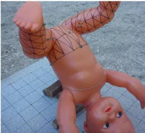
The dool’s hip and legs were marked with traces randomly using a permanent black marker. The doll was placed on a stand, for elevation of the hip region, in supine position with her legs up (Figure 2).
This stand was placed above a non-relective board
used as external reference with traces forming square 5 × 5 cm. The camera of a smartphone Sony Xperia R800 (Sony, 2015) with resolution of 5.1 Megapixels,
2592 × 1944 pixel, VGA, autofocus, LED lash, video
720p, CMOS sensor (Complementary Metal Oxide Semiconductor) technology without anti-vibration option was used for the acquisition of the photos. A set of 55 photos of the region of the dool’s hip and legs
were taken indoors with artiicial light and without use of lash or zoom resources. Camera calibration
was not necessary for this procedure. The photos were taken moving the camera around the doll in a spiral trajectory, in order to obtain at least two scenes that
show the same point. The irst sequence of photos
started with the camera positioned approximately 50 cm above the doll, and with 45 degrees of inclination with the horizontal plane. The second sequence of photos
Figure 2. Experimental setup used in the photogrammetric procedure for 3D surface reconstruction of the hip and legs of a plastic doll. The doll with hip and legs marked by random traces was placed on a stand in
were taken by moving the camera each 15 degrees approximately in a spiral trajectory until it reaches the horizontal position (Figure 3). A computer Intel i7 (3.4 Ghz processor and 16 GB-RAM and Windows 8.1 operation system) and two free and open source softwares, Python Photogrammetry Toolbox (PPT-GUI) and MeshLab were used as described bellow: First, the 55 photos were processed using the PPT-GUI user interface. This step is required to provide the
photos pathway where the JPG iles is stored using
the Check Camera Database tab (JPG stands for Joint Photographic Experts Group is a commonly used method of lossy compression for images produced by
digital photography). The ile was saved with a simple
name without symbols or spaces as required to the
pathway. The PPT-GUI read the JPG iles looking for
the camera information to recognize the sensor size, in this case, 3.8 mm width. A point cloud is a set of
data points in the 3D coordinate system deined by X,
Y, and Z coordinates representing the external surface
of an object that was photographed. The Bundler is an algorithm of the PPT-GUI that produces point clouds from a list of unordered images. After run the Bundler a sparse point cloud was calculated requiring the photos
pathway and the scale photos factor inputs that deine
the scale at which the 3D reconstruction should be performed. The use of a scale 1:1 generates a model in full size according to the size of the camera sensor. However this option requires larger use of memory capability and computer processing. The sparse point cloud of the doll was stored in a temporary folder and a Patch-based Multi-view Stereo (PMVS) algorithm without Clustering Views for Multi-view Stereo (CMVS) was used (Furukawa and Ponce, 2009). This last step provided a dense point cloud
in a polygon ile format (PLY) stored into a PMVS
folder inside of the temporary folder created earlier.
A PLY ile supports a relatively simple description of a single object as a list of nominally lat polygons. The PLY point cloud ile was imported into Meshlab
software and some unnecessary points were removed. The point cloud was resized to a real measure and an useful mesh of the 3D hip and legs model of the doll was create. Using a metric tool into the software Meshlab it was possible to measure the trace dimension 5 × 5 cm in the board used as external reference plate and perform the model resizing by a scale factor. The scale factor is the ratio between the real and the current measure on Meshlab metric system. The scale
factor was added into a transform scale inside the ilters
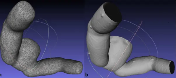
and normal curves and orientation (the number 1 is equal to 1 mm). After that, the reference plate was discarded and the point cloud of the dool’s hip and legs was converted into a 3D mesh using a Poisson reconstruction approach (Figure 4a).
Figure 3. Photogrammetry scheme used: The sphere represents the target object, in this case the doll, over the reference plate. The cubes represent the camera of a smartphone moving in a spiral trajectory around the doll.
Stage 2: idealization and modeling of a hip orthosis for DDH
Based on the 3D reconstruction of the dool’s hip and legs by photogrammetry, a Computer Aided Design (CAD) based orthesis model was designed with the Blender software. The hip orthesis was designed in two separated parts (anterior and posterior) with an opening in the genital area for physiological needs
and cleaning. For each part, irst the left side of the
orthosis was modeled then to save time and computer memory this side was mirrored in the sagittal plane. The two parts of the CAD model were then converted
into a stereolithography (STL) ile format using a
mesh to solid converter tool. A STL format contains triangular facet representation of surfaces and have become standard data inputs of RP and manufacturing systems.
Stage 3: manufacture of the hip orthosis by RP
The free software MatterControl was used to
generate a G-code of the STL ile for 3D printing.
G-code is used in computer-aided manufacturing to control the 3D machine tools. The MatterControl takes the CAD model, slices it into layers, and output the G-code required for each layer. Both parts, anterior and posterior, of the hip orthosis were produced in a 3DCloner machine with FDM technology (Microbras, Santa Terezinha de Itaipu, Brazil). This is a low cost and affordable 3D printer available on market for the price of 1,200 dollars. The 3D printing volume of the 3DCloner is 320 mm (width), 210 mm (depth) and 160 mm (height). The biodegradable polymer polylactic acid (PLA) in the form of a spool of
1.75 mm diameter ilament was used. To produce an
orthosis, 400 grams of PLA including extra pillars of polymer to support hanging structures was used.
The printer coniguration used 0.3 mm layer thickness and 30% ill density in the actual scale of 1:1. The hip
orthosis was built from the bottom up - one layer at a time. The build time to produce the orthosis was about 30 hours.
Stage 4: evaluation - photogrammetry x 3D laser scanning
To evaluate the eficiency of the photogrammetry
to 3D reconstruction of the dool’s hip and legs, the volume and shape of the 3D model obtained was compared with a 3D model of the same doll obtained with a comercial Artec MHTTM scanner (Artec Group
Inc, Luxemburgo). This scanner has a video camera technology and projects structured light using a triangle between the scanner lens, laser, and an object being
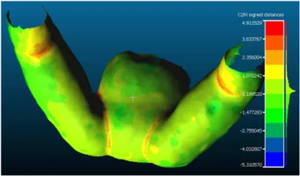
scanned. The frames are combined automatically in the scanner software into a single 3D mesh, in the 3D reconstruction phase (Ciobanu et al., 2013b). In this case, a mesh was generated in an automatic sequence with no need of additional steps as performed by the photogrammetry (Figure 4b). To compare both meshes, the open software CloudCompare was used. This is a 3D point cloud processing software to handle triangular meshes and calibrated images. The meshes were partially aligned and a topology mapping with the difference found between the two meshes was created.
Stage 5: evaluation - digital model x physical model
After production of the hip orthosis by RP its physical dimensions were compared with the 3D model reconstructed by photogrammetry. The result of this evaluation is used as an indicator of the good
it of the orthesis in doll’s body and the accuracy of
the PR to produce a hip orthosis. A custom orthosis
that its well the user’s body shapes allows more
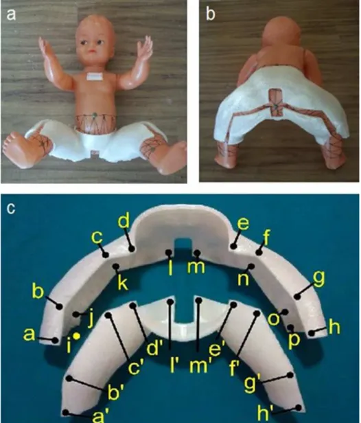
comfort. In both CAD model of the orthosis and in the 3D printed orthosis, 16 reference points were
deined for the anterior (a’, b’, c’, d’, e’, f’, g’, h’, i’,
j’, k’, l’, m’, n’, o’ and p’) and 16 reference points for the posterior part (a, b, c, d, e, f, g, h, i, j, k, l, m, n, o and p) (Figure 5c). Based on these points, 16 linear
parameters were deined for the anterior (a-b, b-c,
c-d, d-e, e-f, f-g, g-h, a-i, i-j, j-k, k-l, l-m, m-n, n-o, o-p and p-h) and 16 parameters for thee posterior part (b’, b’-c’, c’-d’, d’-e’, e’-f’, f’-g’, g’-h’, a’-i’, i’-j’, j’-k’, k’-l’, l’-m’, m’-n’, n’-o’, o’-p’, p’-h’). The measurements of the 32 parameters in both CAD model of the orthosis and the 3D printed orthosis were performed by a single observer. In the CAD model other measurements of the linear parameters were performed using milimeters in the software Blender. In the physical model, the measurements were performed in milimeters with a Mitutoyo digital caliper (Mitutoyo Corp, Kanogawa, Japan). The difference between the linear dimensions of the digital and Physical orthosis was computed. Each set of measurements was carried out by the same observer three times. Intraobserver reliability was examined by repeating the measurement of all parameters in both digital and physical orthosis.
Results
9,002 vertices and 18,000 faces (Figure 4a) and a mesh by 3D laser scanner with 268,139 vertices and 524,044 faces (Figure 4b). The number of vertices and faces using both techniques can vary due to the selected limits of the legs and waist. The number of vertices and faces also show that the resolution of the 3D model generated by photogrammetry was lower than the resolution of the 3D model generated by the laser scanner. This fact is not a problem because the
it of the prosthesis does not require high precision.
Instead the low resolution requires less computer time and it is desirable that the post-processing is faster. Regardless of the number of vertices and faces, the comparison between the both meshes performed by
CloudCompare software presented a difference of 10 mm (Figure 6). This implies that considering the topology of the whole model, the largest difference between a peak and a valley was found to be 10 mm and that the average difference for the whole model was 4 mm. This amplitude is presented in a topographic mapping composed of peaks and valleys superposing the difference between the two meshes, where the transition from green to red represents an elevation and the transition from a green to a blue region represents a depression. The Figure 7 shows both parts of the hip orthosis model in three diferent views (anterior, superior and posterior). The manufacturing process of the orthesis by RP is avaliable online (Biomecanica
e Forense UNIFESP, 2015). Figures 5a, b show anterior and posterior view of the doll using the hip orthosis produced by RP. The manufacture method
to produce the orthosis was accurate in itting to the
contours of the doll’s hip and legs. The comparison of the linear parameters of the digital and physical model of the orthosis has shown that a low difference was found between the parameters, the greatest difference value was 5 mm (Figure 5c). It means that the contours of the dool’s hip and legs were acquired with a good accuracy through the photogrammetry. The intraobserver reliability test (to compare indirect and direct measurements of the linear parameters
deined in the digital and physical model) showed that
measurements of the linear parameters were better repeated when obtained directly from the digital model.
Discussion
This work presented a digital approach for design and fabrication by RP of an orthosis that can be considered as an alternative for treatment of DDH.
The anatomical structure of dool’s hip and legs was digitalized in surface data, used as input for modeling of an orthosis in a CAD software, and later production in a RP machine.
Treatment of DDH in children from irst month
of age up to four years presents many challenges (Vitale and Skaggs, 2001). The overall medical cost of hip spica casting, including supplies and overall hospitalization time is low, the patients return home
quickly, but the physical and inancial burden of care
is transfered to the family (Ferreira and Thomson, 2012). In this study, we started from the principle that a hip orthosis produced from a polymeric material by RP could replace the hip spica cast currently used for DDH treatment with the advantage of reducing some problems related to the use of this devices. In the most cases of DDH a hip spica cast is used for months in children undergoing nonsurgical and surgical treatment (Dezateux et al., 2003). Although plaster casts and splints remain an important treatment method for acute and chronic orthopaedic conditions, there are known risks associated with cast
Figure 6. Topographic map generated with the CloudCompare software after comparison between the meshes from photogrammetry and laser scanning. The map is composed of peaks and valleys superposing the difference between the meshes. The transition from green to red represents an elevation and from a green to a blue region represent a depression. For the whole model, the largest difference was found to be 10 mm and the average difference was 4 mm.
application, immobilization, and removal (Halanski and Noonan, 2008). For instance, casting materials create an exothermic reaction and skin’s burns that are associated with water temperatures above 24 °C, more than eight layers and inadequate ventilation.
The plaster has been the deinitive management for
immobilization of the skeleton for over 100 years and now there is a trend that it will be replaced by modern techniques (Daines et al., 2014; DeMaio et al., 2012). A hip orthesis produced by RP can be fully ventilated, light, shower friendly, hygienic and stylish. Such orthesis could be used not only to DDH treatment but also to treat fractures and other musculoskeletal conditions for children when rest, immobilization, or correction of deformity is required (DeMaio et al., 2012). The material used to produce the orthosis, PLA, is a polymer made from renewable agricultural raw materials, biodegradable through composting, with good stiffness and strength. Unlike plaster, a
hip orthosis produced in PLA will not absorb luids
(pus, blood, or sweat). Besides such orthosis could be washed in water, it is lightweight, and could allow the skin to ventilate reducing the risk of skin
lesion. As the orthosis would be placed by itting
the newborn, this procedure can prevent the need for newborn being anesthetized for each application procedure and removal of cast. However, PLA has low resistance to temperature changes (from 60°C) being necessary to avoid direct exposure of the orthosis made of this material to sunlight or hot environments for a long time.
There is no standard method for collecting topographical surface data for hip and leg of newborns and it is important that new methods can be used and evaluated. A suitable anatomical data acquisition method for anatomical structures should consider accuracy, resolution, patient comfort and safety. The current work showed the production viability of a customized hip orthosis using cost effective scanning by photogrammetry, modeling using open software and production with a low cost RP machine. The photogrammetry, is cost-effective (about ten times cheaper than the laser scaner), has the advantage of being performed with open software using a digital photo camera, even a smartphone, for digitalization of an anatomical structure. In this study, many traces were
done on the doll to facilitate the identiication. In a real
child it would be possible to use a smaller number of traces that can be done with a non-allergic pen easily removable with water. For acquisition of the photos it is necessary that the child does not change position during the process. In a real case the child could be immobilized
by their parents or in more dificult cases the child could
be sedated. This is better than the usual procedure of anesthesia that occurs in the application of spica cast. Despite photogrammetry having the disadvantage of requiring considerable time during the 3D reconstruction
phase, new free software such as Autodesk 123D Catch faster and easier to use were released after the realization of this study. The laser scanner has the main advantage of fast processing for 3D reconstructions but is more expensive than photogrammetry (Ciobanu et al., 2013b). Both techniques allow the production of hip orthosis with less direct contact with the patient causing less stress with good repeatability (Palousek et al., 2014). Our results showed that both photogrammetry and 3D laser scanners provided adequate quality for 3D reconstruction of dool’s hip and legs with suitable accuracy and resolution (Figure 4a, b). The difference of 10 mm found between the two 3D reconstructions is not relevant when compared to the size of the dool’s hip and legs. However, in both cases post processing is required to create a digital model suitable for RP (Paterson et al., 2010). Similarly, the 5 mm of difference found between the digital model and the physical orthosis (Figure 5c) do not imply a bad adjustment of the orthosis in doll hip.
The rapidly prototyped orthosis showed a good it on the
plastic doll used as subject (Figure 5a, b). Generally, the production of orthoses as braces or a hip spica cast require the presence of well-trained occupational therapists or expert, which can not be found in every clinic set-up. The approach using RP do produce a hip orthosis for DDH tratment has potential for decreasing fabrication time and cost, especially when a replacement of the plaster cast is required.
an individual child’s hip and all bony prominences and the anatomical contours of the hip and legs are takes into account. This procedure will give more stability to the part of the patient’s body where the
orthosis is ixed. Improving the comfort of the child relected in reduce the dificulties already reported by
parents to perform day to day tasks associated with taking care for a child with DDH.
A new manufacturing methodology for a hip orthosis that could replace the use of hip spica cast was described. The fabrication of hip orthosis using photogrammetry and RP is fully feasible. These techniques represent an exciting advance for the development of hip orthosis that could greatly facilitate orthopedic research. In a future approach, material and mechanical tests, design optimization and physical tests with patients should be carried to test the effectiveness of this new orthosis for DDH treatment in newborns.
Acknowledgements
The authors would like to acknowledge the Microbras company for donation of the 3DCloner printer used in this research, Thabata Alcantara for illustrations and Dr. Rodrigo Dornelles for the availability of the laser scanner.
References
Aroeira RMC, Leal JS, Pertence AEM. New method of scoliosis assessment: preliminary results using computerized photogrammetry. Spine. 2011; 36(19):1584-91. http://dx.doi. org/10.1097/BRS.0b013e3181f7cfaa. PMid:21245778.
Biomecanica e Forense UNIFESP. 3D printing: orthosis for developmental dyplasia of hip [internet]. 2015. [cited 2015 Jun 12]. Available from: https://youtu.be/EZMblbd7ePM.
Brncick M. Computer automated design and computer automated manufacture. Physical medicine and rehabilitation clinics of North America. 2000; 1(3):701-13. PMid:10989487.
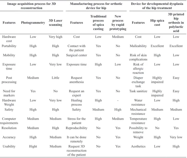
Table 1. Comparative summary of the main features of the techniques explored in this study (acquisition of images for 3D reconstruction and manufacturing of orthotic devices for hip). The summary also includes a qualitative comparison between the use of a hip spica cast and a hip orthosis produced by rapid prototyping in developmental dysplasia of the hip treatment (a quantitative comparison requires further study with volunteers).
Image acquisition process for 3D reconstruction
Manufacturing process for orthotic device for hip
Device for developmental dysplasia of the hip treatment
Features Photogrammetry 3D Laser
scanning Features
Traditional process of spica casting
New process by rapid prototyping
Features Hip spica cast
3D printed hip orthosis in
polylactic acid
Hardware cost
Low Very high Cost Low Medium Cost Low Low
Portability High High Contact with
the patient
Yes No Malleability Excellent Excellent
Mobility High High Surgical center Yes No Risk of skin
complications
High Low
Exposure time
Low Very low Exposure time High Low Risk of
allergic reaction
Low Low
Post processing
Medium Little Request
anesthesia
Yes No Diaper
exchange task
Highly impaired
Easy
Need for markers
Yes No Request an
expert
Yes No Task sanitize Highly
impaired
Easy Hardware
Weight
Low Very low Healing
process
High - Water
resistance
Low High
Safety High High Safety Medium High Mechanical
resistance
Medium Medium
Computer requirements
Medium Medium Stress for the patient
High Medium Temperature
resistance
High Low
Resolution Medium High Reproducibility No Yes Possibility to
remove
No Yes
Accuracy High Medium It can be done
remotely
No Yes Weight High Very low
Usability Hight Medium Request 3D
reconstruction of the patient
Ciobanu O, Ciobanu G, Rotariu M. Photogrammetric scanning technique and rapid prototyping used for prostheses and ortheses fabrication. Applied Mechanics and Materials. 2013a; 371:230-4. http://dx.doi.org/10.4028/www.scientific. net/AMM.371.230.
Ciobanu O, Rotariu M. Photogrammetric scanning and applications in medicine. Applied Mechanics and Materials. 2014; 657:579-83. http://dx.doi.org/10.4028/www.scientific. net/AMM.657.579.
Ciobanu O, Xu W, Ciobanu G. The use of 3D scanning and rapid prototyping in medical engineering. Fiability & Durability. 2013b; 1:241-7.
Daines SB, Aronsson DD, Beynnon BD, Sturnick DR, Lisle JW, Naud S. What is the best material for molding casts in children? Journal of Pediatric Orthopedics. 2014; 34(7):743-8. http://dx.doi.org/10.1097/BPO.00000000000001734(7):743-8. PMid:24590347.
DeMaio M, McHale K, Lenhart M, Garland J, McIlvaine C, Rhode M. Plaster: our orthopaedic heritage. The Journal of Bone & Joint Surgery. 2012; 94(20):e152. http://dx.doi. org/10.2106/JBJS.L.00183. PMid:23079886.
Demir SG, Erden S, Bulut H, Carboga B, Elbas NO. The problems experienced by parents providing postoperative home care following their child’s surgery for developmental dysplasia of the hip. Orthopaedic Nursing. 2015; 34(5):280-86. http://dx.doi.org/10.1097/NOR.0000000000000178. PMid:26375837
Dezateux C, Brown J, Arthur R, Karnon J, Parnaby A. Performance, treatment pathways, and effects of alternative policy options for screening for developmental dysplasia of the hip in the United Kingdom. Archives of Disease in Childhood. 2003; 88(9):753-9. http://dx.doi.org/10.1136/ adc.88.9.753. PMid:12937091.
DiFazio R, Vessey J, Zurakowski D, Hresko MT, Matheney T. Incidence of skin complications and associated charges in children treated with hip spica casts for femur fractures. Journal of Pediatric Orthopedics. 2011; 31(1):17-22. http:// dx.doi.org/10.1097/BPO.0b013e3182032075. PMid:21150727. Dyskin E, Ferrick M. Semirigid abduction bracing is effective treatment of reducible developmental dysplastic hips after failure of Pavlik harness. Annals of Orthopedics & Rheumatology. 2015; 3(2):1045.
Ferreira JV, Thomson JD. Socioeconomic Implications of Spica Casting. The Journal of Bone & Joint Surgery. 2012; 94(14):e107. http://dx.doi.org/10.2106/JBJS.L.00500. PMid:22695991.
Furukawa Y, Ponce J. Accurate camera calibration from multi-view stereo and bundle adjustment. International Journal of Computer Vision. 2009; 84(3):257-268. Giannatsis J, Dedoussis V. Additive fabrication technologies applied to medicine and health care: a review. International Journal of Advanced Manufacturing Technology. 2009; 40(1-2):116-27. http://dx.doi.org/10.1007/s00170-007-1308-1. Guarniero R. Displasia do desenvolvimento do quadril: atualização. Revista Brasileira de Ortopedia. 2010; 45(2):116-21. http://dx.doi.org/10.1590/S0102-36162010000200002.
Halanski M, Noonan KJ. Cast and splint immobilization: complications. The Journal of the American Academy of Orthopaedic Surgeons. 2008; 16(1):30-40. PMid:18180390.
Hedequist D, Kasser J, Emans J. Use of an abduction brace for developmental dysplasia of the hip after failure of Pavlik harness use. Journal of Pediatric Orthopedics. 2003; 23(2):175-7. http://dx.doi.org/10.1097/01241398-200303000-00008. PMid:12604946.
Ibrahim DA, Skaggs DL, Choi PD. Abduction Bracing After Pavlik Harness Failure: An Effective Alternative to Closed Reduction and Spica Casting? Journal of Pediatric Orthopedics. 2013; 33(5):536-9. http://dx.doi.org/10.1097/ BPO.0b013e31827d7e2a. PMid:23752152.
Igathinathane C, Davis JD, Purswell JL, Columbus EP. Application of 3D scanned imaging methodology for volume, surface area, and envelope density evaluation of densified biomass. Bioresource Technology. 2010; 101(11):4220-7. http:// dx.doi.org/10.1016/j.biortech.2009.12.140. PMid:20137915. Jumani MS, Shakil S, Sadiq AS. Fused deposition modelling technique (FDM) for fabrication of custom-made foot orthoses: a cost and benefit analysis. Scientific International (Lahore). 2014; 26(5):2571-6.
Majumdar R, Laxton P, Thuesen A, Nester C, Richards B. Design, development and biomechanical evaluation of a prefabricated anti pronation foot orthosis. Journal of Foot and Ankle Research. 2012; 5(Suppl 1):S22-5. http://dx.doi. org/10.1186/1757-1146-5-S1-P22.
Mavroidis C, Ranky RG, Sivak ML, Patritti BL, DiPisa J, Caddle A, Gilhooly K, Govoni L, Sivak S, Lancia M, Drillio R, Bonato P. Patient specific ankle-foot orthoses using rapid prototyping. Journal of Neuroengineering and Rehabilitation. 2011; 8(1):1-11. http://dx.doi.org/10.1186/1743-0003-8-1. PMid:21226898.
Milusheva SM, Tosheva EY, Hieu LC, Kouzmanov LV, Zlatov N, Toshev YE. Personalised ankle-foot orthoses design based on reverse engineering. In: Pham DT, Eldukhri EE, Soroka AJ, editors. Proceedings of the 5th Virtual International Conference on Intelligent Production Machines and Systems; 2006 June 10; Sakarya, Turkey. Oxford: Elsevier; 2006. p. 12-14.
Moghaddam MB, Brown TM, Clausen A, Silva T, Ho E, Forrest CR. Outcome analysis after helmet therapy using 3D photogrammetry in patients with deformational plagiocephaly: the role of root mean square. Journal of Plastic, Reconstructive & Aesthetic Surgery; JPRAS. 2014; 67(2):159-65. http://dx.doi.org/10.1016/j.bjps.2013.09.036. PMid:24411583.
Mulpuri K, Song KM, Goldberg MJ, Sevarino K. Detection and nonoperative management of pediatric developmental dysplasia of the hip in infants up to six months of age. The Journal of the American Academy of Orthopaedic Surgeons. 2015; 23(3):202-5. http://dx.doi.org/10.5435/ JAAOS-D-15-00006. PMid:25656273.
Noordin S, Umer M, Hafeez K, Nawaz H. Developmental dysplasia of the hip. Orthopedic Reviews. 2010; 2(2):73-8. http://dx.doi.org/10.4081/or.2010.e19. PMid:21808709. Palousek D, Rosicky J, Koutny D, Stoklasek P, Navrat T. Pilot study of the wrist orthosis design process. Rapid Prototyping Journal. 2014; 20(1):27-32. http://dx.doi. org/10.1108/RPJ-03-2012-0027.
Patar A, Jamlus N, Makhtar K, Mahmud J, Komeda T. Development of dynamic ankle foot orthosis for therapeutic application. Procedia Engineering. 2012; 41:1432-40. http:// dx.doi.org/10.1016/j.proeng.2012.07.332.
Paterson A. Digitisation of the splinting process: exploration and evaluation of a computer aided design approach to support additive manufacture [thesis]. Leicestershire: Loughborough University; 2013.
Paterson AM, Bibb R, Campbell RI, Bingham G. Comparing additive manufacturing technologies for customised wrist splints. Rapid Prototyping Journal. 2015; 21(3):230-3. http://dx.doi.org/10.1108/RPJ-10-2013-0099.
Paterson AM, Donnisson E, Bibb RJ, Campbell RI. Computer-aided design to support fabrication of wrist splints using 3D printing: A feasibility study. Hand Therapy. 2014a; 19(4):102-3. http://dx.doi.org/10.1177/1758998314544802. Paterson AMJ, Bibb RJ, Campbell RI. A review of existing anatomical data capture methods to support the mass customisation of wrist splints. Virtual and Physical Prototyping. 2010; 5(4):201-7. http://dx.doi.org/10.1080/ 17452759.2010.528183.
Paterson AMJ, Bibb RJ, Campbell RI. Orthotic rehabilitation applications. In: Bibb R, Eggbeer D, Paterson AMJ, editors. Medical modelling: the application of advanced design and rapid prototyping techniques in medicine. New York: Woodhead Publishing; 2014b. p. 283.
Rampal V, Sabourin M, Erdeneshoo E, Koureas G, Seringe R, Wicart P. Closed reduction with traction for developmental dysplasia of the hip in children aged between one and five years. Journal of Bone & Joint Surgery. 2008; 90(7):858-63. http://dx.doi.org/10.1302/0301-620X.90B7.20041. PMid:18591592.
Remondino F. From point cloud to surface: the modeling and visualization problem. The International Archives of the Photogrammetry, Remote Sensing and Spatial Information Sciences. 2003; 34(5):1-11.
Santili C, Akkari M, Waisberg G, Alcantara T, Abulasan T, Barreto SL, Prado JC. Tratamento incruento das fraturas diafisárias do fêmur nas crianças. Acta Ortopedica Brasileira. 2005; 13(5):249. http://dx.doi.org/10.1590/ S1413-78522005000500008.
Schwend RM, Pratt WB, Fultz J. Untreated acetabular dysplasia of the hip in the Navajo. A 34 year case series followup. Clinical Orthopaedics and Related Research.
1999; 364:108-16. http://dx.doi.org/10.1097/00003086-199907000-00015. PMid:10416399.
Schwend RM. Developmental dysplasia of the hip: global orthopedics. New York: Springer; 2014.
Sony Mobile Communications. [internet]. Sony; 2015. [cited 2015 Dec 12]. Available from: http://www.sonymobile. com/global-en/xperia/.
Steps. A parents’ guide to caring for a child in a hip spica cast [internet]. Cheshire: Steps; 2010. [cited 2015 Dec 01]. Available from: http://www.steps-charity.org.uk/How-We-Help/for-babies-and-toddlers.html.
Storer SK, Skaggs DL. Developmental dysplasia of the hip. American Family Physician. 2006; 74(8):1310-6. PMid:17087424.
Teng JB, Yu CW, Wang YZ, Mu KX. Sonographic detection of unilateral hip dislocation in a spica cast after closed reduction for developmental dysplasia of the hip. Journal of Ultrasound in Medicine. 2012; 31(6):827-31. PMid:22644678. The Royal Children’s Hospital Melboure – RCHM. Clinical guidelines: hip spica nursing care [internet]. Victoria: RCHM; 2015. [cited 2015 Dec 01]. Available from: http:// www.rch.org.au/rchcpg/hospital_clinical_guideline_index/ Hip_Spica_ Nursing_Care/.
Uras I, Yavuz OY, Uygun M, Yldrm H, Kömürcü M. The efficacy of semirigid hip orthosis in the delayed treatment of developmental dysplasia of the hip. Journal of Pediatric Orthopaedics B. 2014, 23(4):339-42. http://dx.doi.org/10.1097/ BPB.0000000000000052. PMid:24811088.
Vitale MG, Skaggs DL. Developmental dysplasia of the hip from six months to four years of age. J Journal of the American Academy of Orthopaedic Surgeons. 2001; 9(6):401-11. PMid:11730331.
Wahlen R, Zambelli P. Treatment of the developmental dysplasia of the hip with an abduction brace in children up to 6 months old. Advances in orthopedics. 2015; 2015:ID-103580. http://dx.doi.org/10.1155/2015/103580. PMid:25815214.
Wilkinson AG, Sherlock DA, Murray GD. The efficacy of the Pavlik harness, the Craig splint and the von Rosen splint in the management of neonatal dysplasia of the hip: a comparative study. Journal of Bone & Joint Surgery. 2002; 84(5):716-9. PMid:12188491
Yang S, Quanjun C. Total hip arthroplasty in developmental dysplasia of the hip: Review of anatomy, techniques and outcomes. World Journal of Orthopedics. 2012; 3(5):42-48. http://dx.doi.org/10.5312/wjo.v3.i5.42. PMid:22655221. Yazdanpanah A, Amoabediny G, Shariatpanahi P, Nourmohammadi J, Tahmasbi M, Mozafari M. Synthesis and characterization of polylactic acid tubular scaffolds with improved mechanical properties for vascular tissue. Engineering Trends in Biomaterials and Artificial Organs. 2014; 28(3):99-105.
Authors
Rodrigo Munhoz1, Cícero André da Costa Moraes2, Harki Tanaka1, Maria Elizete Kunkel3*
1 Centro de Engenharia, Modelagem e Ciências Sociais Aplicadas – CECS, Universidade Federal do ABC – UFABC,
Santo André, SP, Brasil.
2 Arc-Team, Cles, Italia.
3 Instituto de Ciência e Tecnologia – ICT, Universidade Federal de São Paulo – UNIFESP, Rua Talim, 330, CEP 12231-280,