ARTICLE
Central nervous system lymphoma: magnetic
resonance imaging features at presentation
Linfoma do sistema nervoso central: características da imagem de ressonância
magnética à apresentação
Ricardo Schwingel1, Fabiano Reis2, Veronica A. Zanardi2, Luciano S. Queiroz3, Marcondes C. França Jr.4
Intracranial lymphoma may present essentially as prima-ry central nervous system (CNS) B-cell non-Hodgkin lympho-ma (B-cell PCNSL), metastatic CNS lympholympho-ma, intravascu-lar lymphomatosis, T-cell PCNSL, and intracranial Hodgkin’s
lymphoma1,2. he tumor has an iniltrative nature, and early
di-agnosis is important to guide proper treatment. Surgical
remov-al has a limited role because it does not improve prognosis3,4.
Magnetic resonance imaging (MRI) may provide rele-vant diagnostic information for patients with suspected CNS
lymphoma2. he radiological presentation may have a pattern
of parenchymal, meningeal, or cranial nerve involvement2.
Many inlammatory and neoplastic conditions may mimic these MRI patterns, so that radiologists should be aware of
the diverse MRI features of CNS lymphoma5,6. Despite this,
there are few studies speciically devoted to describe MRI ab-normalities in this disease2,4,5,7-11. herefore, we presented a
detailed description and analysis of MRI data of a large series of Brazilian patients with histological conirmation of CNS
School of Medical Sciences, State University of Campinas, Campinas, São Paulo SP, Brazil.
1 Medical Student;
2 MD, PhD, Professor, Department of Radiology; 3 MD, PhD, Professor, Department of Pathology; 4 MD, PhD, Professor, Department of Neurology.
Correspondence: Fabiano Reis; Faculdade de Ciências Médicas, Universidade Estadual de Campinas, Departamento de Radiologia; Rua Tessália Vieira de Camargo 126 / Caixa Postal: 6111 / Cidade Universitaria Zeferino Vaz; 13083-887 Campinas SP - Brasil; E-mail: [email protected]
Support: FAPESP process no. 2010/01939-0.
Conflict of interest: There is no conflict of interest to declare.
Received 05 August 2011; Received in final form 04 November 2011; Accepted 11 November 2011 ABSTRACT
Objective: This paper aimed at studying presentations of the central nervous system (CNS) lymphoma using structural images obtained by
magnetic resonance imaging (MRI). Methods: The MRI features at presentation of 15 patients diagnosed with CNS lymphoma in a university hospital, between January 1999 and March 2011, were analyzed by frequency and cross tabulation. Results: All patients had supratentorial lesions; and four had infra- and supratentorial lesions. The signal intensity on T1 and T2 weighted images was predominantly hypo- or isoin-tense. In the T2 weighted images, single lesions were associated with a hypointense signal component. Six patients presented necrosis, all of them showed perilesional abnormal white matter, nine had meningeal involvement, and five had subependymal spread. Subependymal spread and meningeal involvement tended to occur in younger patients. Conclusion: Presentations of lymphoma are very pleomorphic, but some of them should point to this diagnostic possibility.
Key words: lymphoma, central nervous system, magnetic resonance imaging.
RESUMO
Objetivo: Este trabalho teve como objetivo estudar as apresentações do linfoma do sistema nervoso central (SNC) por meio de imagens
es-truturais, obtidas por ressonância magnética (RM). Métodos: Foram analisadas as características das imagens por RM, à apresentação, de 15 pacientes diagnosticados com linfoma do SNC em um hospital universitário, entre janeiro de 1999 e março de 2011, pela frequência e por tabulação cruzada. Resultados: Todos os pacientes apresentaram lesões supratentoriais; em quatro (27%) havia lesões infra e supratento-riais. A intensidade do sinal em T1 e T2 foi predominantemente hipo ou isointensa. Lesões únicas foram associadas ao componente de sinal hipointenso nas imagens ponderadas em T2. Seis pacientes apresentaram necrose. Foram encontrados: alteração desinal da substância branca perilesional em todos os pacientes, acometimento meníngeo em nove e disseminação subependimária em cinco. Disseminação su-bependimária e acometimento meníngeo tenderam a ocorrer nos pacientes mais jovens. Conclusão: As apresentações do linfoma no SNC são pleomórficas, mas algumas delas podem apontar para essa possibilidade diagnóstica.
lymphoma, in order to study the radiological characteristics of the disease at presentation.
METHODS
We analyzed the MRI indings at presentation of 15 im-muno- and non-immunocompetent patients (4 men and 11 women), with a mean age of 42 years-old [19 to 77 years-old; standard deviation (SD): 17.9 years-old], diagnosed with pri-mary or secondary CNS lymphoma. he images were per-formed at the State University of Campinas (UNICAMP) hospital between January, 1999 and March, 2011. he diag-nosis was conirmed after surgical biopsy or tumor excision of all patients, according to clinical indication. he surgical specimens were processed for routine histopathology, and the classiication of tumor type followed the World Health Organization (WHO) guidelines. he project was submitted to the Ethics Committee of our service, which approved the research protocol.
MRI scans were performed using a 2T scanner (Elscint
Prestige®, Haifa, Israel), with T1 and T2 acquisitions in three
orthogonal planes, including T1-weighted SE gadolinium-enhanced images. MRI acquisition parameters were: sagit-tal T1 spin echo, 6 mm thick; lip angle=180°; repetition time (TR)=430 milliseconds; echo time (TE)=12 milliseconds;
ma-trix 200×350; ield of view (FOV)=25×25 cm2; T2-weighted
and “fast spin echo” (FSE), 3 mm thick; lip angle=160°; TR=4,800 milliseconds; TE=108 milliseconds; matrix 256×256;
FOV=22×22 cm2; axial T1-weighted spin echo (SE): TR=540
milliseconds; TE=28 milliseconds. T1-weighted SE gadolini-um-enhanced images were obtained in the three orthogonal planes.
We analyzed the following variables: age, sex, HIV infec-tion, uniqueness or multiplicity of the lesion, tumor locainfec-tion, signal on T1 and T2-weighted images, necrosis, contrast en-hancement, perilesional white matter changes, meningeal involvement, subependymal spread, primary site of the lym-phoma, and histological subtype.
For the images’ evaluation, necrosis was deined as an area within a contrast-enhancing lesion that did not enhance on T1-weighted images and produced a hyperintense signal on T2-weighted images. Further, white matter changes were classiied as mild, moderate or pronounced, depending on whether the hyperintense perilesional area on T2-weighted images was smaller, similar or larger than the enhanced le-sion area on T1-weighted images after contrast injection, respectively.
For statistical analysis, indings were grouped into cat-egories, according to the variability of results. he variables were then compared by means of cross tabulation and the application of Fisher’s exact, Mann-Whitney’s, and Kruskal-Wallis’ tests.
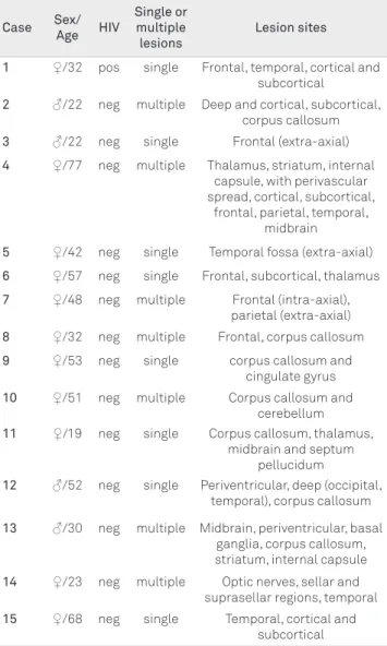
Table 1. Patients and magnetic resonance imaging lesions
data.
Case Sex/
Age HIV
Single or multiple lesions
Lesion sites
1 ♀/32 pos single Frontal, temporal, cortical and subcortical
2 ♂/22 neg multiple Deep and cortical, subcortical, corpus callosum
3 ♂/22 neg single Frontal (extra-axial)
4 ♀/77 neg multiple Thalamus, striatum, internal capsule, with perivascular spread, cortical, subcortical,
frontal, parietal, temporal, midbrain
5 ♀/42 neg single Temporal fossa (extra-axial)
6 ♀/57 neg single Frontal, subcortical, thalamus
7 ♀/48 neg multiple Frontal (intra-axial), parietal (extra-axial)
8 ♀/32 neg multiple Frontal, corpus callosum
9 ♀/53 neg single corpus callosum and cingulate gyrus
10 ♀/51 neg multiple Corpus callosum and cerebellum
11 ♀/19 neg single Corpus callosum, thalamus, midbrain and septum
pellucidum
12 ♂/52 neg single Periventricular, deep (occipital, temporal), corpus callosum
13 ♂/30 neg multiple Midbrain, periventricular, basal ganglia, corpus callosum, striatum, internal capsule
14 ♀/23 neg multiple Optic nerves, sellar and suprasellar regions, temporal
15 ♀/68 neg single Temporal, cortical and subcortical
pos: positive; neg: negative.
RESULTS
Tables 1 to 3summarize the patients’ data. Fourteen out
of the 15 patients were seronegative for HIV. We found mul-tiple parenchymatous lesions (intra-axial) on MRI scans in seven patients (47%), as seen in Fig 1. Single lesions were identiied in eight patients (53%). Out of these eight patients with single lesions, six had intra-axial lesions, and two had extra-axial lesions.
On T1-weighted images, we observed four cases (27%) with hypointense signal, six (40%) with isointense signal, one (7%) with hyperintense and four cases (27%) with mixed ones. On T2-weighted images, there were seven cases (47%) with hy-pointense signal, three (20%) with isointense signal, one (7%) with hyperintense signal, and four (27%) with mixed signals. here was a signiicant diference (p<0.05) between the T2 weighted image signal of the group with single lesions and the group with multiple ones. In 62% of the cases with a single le-sion, these presented with a hypointense signal component in
the T2 weighted image (↓ or ↓ and =, Table 2), while in 42.9% of
the cases, in which there were multiple lesions, it was possible to ind the association of lesions with hypointense and
hyper-intense signals on the T2 weighted image (↓ and ↑, Table 2).
Necrosis was observed in six patients (40%). Contrast enhancement was observed in 14 patients (93%); in 6 cases, it was homogeneous. he perilesional white matter signal change was pronounced in four, moderate in ive and mild in six cases. here was a signiicant association (p<0.05) be-tween the signal of the lesion in the T1 weighted image and the degree of perilesional white matter change. he group with lesions presenting a hyperintense signal component on the T1 weighted image (= and ↑ or ↑, Table 2), and the area with perilesional signal changes tended to be more pro-nounced, while in the group with lesions presenting a
hy-pointense signal component on the T1 weighted image (↓ or
↓ and =, Table 2), the area with abnormal perilesional signal
tended to be smaller.
Meningeal involvement was identiied in ten patients (59%): leptomeningeal in four (27%), dural in four (27%), and one patient had both dural and leptomeningeal involve-ment. here was a trend of association (p=0.066) of age and
meningeal involvement. In the group of patients with men-ingeal involvement, the mean age was lower (35.1 years-old, SD=14.6 years-old) than in the group of patients without meningeal involvement (52.0 years-old, SD=18.8 years-old).
Subependymal dissemination was found in ive patients (33%). We found a signiicant association (p<0.05) of age and subependymal spread. he group of patients with subependy-mal spread had a lower mean age (27.4 old, SD=13.2 old) than the one without subependymal spread (49.1 years-old, SD=15.7 years-old). here was no statistical signiicance (p<0.05) in the analysis of cross tabulation of other variables.
Information on the primary site of lymphoma and pathol-ogy are shown in Table 3.
Fig 1. Case 2: Coronal T1 post-contrast (A and B) and axial T1 post-contrast (C) showing multiple and deep, cortical, and
subcortical lesions that reach the genu of the corpus callosum. The second image (B) shows contrast enhancing nodules in the cerebellar parenchyma and leptomeningeal enhancement. There is also subependymal spread. The central nervous system involvement was secondary to non-Hodgkin lymphoma of B-cell phenotype.
A
B
C
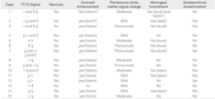
Fig 2. Case 10: Axial T1-weighted MRI after contrast (A) shows
lesions in the genu and splenium of the corpus callosum. The contrast enhancement is intense and subependymal spread is observable. The T2 weighted image (B) shows a heterogeneous lesion with iso/hypointense signal. There is a hyperintense signal in the surrounding white matter. In this case, the central nervous system was primarily affected and the pathology showed a diffuse large B-cell non-Hodgkin lymphoma (centroblastic).
DISCUSSION
Most of the analyzed patients were immunocompetent at diagnosis. Our inding of single lesions in 53% of patients is as
expected according to the literature.his ratio ranged from
33%, in a series analyzing mixed groups of immunocompe-tent and immunodeicient patients, to 81%, in a series that
analyzed only immunocompetent patients4,5,7-11.
Furthermore, our data are in line with works that show that the supratentorial region is usually more afected than
the infratentorial region1,2,6,9,12. Midbrain and cerebellum were
the afected sites in patients with infratentorial lesions in our
paper, butthey are two unusual sites afected by lymphoma
in the CNS4,7,8.
he signal features in MRI conventional sequences de-pend on the tissue characteristics. More solid lesions with little or no necrosis appear as hypo- or isointense images on T2-weighted sequences, and with isointense signal on T1-weighted images. hese usually present strong enhancement
after contrast injection11. he absence of necrosis in a bulky
and iniltrative T2-hypointense lesion in the corpus callosum is not a frequent inding, but is rather speciic and thus highly
suggestive of lymphoma7. On the opposite, if a lesion has a
large component of necrosis, which is usually described in immunocompromised patients, this more liqueied compo-nent tends to have a hypointense signal on T1-weighted im-ages, a hyperintense signal on T2-weighted imim-ages, and a
sur-rounding ring enhancement2,5,6,8,11,12.
Table 2. Magnetic resonance imaging lesions characteristics.
Case T1/T2 Signal Necrosis Contrast
enhancement
Perilesional white matter signal change
Meningeal involvement
Subependimal dissemination
1 = and ↑/↓ Yes Yes (heteroA) Moderate Yes (dural) and (leptoC)
No
2 =/↓ and ↑ No yes (homoB) Mild Yes (lepto) Yes 3 = and ↑/↓ No yes (hetero) Pronounced Yes (dural) Yes
4 ↓/= and ↑ Yes yes (hetero) Mild No No
5 =/= No yes (homo) Moderate Yes (dural) No
6 ↑/↓ No yes (hetero) Pronounced Yes (dural) No 7 ↓ and =/
↓ and ↑
Yes yes (hetero) Pronounced Yes (dural) No
8 =/↓ Yes yes (hetero) Moderate No No
9 ↓ and =/↓ No yes (homo) Pronounced No No
10 =/↓ and ↑ Yes yes (hetero) Moderate Yes (lepto) Yes
11 ↓/= No yes (homo) Mild Yes (lepto) Yes
12 ↓/= Yes yes (hetero) Mild No No
13 =/↑ No no Mild No No
14 ↓/↓ No yes (homo) Mild Yes (lepto) Yes
15 =/↓ No yes (homo) Moderate No No
= isointense signal; ↓hypointense signal; ↑hyperintense signal; Aheterogeneous; Bhomogeneous; Cleptomeningeal.
Table 3. Pathological information.
Case Primary site of lymphoma Biopsy site Histopathological diagnosis
1 CNS CNS Diffuse large B-cell NHL
2 BM BM B-cell NHL. Bone marrow fibrosis.
3 RES Cervical lymph node Hodgkin’s disease, nodular sclerosis type II
4 BM / CNS BM Low-grade NHL (lymphoplasmacytic)
5 Maxillary sinus Maxillary sinus Diffuse large B-cell NHL
6 Bronchus Bronchus Large B-cell NHL
7 CNS CNS Classical CNS HL
8 CNS CNS B-cell NHL
9 CNS CNS Diffuse large B-cell NHL (centroblastic)
10 CNS CNS Diffuse large B-cell NHL (centroblastic)
11 BM / CNS BM High-grade B-cell NHL
12 RES Inguinal lymph node Mixed cell HL
13 Testis / CNS Testis Diffuse large B-cell NHL
14 CNS CNS Diffuse large B-cell NHL
15 CNS CNS Diffuse large B-cell NHL
Some studies report that homogeneous enhancement by contrast is a frequent inding and the absence of contrast en-hancement, as we observed in one patient, as rare in
intrac-ranial lymphoma4,7-11. he perilesional white matter signal
change is common in CNS lymphoma ranging from 77% to
more than 90% in other series2,4,5,7,8, and in our paper it was
found in all patients, but in varying degrees.
We found a higher proportion of patients with meningeal
involvement at presentation thanother authors2,5. Also, it is
noteworthy thatin two cases of single dural lesion, the tumor
radiologically mimicked a meningioma.
In non-Hodgkin’s lymphoma (NHL), secondary spread involves the CNS, usually in the form of leptomeningeal in-iltrates. Parenchymal lesions, when present, typically result from secondary involvement from the leptomeninges via
in-iltration of the perivascular spaces1. We emphasize the
ind-ing of three cases of intracranial Hodgkin’s lymphoma (Fig 3), since most cases of secondary CNS involvement by Hodgkin’s lymphoma are late manifestations of disseminated disease and primary CNS involvement by this type of lymphoma is very rare1,13,14.
In conclusion, lymphoma presentations are very pleo-morphic in the CNS, but some of them (single or associated) should point to this diagnostic possibility. he diferential di-agnosis of CNS lymphoma includes gliomas, metastasis, and inlammatory disorders. In our group of patients, the signal intensity on T1 and T2 weighted images was predominantly hypo- or isointense. In the T2 weighted images, single lesions
were associated with a hypointense signal component. Later, we found an association of subependymal spread and men-ingeal involvement, indings that tended to occur in younger patients. Our analysis also showed the correlation of the hy-perintense signal in the T1 weighted image with more pro-nounced perilesional changes. hese radiological features, along with other indings such as location of the lesion, con-trast enhancement (in the lesion and in subependymal and meningeal sites, which may be a potential clue to lymphoma-tous dissemination), can help this diferentiation.
1. Slone HW, Blake JJ, Shah R, Guttikonda S, Bourekas EC. CT and MRI findings of intracranial lymphoma. AJR 2005;184:1679-1685.
2. Besada C, Schvartzman P, Paganini L, Santa Cruz D, Fues J. Neuroimágenes estructurales y funcionales en la caracterización del linfoma Del SNC. Rev Argent Radiol 2010;74:147-153.
3. Sierra del Rio M, Rousseau A, Soussain C, Ricard D, Hoang-Xuan K. Primary CNS lymphoma in immunocompetent patients. The Oncologist 2009;14:526-539.
4. Küker W, Nägele T, Korfel A, et al. Primary central nervous system lymphomas (PCNSL): MRI features at presentation in 100 patients. J Neurooncol 2005;72:169-177.
5. Koul R, Dufan T, Dubey A, et al. Imaging features of primary central nervous system lymphoma at presentation. Case series from a regional cancer centre. Oncol Exchange 2008;7:24-27.
6. Johnson BA, Fram EK, Johnson PC, Jacobowitz R. The variable MR appearance of primary lymphoma of the central nervous system: comparison with histopathologic features. AJNR Am J Neuroradiol 1997;18:563-572.
7. Bühring U, Herrlinger U, Krings T, Thiex R, Weller M, Küker W. MRI features of primary central nervous system lymphomas at
presentation. Neurology 2001;57:393-396.
8. Haldorsen IS, Kråkenes J, Krossnes BK, Mella O, Espeland A. CT and MR imaging features of primary central nervous system lymphoma in Norway, 1989-2003. AJNR Am J Neuroradiol 2009;30:744-751.
9. Zali A, Shahzadi S, Mohammad-Mohammadi A, Taherzadeh K, Parsa K. Cerebral lymphoma: clinical and radiological findings in 90 cases. Arch Iranian Med 2007;10:194-198.
10. Zhang D, Hu LB, Henning TD, et al. MRI findings of primary CNS lymphoma in 26 immunocompetent patients. Korean J Radiol 2010;11:269-277.
11. Koeller KK, Smirniotopoulos JG, Jones RV. Primary central nervous system lymphoma: radiologic-pathologic correlation. Radiographics 1997;17:1497-1526.
12. Erdag N, Bhorade RM, Alberico RA, Yousuf N, Patel MR. Primary lymphoma of the central nervous system: typical and atypical CT and MR imaging appearances. AJR 2001;1176:1319-1326.
13. Gerstner ER, Abrey LE, Schiff D, et al. CNS Hodgkin lymphoma. Blood 2008;112:1658-1661.
14. Re D, Fuchs M, Schober T, Engert A, Diehl V. CNS involvement in Hodgkin’s lymphoma. J Clin Oncol 2007;25:3181.
References
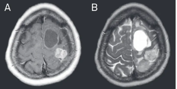
Fig 3. Case 7: Axial T1-weighted image after contrast (A)
showing intra-axial lesion in the left superior frontal gyrus, with peripheral contrast enhancement. In the T2-weighted image (B), the lesion is predominantly hyperintense. The images also show an extra-axial solid lesion in the left parietal convexity, with intense contrast enhancement and isointense in the T2-weighted image. The pathology revealed primary CNS classical Hodgkin lymphoma.