Insights into the epigenetic regulation of the rice genome:
the role of DNA methylation and histone modifications in salt
stress responses
Liliana de Jesus Duarte Ferreira
Dissertation presented to obtain the PhD degree in Biology
Instituto de Tecnologia Química e Biológica António Xavier Universidade Nova de Lisboa
Work performed at:
Plant Functional Genomics Lab - GPlantS
Instituto de Tecnologia Química e Biológica António Xavier Universidade Nova de Lisboa
Av. da República 2780-157 Oeiras Portugal
Supervisors:
Dr. Ana Paula Santos
Senior Researcher at GPlantS Unit (ITQB-UNL)
Prof. M. Margarida Oliveira Head of GPlantS Unit (ITQB-UNL)
Associate Professor with Habilitation (“Agregação”) at ITQB-UNL
Aos meus pais, pelo amor e apoio incondicionais
If you can't explain it simply, you don't understand it well enough.
Acknowledgments
First of all, I would like to deeply thank my supervisor Ana Paula Santos for all the help and motivation during all this years. Thank you for introducing me in the science world and to believing in me and in my work. I would like also to profoundly thank Professor Margarida Oliveira for giving me the opportunity to be part of her group and to do my PhD in her laboratory. Thank you for your guidance and support.
Secondly, I would like to give a big “thank you” to the members of the GPlantS lab: to the “seniors” Nelson Saibo, Isabel Abreu and Tiago Lourenço, for always sharing your knowledge and for the precious comments and suggestions; to my partners in suffering during our PhDs, Duarte Figueiredo, Pedro Barros, Tânia Serra, Diego Almeida, Cecília Pina, André Cordeiro, Mafalda Rodrigues, Nuno Gonçalves, Helena Sapeta, Margarida Rosa, Alicja Gorska, Rita Borba, for being always so nice with me, for helping with everything I need and for being truly exemples of perseverance; to Vanessa Azevedo for the friendship and the help in the mutants “saga”; to the enthusiastic Sebastião Ravasco, for making difficult questions and forcing me to think “out of the box”; to all the other members, Natacha Vieira, João Fradique, Paulo Gouveia, Bruno Alexandre, Ana Rita Leal and Inês Luís, for making the lab a really nice place to work at.
I also would like to thank Rob Martienssen from Cold Spring Harbour Laboratory (USA) for having accepted me in his lab for a short stay. It was a truly wonderful experience. Thanks also to Mark Donoghue and Filipe Borges for the precious help in bioinformatics.
Gostava de agradecer profundamente aos meus pais, por sempre estarem lá para mim e por acreditarem em mim. Tudo o que sou é graças a vocês. Amo-vos muito. Gostava também de agradecer ao meu pequenino, que mesmo sem saber, deu-me muita força para a conclusão deste trabalho. Salvador, a mãe adora-te.
Por fim, gostava também de agradecer ao Bruno a compreensão demonstrada ao longo dos anos e aos meus familiares e amigos pelo interesse que sempre demonstraram pelo meu trabalho.
The author of this thesis, Liliana de Jesus Duarte Ferreira, hereby declares to have had active participation in the following research papers:
Ferreira LJ, Oliveira MM, Santos AP. Chromatin and epigenetics flexibility in plant responses to environmental stresses. (submitted)
This manuscript includes part of the work described in Chapter 1.
Ferreira LJ, Azevedo V, Maroco J, Oliveira MM, Santos AP (2015) Salt tolerant and sensitive rice varieties display differential methylome flexibility under salt stress. PloS ONE 10(5):e0124060.
This manuscript includes the work described in Chapter 2.
Ferreira LJ, Donoghue MTA, Borges F, Saibo NJ, Martienssen R, Oliveira MM, Santos AP. Identification of Differentially Methylated Regions in a salt tolerant rice variety and their role in gene expression regulation. (in preparation)
This manuscript includes the work described in Chapter 3.
Ferreira LJ, Ravasco S, Figueiredo D, et al. Deciphering histone modifications in rice by chromatin immunoprecipitation (ChIP) and in situ immunofluorescence. (accepted for publication in the book under the working title Rice)
List of abbreviations
3D – Three-dimensional 5-AC – 5-azacytidine 5-mC – 5-methylcytosine
ERF – Ethylene Response Factor ATP – Adenosine triphosphate bp – Base pair
BS - Bisulfite
BSA – Bovine serum albumin cDNA – Complementary DNA
ChIP – Chromatin immunoprecipitation DNA – Deoxyribonucleic acid
DMR – Differentially methylated region dS - Decisiemens
DTT – Dithiothreitol
EDTA – Ethylenediaminetetraacetic acid
EGTA - Triethylene glycol diamine tetraacetic acid ELISA – Enzyme-linked immunosorbent assay FISH – Fluorescence in situ hybridization HAT – Histone acetyltransferase
HDAC – Histone deacetylase HDM – Histone demethylase HMT – Histone methyltransferase
M – Molar
Mb – Mega base pair mM – Millimolar
mRNA – messenger RNA
MSAP – Methylation-sensitive amplified polymorphism MTase – DNA methyltransferase
PCR – Polymerase chain reaction
PIPES - Potassium piperazine-1,4-bis(2-ethane)sulfonic acid PTM – Post-translational modification
qPCR – Real time quantitative PCR rDNA – Ribosomal DNA
RNA – Ribonucleic acid rRNA – Ribosomal RNA
ROS – Reactive oxygen species RT-PCR – Reverse transcription PCR
SDS-PAGE – Sodium dodecyl sulfate polyacrylamide gel electrophoresis T-DNA – Transfer DNA
TF – Transcription factor TSA – Trichostatin A v/v – Volume / volume w/v – Weight / volume WB – Western blot WT – Wild type μM – Micromolar
Summary
Plants exhibit complex and integrated strategies to respond to stressful events involving particular signalling pathways, transcription factors and a number of specific stress-responsive genes. In addition, the regulation of genome accessibility is ensured by an efficient network of chromatin organization and epigenetic mechanisms which imply a heritable influence on gene activity that is not associated with changes in the DNA sequence. However, the functional role of these factors in plant response to stressful conditions is still largely unknown and so far direct connections between epigenome regulation and phenotypic plasticity are still poorly explained.
The present thesis addresses the role of the epigenetic regulation of rice responses to salt stress, focusing on DNA methylation and histone modifications. Rice (Oryza sativa L.) is the staple food for more than half of the world population and faces specific problems with cultivation under salinity stress, posing a real threat to the sustained production of this cereal.
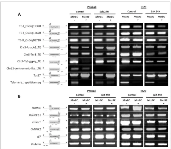
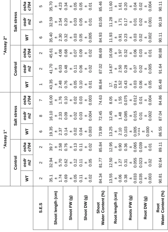
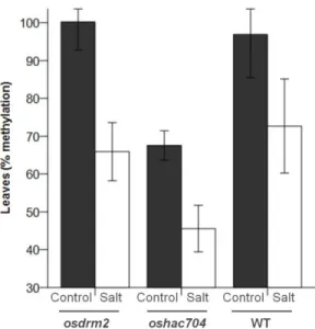
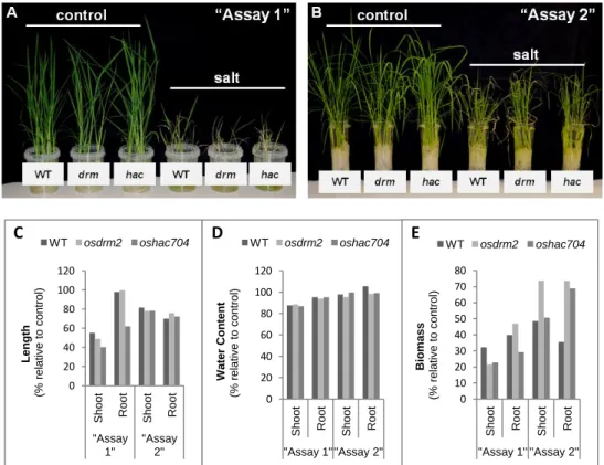
DNA methylation has been referred as an important player in plant genomic responses to environmental stresses. The analysis of global DNA methylation in different tissues of rice varieties with distinct salt susceptibility showed a global demethylation upon high salt imposition which was particularly evident in leaves. Moreover, the salt tolerant and sensitive rice varieties exhibited differential methylome flexibility, with the salt-tolerant variety Pokkali showing a remarkable ability to quickly relax DNA methylation in salt stress response. The phenotypic evaluation of some parameters related to salinity tolerance, such as root length and biomass, revealed a better performance of the osdrm2 mutant (defective in a DNA methyltransferase) under stress. All together, these findings emphasize a tissue- and genotype-specificity concerning global DNA methylation levels and suggest that higher methylome flexibility is an important player in salinity tolerance.
A deeper analysis into the methylome profile of leaves of the salt tolerant variety Pokkali, achieved by the MeDIP-seq method, led to the identification of salt stress-specific Differentially Methylated Regions (sDMRs) between control and salt stress samples. The methylation pattern analysis of these sDMRs revealed a loss of methylation in response to salt stress and occasionally the position of the sDMRs, namely their proximity to genes, was correlated with salt stress gene induction, suggesting that sDMRs may have a role in gene expression regulation.
The transcription control of stress responsive genes involves a multilayer regulatory process that includes chromatin structure, histone modifications and transcription factors. Here, we investigated the role and dynamics of specific histone modifications in the regulation of rice ROOT MEANDER CURLING (OsRMC, a gene highly induced by salt stress, and very conserved among rice varieties). The analysis of specific histone modification marks by Chromatin Immunoprecipitation (ChIP) in rice leaves revealed a differential enrichment of euchromatic marks depending on the promoter region. Upon salt stress, the chromatin domain where transcription factors bind was particularly enriched in histone modification marks related to euchromatin structure, suggesting a nucleosome repositioning associated with salt stress activation of OsRMC.
The knowledge gathered in this work contributes for a better understanding of the mechanisms controlling the plasticity of chromatin and epigenetic networks in plant response/tolerance mechanisms to salinity stress. Research in the field of environmental epigenetics will enhance our knowledge on genome and phenotype regulation and help designing strategies to improve plant adaptation and performance in sub-optimal conditions. This is especially needed, considering the increasing global population and accelerating climate changes, with higher and more variable temperatures, increased soil salinity, severe droughts and flooding.
Sumário
As plantas possuem múltiplas estratégias de grande complexidade para responder a condições de stress envolvendo determinadas vias de sinalização, fatores de transcrição e uma panóplia de genes específicos de resposta a stress. A regulação da acessiblidade do genoma depende da organização estrutural da cromatina e de mecanismos epigenéticos que influenciam a atividade de genes sem que haja alterações na sequência do ADN. No entanto, conexões funcionais entre regulação epigenética, plasticidade fenotípica e resposta ao stress são ainda pouco compreendidas.
Esta dissertação aborda o papel da regulação epigenética do genoma de arroz em resposta ao stress salino, focando-se em particular na análise da metilação do ADN e das modificações das histonas. O arroz (Oryza sativa L.) é a base da alimentação de mais de metade da população mundial e apresenta problemas específicos no cultivo sobre stress salino, ameaçando a sustentabilidade da produção deste cereal.
A metilação do ADN é extensamente referida como um importante fator nas respostas genómicas das plantas face a stresses ambientais. A análise da metilação global do ADN em diferentes tecidos de variedades de arroz com suscetibilidades distintas à salinidade mostrou a ocorrência de uma desmetilação global após imposição de stress salino, particularmente evidente nas folhas. Além disso, as variedades tolerantes e sensíveis ao sal evidenciaram diferentes capacidades de flexibilização do metiloma. A variedade tolerante ao sal Pokkali evidenciou uma capacidade notável de rapidamente diminuir a metilação do ADN em resposta ao stress salino. A avaliação fenotípica de alguns parâmetros particularmente relacionados com a tolerância à salinidade, nomeadamente o comprimento da raíz e a biomassa, revelaram uma melhor performance do mutante osdrm2 (knockout numa metiltransferase do ADN) em condições de stress. Em
conjunto, estes resultados realçam uma especificidade a nível de tecido e de genótipo no que diz respeito aos níveis de metilação globais, sugerindo que a flexibilidade do metiloma possa ser um factor relevante na tolerância à salinidade.
Uma análise mais aprofundada do metiloma de folhas da variedade de arroz tolerante ao sal por MeDIP-seq, permitiu a identificação de Regiões Diferencialmente Metiladas específicas do stress salino (sDMRs) entre amostras de controlo e stress salino. A análise do padrão de metilação destas sDMRs revelou uma perda de metilação em condições de stress salino e, em alguns casos, a posição das sDMRs, nomeadamente a sua proximidade de genes, foi correlacionada com a indução dos genes pela salinidade, sugerindo um papel regulatório das sDMRs ao nível da expressão génica.
O controlo da transcrição de genes de resposta ao stress envolve múltiplos processos reguladores que incluem a estrutura da cromatina, fatores de transcrição e modificações histónicas. Neste contexto, investigou-se o papel de modificações de histonas específicas na regulação transcricional do gene de arroz ROOT MEANDER CURLING (OsRMC, altamente induzido pelo sal e bastante conservado entre diferentes variedades de arroz). A análise de marcas de modificações de histonas específicas, por imunoprecipitação da cromatina (ChIP) em folhas de arroz revelou um enriquecimento diferencial em marcas eucromáticas dependendo da região do promotor. A região da cromatina onde se liga o fator de transcrição mostrou um enriquecimento em marcas associadas a eucromatina após o stress salino, sugerindo que possa ocorrer um reposicionamento dos nucleossomas aquando da ativação do gene OsRMC pelo stress salino.
Este trabalho contribui para uma melhor compreensão da plasticidade da organização estrutural da cromatina e marcas epigenéticas na resposta/tolerância das plantas ao stress salino. Por ajudar a compreender
melhor a regulação genómica e fenotípica, a investigação na área da epigenética ambiental pode ajudar a delinear estratégias para o melhor desempenho das plantas em condições desfavoráveis. Esta investigação é especialmente necessária tendo em conta o aumento da população global e a rápida evolução das alterações climáticas, nomeadamente temperaturas mais altas e variáveis, aumento da salinidade do solo, secas severas e inundações.
Table of Contents
Acknowledgments ... ix List of abbreviations ... xiii Summary ... xv Sumário ... xvii Table of Contents ... xxi List of Figures and Tables ... xxv Chapter 1 ... 1
1.1. Chromatin and nuclear architecture have a role in genome regulation . 3 1.2. Epigenetics: mechanisms and functions ... 5
1.2.1. DNA methylation ... 5 1.2.2. Histone modifications ... 10 1.2.3. Interplay between histone modifications and DNA methylation ... 12 1.2.4. Methods to decipher epigenetic marks ... 13 1.3. Epigenetic regulation of stress responses in plants ... 17 1.4. Salinity effects on plants ... 19
1.4.1. Plant responses to salinity ... 20 1.4.2. Transducing stress signals ... 21 1.4.3. Salinity tolerance ... 23 1.4.4. Susceptibility to salt stress: contribution of the epigenetic background ... 24 1.5. The model plant Oryza sativa ... 25 1.6. Thesis objectives and outline ... 26 1.7. References ... 27 Chapter 2 ... 41
2.1. Abstract ... 43 2.2. Introduction ... 44
2.3. Material and methods ...45 2.3.1. Plant material, growth conditions and salt stress treatments ...45 2.3.2. Quantification of global DNA methylation ...47 2.3.3. Imaging of 5-methylcytosine in interphase nuclei of tissue sections ...48 2.3.4. Expression studies of DNA methyltransferases and demethylases by real-time quantitative PCR (qPCR) ...49 2.3.5. DNA methylation analysis by McrBC digestion ...50 2.3.6. Genotyping rice T-DNA insertion lines ...50 2.3.7. Phenotypic evaluation of rice plants with mutations for epigenetic regulators ...51 2.3.8. Statistical data analysis ...51 2.4. Results ...52
2.4.1. Salt stress induced DNA demethylation ...52 2.4.2. Salt-stress effects on DNA methyltransferases and demethylases expression patterns were genotype specific ...55 2.4.3. DNA methylation of stress related targets ...58 2.4.4. Mutations of epigenetic modulators affected phenotypic parameters related to salinity tolerance ...58 2.5. Discussion ...64 2.6. Acknowledgments ...68 2.7. References ...68 2.8. Supporting information ...72 Chapter 3 ...77 3.1. Abstract ...79 3.2. Introduction ...79 3.3. Material and methods ...81
3.3.1. Plant material, growth conditions and salt stress treatment ...81 3.3.2. Methylated DNA immunoprecipitation sequencing (MeDIP-Seq) ..82 3.3.3. Mapping and processing the MeDIP-Seq reads ...82
3.3.4. Identification of Differentially Methylated Regions (DMRs) ... 83 3.3.5. Bisulfite Sequencing (BS) ... 83 3.3.6. Gene expression studies by quantitative real-time PCR ... 84 3.3.7. Gene ontology analysis ... 84 3.4. Results ... 85
3.4.1. The leaf methylome of the rice tolerant variety Pokkali ... 85 3.4.2. Differentially Methylated Regions (DMRs) showed decreased methylation after salt stress ... 85 3.4.3. Differentially Methylated Regions (DMRs) may have a role in gene regulation upon salt stress ... 92 3.5. Discussion ... 95 3.6. Acknowledgments... 97 3.7. References ... 98 3.8. Supporting information ... 102 Chapter 4 ... 113 4.1. Abstract ... 115 4.2. Introduction ... 116 4.3. Materials and methods ... 118 4.3.1. Plant material, growth conditions and stress treatments ... 118 4.3.2. Imaging of H3K4me2 in interphase nuclei of root sections ... 119 4.3.3. Protein extraction and immunoblotting analysis ... 120 4.3.4. Gene expression analysis ... 121 4.3.5. Chromatin immunoprecipitation (ChIP) assay ... 122 4.4. Results ... 126
4.4.1. The OsRMC gene activation in response to salt is epigenetic modulated ... 126 4.4.2. In situ imaging of histone modification marks revealed an enrichment in euchromatic marks in response to salt stress ... 128 4.4.3. The enrichment of euchromatic histone marks correlates with the OsRMC activation in response to salt stress ... 129
4.5. Discussion ... 131 4.6. Acknowledgments ... 134 4.7. References ... 134 4.8. Supporting information ... 137 Chapter 5 ... 143 5.1. References ... 150
List of Figures and Tables
Chapter 1:
Figure 1: Chromatin structure and epigenetic modifications. ... 6 Figure 2: Regulatory network of plant stress responses. ... 18 Chapter 2:
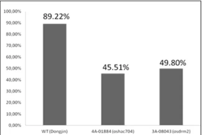
Figure 1: Global DNA methylation levels in salt tolerant and sensitive rice varieties. ... 52 Figure 2: 3D imaging of DNA methylation in single interphase nuclei. ... 55 Figure 3: DNA demethylases (DNG701 and DNG710) and DNA methyltransferase (OsDRM2) expression studies ... 57 Figure 4: McrBC based methylation analysis ... 59 Table 1: Phenotypic evaluation of rice mutants ... 60 Figure 5: Global DNA methylation levels in rice mutants. ... 62 Figure 6: Phenotypic evaluation of epigenetic rice mutants under salt stress ... 63 Figure S1: Schematic representation of the rice T-DNA insertion lines ... 72 Figure S2: Expression studies of OsDRM2 and OsHAC704 in the T-DNA rice mutant lines. ... 73 Figure S3: Spikelet fertility in WT (Dongjin) and T-DNA rice mutant lines. ... 73 Figure S4: Threshold cycle (CTs) values for the ubiquitin-conjugating enzyme E2 (UBC2) and elongation factor (eEF) genes under salt stress conditions. ... 73 Table S1: List of Primers used for expression studies of DNA demethylases and DNA methyltransferase. ... 74 Table S2: List of Primers used in the McrBC methylation analysis. ... 74 Table S3: List of Primers used for genotyping rice T-DNA insertion lines. ... 75 Table S4: Statistical analysis underlying phenotypic evaluation of rice mutants. ... 75
Chapter 3:
Figure 1: Identification of DMRs between control and salt stress samples in a salt tolerant rice variety. ...86 Table 1: Summary of MeDIP-seq data analysis. ...88 Table 2: List of Differentially Methylated Regions (DMRs) between control and salt stress conditions. ...89 Figure 2: Classification of DMRs according to genomic features. ...91 Figure 3: Bisulfite sequencing (BS) analysis for DMR2 and DMR15...93 Figure 4: Expression studies of genes nearby DMRs by quantitative real-time qPCR. ...94 Figure S1: Chromosome-level view of DNA methylation in control (A) and salt stress (B) conditions ... 102 Figure S2: Methylation status of all DMRs identified between control and salt stress conditions ... 104 Figure S4: Gene Ontology (GO) analysis. ... 111 S1 Table: List of primers used for BS-PCR analysis. ... 112 S2 Table: List of primers used for expression studies of genes located nearby DMRs ... 112 Chapter 4:
Figure 1: OsRMC expression studies. ... 127 Figure 2: Genome wide detection of H3K4me2 is response to salt stress. 128 Figure 3: Dynamics of H3K4ac, H3K9ac and H4K20me3 marks at OsRMC gene promoter after salt stress ... 130 Figure 4: A proposed schematic model for explaining the role of epigenetic factors and chromatin dynamics on salt stress induction of OsRMC under salt stress ... 133 Figure S1: Visual description of the crosslink step in the ChIP process ... 137 Figure S2: FAIRE assay to test chromatin crosslink efficiency. ... 138 Table S1: List of primers used for gene expression analysis of OsRMC gene. ... 139
Table S2: ChIP buffers ... 139 Table S3: List of primers used for analysis of immunoprecipitated DNA by qPCR. ... 141 Chapter 5:
Chapter 1
________________________________________________
General Introduction and Research Objectives
Liliana J. Ferreira performed the bibliographic search and wrote this chapter, part of which was submitted for publication as:
Ferreira LJ, Oliveira MM, Santos AP. Chromatin and epigenetics flexibility in plant responses to environmental stresses.
Chapter 1:
General Introduction and Research Objectives
________________________________________________
1.1. Chromatin and nuclear architecture have a role in genome regulation
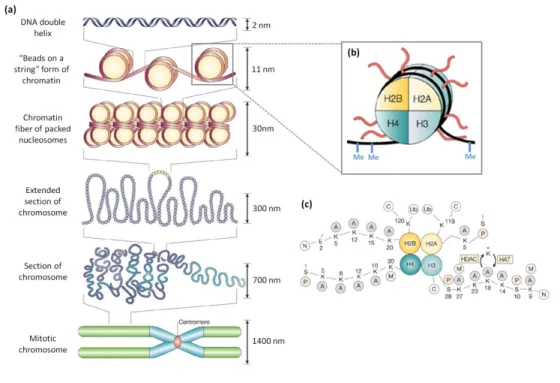
In eukaryotes, the genetic information is encoded by DNA which can have several meters and thus needs to be packaged in order to fit into a physically restricted space that is the nucleus. A high level of compaction and organization is achieved through association of DNA with proteins named histones, forming chromatin (Figure 1). The nucleosome is the fundamental unit of chromatin and is composed by approximately 146 bp of DNA wrapped around an octamer of histones, containing two copies of each of the four core histones (H2A, H2B, H3 and H4) (Kornberg, 1974). Each nucleosome is linked to the next nucleosome through a portion of linker DNA, creating the 10 nm fiber known as the “beads on a string” model (Luger et al., 1997). The length of linker DNA ranges between approx. 20–90 bp and varies among different species, tissues, and even fluctuates within a single cellular genome (van Holde, 1988). The linker histone H1 drives the package of the 10 nm fibers into a solenoid of 30 nm diameter, forming a super helix with 6 nucleosomes per turn (Finch and Klug, 1976; Robinson et
al., 2006). In an alternative model the 30 nm fiber is composed by a “zigzag”
nucleosome array and the spacer DNA frequently passes through the central axes of the fibre (Schalch et al., 2005; Woodcock and Ghosh, 2010). Higher levels of DNA compaction acting on the 30 nm fibers are normally referred as the large-scale level of chromatin organization which is still a matter of discussion (Woodcock and Ghosh, 2010). The knowledge of the spatial arrangement of chromatin in the 3D interphase nucleus greatly benefited from the emergence of the in situ hybridization technique with specific DNA
probes and from the improvement of microscope resolution. Fluorescence in situ hybridization (FISH) with chromosome-specific probes started to be successfully applied in animal cells showing that interphase chromosomes occupy distinct territories in the interphase nucleus (Manuelidis, 1984; Lamond and Earnshaw, 1998). More recently, specific regions of chromatin as the lamina-associated domains (LADs), and the topological association chromatin domains (TADs), which are bound together by particular protein complexes, were found in specific nuclear territories (Nicodemi and Pombo, 2014).
The chromatin structure is affected by the incorporation of histone variants such as H2A.Z and CENH3 that greatly influence gene activity and genome structure (Zhang et al., 2005; Deal and Henikoff, 2011; Coleman-Derr and Zilberman, 2012). For example, the histone H3, predominantly present during replication, can be replaced by the histone variant H3.3 in a replication-independent manner in active chromatin regions (Chen et al., 2014). Another divergent form of histone H3, the CENP-A, is exclusively found in centromeric regions, having important functions in regulating the proper segregation of chromosomes (Howman et al. 2000; Régnier et al. 2005). Similarly, the histone H2A.Z, variant of H2A, is found around gene promoters (Redon et al. 2002; Zhang et al. 2005).
Chromatin has not only a role in the structural organization of DNA but is also involved in gene expression regulation and in preventing and repairing genomic lesions (Lukas et al., 2011). At cytological level, chromatin can be defined as euchromatin or heterochromatin, which differs mainly on their compaction state and transcriptional potential. Chromatin compaction has been negatively correlated with transcriptional competence since the ability of a gene to be transcribed depends on its accessibility to the transcription machinery. The heterochromatin is highly condensed, transcriptionally inactive and rich in repetitive sequences while euchromatin presents less compaction with irregularly spaced nucleosome arrays, being gene rich and
transcriptionally competent (Heitz, 1928; Fransz et al. 2002; Berger, 2007). The chromatin accessibility is mainly determined by the recruitment of non-histone chromatin binding proteins, which recognizes modified non-histone motifs and exerts its regulatory functions. These functions involve post-translational modifications of histone tails, incorporation of histone variants, nucleosome sliding and remodelling by ATP-dependent remodelling complexes (Rosa and Shaw, 2013).
1.2. Epigenetics: mechanisms and functions
All cells of an organism carry the same DNA sequence but a chromatin-based selective read out of the genome is capable to originate distinct cell types. The study of the transmission of chromatin states without changes of the underlying DNA sequence is the target issue of epigenetic research. The word “epigenetics” was used for the first time by Conrad Waddington (1905– 1975) to describe “the branch of biology which studies the causal interactions between genes and their products, which bring the phenotype into being” (Waddington, 1942). In a broad sense, epigenetics can be understood as a bridge between the genotype and phenotype, a group of molecular mechanisms that modulates the final outcome of a locus or chromosome without any change on the underlying DNA sequence. Some of the core molecular actors playing epigenetic roles are described below and are represented in Figure 1.
1.2.1. DNA methylation
DNA methylation is the best characterized chemical modification of chromatin. Conventionally, it consists on the binding of a methyl group (CH3), provided by S-adenosylmethionine (SAM), to the 5- carbon of a
Figure 1: Chromatin structure and epigenetic modifications. Schematic
representation of different levels of chromatin organization, from the basic unit of chromatin, the nucleosome, to the highly condensed mitotic chromosome (a). The nucleosome, the fundamental repeating units of chromatin, is schematically shown in (b). The core proteins of nucleosomes are designated H2A, H2B, H3 and H4. Each histone is present in two copies, so the DNA (black) wraps around an octamer of histones. DNA can be epigenetically modified by the addition of a methyl group (Me) to cytosine residues (b). The amino-terminal tails of core histones can be subjected to several post-translational modifications (c). Adapted from Marks et al., 2001; Felsenfeld and Groudine, 2003.
(MTases). The presence of 5-mC in DNA was first detected in the tubercle bacillus (Johnson and Coghill, 1925) and has been considered as the “fifth base” because of the additional variation it can generate (Doerfler, 2006). A more recent discovery is the 5-hydroxymethylcytosine (5-hmC), currently accepted as the “sixth base” (Munzel et al., 2011). Other DNA bases can also be methylated, namely the adenines. In bacteria, the N6-methyladenine (m-6A) has an important role as a defence mechanism against bacteriophage infections. The presence of m-6A is not limited to eubacterial
DNA but also occurs in some archaebacteria and eukaryotic cells, although its role remains largely unknown (Ratel et al., 2006). The methylation does not interfere with the Watson/Crick pairing properties of cytosine but the methyl group, positioned in the major groove of the DNA, can be detected by proteins interacting with DNA.
DNA methylation is present in almost all eukaryotes, but their distribution pattern is quite different among different species. In mammals, genomic DNA methylation is found throughout the genome with the exception of short unmethylated regions called CpG islands (Bird, 2002). Other well-studied model systems are devoid of DNA methylation, as for example, the yeast Saccharomyces cerevisiae (Capuano et al., 2014) and the nematode worm Caenorhabditis elegans (though the presence of adenine methylation was recently confirmed) (Greer et al., 2015). In fungi, DNA methylation is restricted to repetitive DNA sequences (Selker et al., 2003) and invertebrates have the so-called “mosaic methylation”, comprising domains of heavily methylated DNA interspersed with methylation-free domains (Tweedie et al., 1997). Plants have the highest levels of DNA methylation among all eukaryotes, with up to 50% of methylated cytosines in some species (Montero et al., 1992). One reason for these high amounts of DNA methylation in plants is that it may occur in any sequence context (CG, CHG, CHH, where H = A, C, or T), while in mammals DNA methylation is confined to CG dinucleotides.
There are three distinct DNA methylation pathways with overlapping functions in plants, maintenance methylation, de novo methylation and demethylation. The maintenance of CG methylation is under the responsibility of the METHYLTRANSFERASE 1 (MET1) and the chromatin remodelling factor (DDM1) (Finnegan et al., 1996; Jeddeloh et al., 1999), among others. The CHG methylation is controlled by the plant-specific CHROMOMETHYLASE 3 (CMT3), involving also the histone methyltransferase responsible for the H3K9 dimethylation (Bartee et al.,
2001; Lindroth et al., 2001; Jackson et al., 2002; Johnson et al., 2007). The DOMAINS REARRANGED METHYLASE 1 and 2 (DRM1/2) maintain DNA methylation at CHH sites (Cao and Jacobsen, 2002; Chan et al., 2004). The DNA methylation pattern can be edited, either by de novo methylation or by demethylation, being a unique way to encode information in a stable but reversible manner. De novo DNA methylation is established through the RNA-directed DNA methylation pathway (RdDM). The biogenesis of the 24-nt small i24-nterfering RNAs (siRNAs) required to target DNA methylation is achieved through the action of the multisubunit plant specific RNA polymerase IV, of the RNA-DEPENDENT RNA POLYMERASE 2 (RDR2) and of the DICER-LIKE 3 (DCL3). Other components of the RdDM pathway are the DNA methyltransferase DRM2, ARGONAUTE 4 (AGO4) and RNA polymerase V, which are needed for the siRNA accumulation (Law and Jacobsen, 2011). DNA demethylation can occur passively when DNA methylation is diluted after DNA replication. In other cases, DNA methylation can be removed through active processes involving the action of 5-methylcytosines glycosylases, namely Repressor of silencing 1 (ROS1), Demeter (DME), DME-like 2 (DML2) and DML3 (in Arabidopsis), normally associated with DNA repair mechanisms (Base Excision Repair-BER), which remove methylated bases and cleave the DNA backbone. The gap is then filled by a DNA polymerase and a DNA ligase (Gehring et al., 2009; He et al., 2011). Although several advances have been achieved on the comprehension of active DNA demethylation, persistent questions remain to be elucidated, namely how DNA glycosylases recognize their targets, since 5-methylcytosine is not a damaged base, pairing perfectly with guanine. In animals, it was recently discovered the Ten Eleven Translocation (TET) family of 5-mC hydroxylases (TET1, TET2 and TET3) that can oxidize and convert mC to three different oxidation products, namely 5-hydroxymethylcytosine (5-hmC), 5-formylcytosine (5-fC) and 5-carboxylcytosine (5-caC) (Tahiliani et al., 2009; Ito et al., 2011). These
findings bring new insights into the process of active DNA demethylation and on their role in gene transcription regulation (Kohli and Zhang, 2013; Scourzic et al., 2015).
DNA methylation is involved in many cellular processes, including silencing of transposable elements, X chromosome inactivation in female mammals, gene imprinting, transgene silencing, and paramutation (He et al., 2011). The methylation of DNA is also a key mechanism to control gene expression. The physical consequences of the methylation of DNA are the obstruction to the binding of transcription factors to the gene and also to the transcription machinery (Tate and Bird, 1993; Choy et al., 2010). Additionally, methylated DNA attracts methyl-cytosine binding proteins, which in turn recruit other chromatin remodelling proteins, including histone deacetylases, leading to chromatin compaction and gene silencing (Nan et al., 1998).
DNA methylation is generally associated with the repression of transposable elements and other repetitive sequences, including the centromeric and pericentromeric regions (Zhang et al., 2006). It is also a powerful mechanism controlling the transcriptional activity of single genes. Similarly to repetitive sequences, DNA methylation in the promoter regions of single genes has been widely correlated with gene repression (Zhang et al., 2006; Li et al., 2007; He et al., 2011). However, the idea that DNA methylation is a hallmark of silenced genes has been consecutively challenged by new discoveries about methylated active genes. For example, 33% of the Arabidopsis genes have CpG methylation in their transcribed regions. Moreover, this gene-body methylation does not shut off gene expression; instead these genes were characterized by moderate level of expression in many tissue types, and many were even classified as ‘housekeeping genes’ (Zhang et al., 2006; Zilberman et al., 2007).
1.2.2. Histone modifications
Histones are highly basic proteins folded into a C terminal globular domain and a flexible relatively unstructured N- tail that protrudes from DNA surface of the nucleosome core particle. Both histone tails and globular domains can be subject to a diverse set of post-translational modifications (PTMs) that modulate their interaction with other chromatin components and hence change the structural and functional properties of chromatin. There are over 60 different residues on histones where modifications have been detected, including methylation of arginine (R); methylation, acetylation, ubiquitination, ADP-ribosylation, and sumoylation of lysines (K); and phosphorylation of serines (S) and threonines (T) (Figure 1). Thus, the PTMs are the basis of a “histone code”, which considerably extends the information of the genetic (DNA) code (Jenuwein and Allis, 2001). However, although the expression “histone code” may help to clarify the need for a specific set of modifications leading to a specific outcome, it hardly reflects a predictable ‘‘code’’ in the strictest sense of the word (Liu et al., 2005). Some authors even claim that post-translational modifications of histones are no different than the post-translational modifications associated with any other proteins in the cell (Schreiber and Bernstein, 2002; Sims and Reinberg, 2008; Lee et al., 2010).
In the last few years, several histone-modifying enzymes have been characterized (Kouzarides, 2007). The histone acetyltransferases have a relatively low specificity, since each enzyme can modify many different lysine residues, although some are specifically limited to certain residues (Kouzarides, 2007). In contrast, histone methyltransferases are highly specific and may be restricted to modifying a single lysine in a single histone (Bannister and Kouzarides, 2005).
The effect of the histone modification is achieved through two ways. One is by affecting higher-order chromatin structure, namely through the disruption of contacts between different histones in adjacent nucleosomes or
between histones and DNA. The other is the recruitment of non-histone proteins carrying specific enzymatic activities, such as remodelling ATPases, which further modify chromatin. Specific histone modifications are associated with various chromatin dependent processes such as the regulation of gene expression and heterochromatin formation. Most studies in plants have focused on the methylation and acetylation of lysine residues on histone H3, namely H3K4me2, H3K4me3, H3K9me2, H3K9me3, H3K9ac, H3K27me, H3K27ac, H3K27me2, H3K27me3, H3K36me3 and H3K56ac (Zhang et al., 2007b; Bernatavichute et al., 2008; Charron et al., 2009; Zhang et al., 2009; Zhou et al., 2010; Roudier et al., 2011). In general, histone acetylation has a quite constant correlation with gene activity, the hypoacetylation being associated with silent genes and the hyperacetylation with active genes. This is explained by the fact that acetylation has a great potential to unfold chromatin, since it neutralizes the basic charge of lysine (Kouzarides, 2007). On the other hand, the methylation of lysine residues can be associated with either gene activation or repression, depending on the residue context. For example, the methylation at H3K9 and H3K36 has a positive effect when it is found on the coding region but can have a negative effect when occurs at gene promoter (Tariq and Paszkowski, 2004; Viejo et al., 2012). Additionally, the H3K9me3 tends to be a mark of active genes in Arabidopsis but in Drosophila and mammals it is mainly associated with heterochromatin (Berger, 2007; Kouzarides 2007; Li et al., 2007). The effect of lysine methylation on gene expression also depends on whether the lysine residue is mono-, di- or trimethylated. For example, in Arabidopsis, the H3K4me1 and H3K4me2 are not directly involved in transcriptional activation, but the H3K4me3-containing genes are highly expressed (Zhang et al., 2009).
An important but largely unanswered question is how histone-modifying complexes are recruited to their targets. One mechanism involves the recruitment by transcriptional activators through the action of their protein
domains. For example, bromodomains preferentially bind peptides with acetylated lysines while chromodomains, MBT repeats and PHD fingers can discriminate among lysines that are mono-, di-, or trimethylated (Smith and Shilatifard, 2010). Coactivators and corepressors, as well as the RNA polymerase II and noncoding RNA, are other pathways allowing the recruitment of histone modifying complexes (Smith and Shilatifard, 2010). Other poorly known question is how histone modifications are maintained during mitotic cell division. It is known that during DNA replication there is the recycling of old histones and incorporation of new unmodified histones (Annunziato, 2005; Probst et al., 2009; Margueron and Reinberg, 2010; Alabert and Groth, 2012). This recycling model not only guarantees the correct location of PTMs on newly replicated DNA, but also ensures that the parental histones serve as a blueprint to modify neighbouring new histones, since modifications like H3K9me3 and H3K27me3 can recruit their cognate enzyme and potentially self-propagate (Aagaard et al., 1999; Hansen et al., 2008; Margueron et al., 2009). However, for several other modifications, mass spectrometry analysis showed that new histones had not acquired modifications in order to become identical to the old parental histones (Scharf et al., 2009; Sweet et al., 2010; Xu et al., 2012). Recently, the importance of histone chaperones as coordinators of the recycling of parental and new histones has been described (Gurard-Levin et al., 2014), but still several questions remain to be elucidated.
1.2.3. Interplay between histone modifications and DNA methylation There is a growing number of evidences for the crosstalk between histone modifications and DNA methylation. For example, H3K9me2 is catalyzed by the histone methyltransferase SUVH4/KYP, which is also required for maintenance of non-CG methylation (Jackson et al., 2002). Simultaneously, the SRA domain of SUVH4 can directly bind to methylated DNA, meaning that DNA methylation is required for recruitment of SUVH4.
Other example of interaction between histone modifications and DNA methylation come from the inability of the DNA methyltransferase CMT3 to bind to the N-terminal tail of histone H3 if the histone tail is not methylated at both H3K9 and H3K27 positions (Lindroth et al., 2004). Therefore, the histone methylation at H3K9 and H3K27 provide a histone code for the recruitment of CMT3 to methylated DNA loci (Lindroth et al., 2004; Johnson et al., 2007). Less is known regarding how the DNA demethylation machinery interacts with histone modifications. There are only a few reports in animals showing that elevated histone acetylation triggers DNA demethylation (Cervoni and Szyf, 2001; D’Alessio et al., 2007). Additionally, methyl-binding proteins are a strong link between DNA methylation and histone modifications. In Arabidopsis, the SRA domain of the histone methyltransferase SUVH4/KYP binds to DNA with methylated cytosines in all contexts, but with a preference for CNG and CNN sequences (Johnson et al., 2007). More recently, it was shown that active DNA demethylation is regulated by a methyl-CpG-Binding domain protein (Li et al., 2015). In summary, all these findings point to the existence of a very complex network of chemical modifications acting on DNA and histones to orchestrate several cellular functions.
1.2.4. Methods to decipher epigenetic marks
In the last decade, epigenetics has become one of the most exciting and rapidly expanding fields in biology along with novel methodologies to elucidate epigenetic signatures. Concerning DNA methylation analysis, most techniques relies on a methylation-dependent pre-treatment of genomic DNA in order to reveal the presence or absence of the methyl group at cytosine residues. Currently, there are three main approaches to study DNA methylation namely, the endonuclease digestion, the affinity enrichment of methylated regions and the bisulfite (BS) DNA treatment. The endonuclease digestion is based on the DNA treatment with methylation-sensitive
restriction enzymes. The digested DNA can then be used to access the methylation status of specific sequences by Southern-blot, PCR amplification and hybridization to high-density oligonucleotide arrays. The main drawbacks of this approach are the incomplete enzyme cleavage and the impossibility to determine the context of the methylation (Laird, 2003). The affinity enrichment of methylated regions approach relies on the use of 5-mC specific antibodies or methyl-binding proteins followed by capture of methylated regions by immunoprecipitation. The captured DNA can then be hybridized to a tiling array (MeDIP-chip) or be sequenced (MeDIP-seq). This approach allows a rapid and efficient genome-wide assessment of DNA methylation but it is biased for CG-rich sequences and does not allow a precise determination of the cytosine context (Lister and Ecker, 2009). The bisulfite conversion approach is based on the differential rate at which cytosine and 5-methylcytosine are deaminated by sodium bisulfite since while cytosine is rapidly converted to uracil, 5-methylcytosine remains unaltered (Frommer et al., 1992). Subsequent PCR amplification and sequencing provides high-resolution detection of DNA methylation content and pattern. The BS approach is currently considered the gold-standard method to precisely identify cytosine methylation. The massive advances made in high-throughput DNA sequencing enabled the mapping of DNA methylated sites at single-base resolution throughout the entire genome. Importantly, the criteria for choosing a specific approach must consider the target biological question. The detection of methylation at maximum resolution may not always be necessary, particularly since some studies have revealed a significant correlation between methylation states of cytosines within 1000 bases-long regions (Cokus et al., 2008). Additionally, deciphering DNA methylation at a specific region may be more relevant than uncovering the methylation status of each individual cytosine (Jones and Liang, 2009).
Histone post-translational modifications (PTMs) are generally detected through the use of specific antibodies in a straightforward approach that involves either western blotting or immunoblotting. The quality and specificity of antibodies should be carefully evaluated with respect to cross-reactivity with alternative histone PTMs before experimental application. Currently, there are over 200 commercially available antibodies against different histone PTMs (Egelhofer et al., 2011). Generally, it is important to associate a specific histone PTM with a protein or genomic region. The co-immunoprecipitation/pulldown experiments can be used to study the interaction of a protein or peptide with a PTM, while peptide microarrays allow screening the interaction of probe proteins with multiple peptides simultaneously. If the goal is to determine the genomic loci of a PTM, the chromatin immunoprecipitation (ChIP) technique is the best option. Further quantification of the relative proportion of different loci with which PTM is associated can be achieved through PCR (ChIP-PCR) or microarray-based techniques (ChIP-chip), depending on the amount of loci under analysis. Large-scale enrichment analysis can also be performed using DNA sequencing (ChIP-seq), generating highly comprehensive data with high resolution. However, the ChIP process generates a limited amount of DNA due to the low yield of antibody pull-down, DNA damage during fragmentation and cleavage of DNA-protein complex, forcing to use a considerable amount of samples. Moreover, the ChIP process is not a functional assay and cannot by itself demonstrate the functional significance of a protein or modified histone located at a genomic region of interest (Carey et al., 2009). Recently, the use of microfluidic devices enables a rapid, semi-automated and highly sensitive ChIP assay from a small amount of tissue such as 1000 mammalian cells (Shen et al., 2015).
Epigenetic marks act in a complex combined mode to affect gene expression and this knowledge has triggered a growing interest in studying multiple epigenetic marks simultaneously. ChIP and bisulfite-sequencing
have been combined to ChIP-BS-seq/BisChIP-seq (Brinkman et al., 2012; Statham et al., 2012), NoME-seq (Nucleosome Occupancy and Methylome sequencing) uses de novo methylation to simultaneously examine nucleosome occupancy and CpG methylation (Kelly et al., 2012), and CATCH-IT (Covalent attachment of tags to capture histones and identify turnover) estimates nucleosome dynamics, namely the rates of assembly, disassembly and turnover of native nucleosomes (Deal et al., 2010).
The vast diversity of methodologies currently available to analyse epigenetic processes enabled the gathering of valuable information about several epigenetic marks. Nevertheless, the majority of those methodologies are applicable on heterogeneous cell populations and on measuring steady-state levels of the epigenetic marks. Since it is well established that epigenetic processes are highly dynamic and cell- and tissue-specific, techniques allowing the integration of single-cell resolution with in vivo analysis should generate more knowledge on the epigenetic regulation of gene expression. In this regard, the immunofluorescence approach presents the advantage of permitting a single-cell resolution analysis of distinct epigenetic marks, e.g. DNA methylation and specific histone modifications. Concerning the in vivo analysis, Fluorescence Recovery After Photobleaching (FRAP) is a very accessible technique for analysing the kinetics of molecules in living cells, including histones and other chromatin associated proteins. Another approach to track histone modifications in vivo are the genetically encoded, generating fluorescent modification-specific intra-cellular antibodies (mintbodies), which were used to determine the kinetics of specific histone modification (H3K9ac) changes upon treatment with a histone deacetylase inhibitor (TSA) (Sato et al., 2013).
1.3. Epigenetic regulation of stress responses in plants
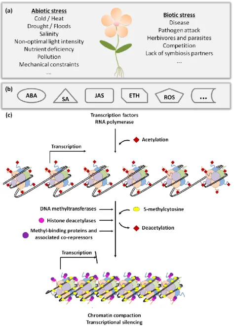
Plants are constantly exposed to potentially stressful conditions and therefore have developed numerous survival strategies, including developmental, morphological and physiological adaptations. Sensing dangerous situations and rapidly initiating effective responses are essential for successful survival. These responses range from signalling cascades to synthesis of defence compounds, including an intricate and highly regulated gene expression network (Figure 2).
While considerable knowledge has been achieved on physiological stress responses involving individual proteins, genes and transcription factors, much less is known about the effect of stress at whole genome level. The concept of large-scale genomic restructuring events in response to unfavourable conditions was introduced by McClintock, more than thirty years ago (McClintock, 1984). Among these events, changes in heterochromatin have the potential to cause large effects on genome function (Madlung and Comai, 2004).
Epigenetic mechanisms are clearly involved in chromatin modifications induced by stress but the precise mechanisms and molecular interactions are still not fully understood. Distinct types of stress namely tissue culture, pathogen attack, interspecific crosses and abiotic stress have been shown to involve epigenetic regulation (Madlung and Comai, 2004). Concerning abiotic stress, several conditions have been analysed, such as suboptimal temperature, water and nutrient availability, and light conditions. For example, the temperature dependent transposon activation in Antirrhinum majus (Coen et al., 1986) was correlated with DNA demethylation under low temperatures (Hashida et al., 2003). Likewise, in Medicago sativa, a cold-induced transcriptional activation of multiple copies of a retrotransposon was observed, although in this case not associated with DNA demethylation (Ivashuta et al., 2002). Therefore, transposon activation and transposition is a widespread phenomenon in plants in response to abiotic stress.
Figure 2: Regulatory network of plant stress responses. Plants can be affected
by several abiotic and biotic stress conditions (a) and to survive they must respond rapidly and effectively. These responses may include signalling cascades, where several hormones can be involved (b). This leads to transcriptional changes either by involvement of transcription factors or through affecting chromatin conformation via DNA methylation, histone tail modifications, histone variant replacements, or nucleosome rearrangements (c) (adapted from Gutzat and Scheid, 2012).
The phenomenon of priming, in which a previous exposure to stress makes a plant more resistant to future stress exposure, is also a general adaptation to stress (Bruce et al., 2007) and recent evidences point to an epigenetic regulation of the priming effect (Luna et al., 2012; Luna and Ton, 2012). Control of gene expression under stress is probably the most studied plant’s response to adverse conditions. Since epigenetic mechanisms, namely chromatin modifications, can be mitotically and meiotically inherited, the epigenetic regulation of gene transcription has the potential to cause more permanent changes of gene expression patterns, being a more efficient strategy to cope with stress (Gutzat and Scheid, 2012). Just to provide some illustrative examples, it was shown in Arabidopsis that the histone deacetylases AtHD2C modulates ABA responsive genes, playing an important role in enhancing plant tolerance to salt and drought (Sridha and Wu, 2006). An increase of H3 acetylation was associated with an increased expression of two stress-responsive genes ADH1 and PDC1 after submergence of rice plants (Tsuji et al., 2006). Also in rice, DNA demethylation occurring in response to osmotic stress was found to facilitate proline accumulation by the up-regulation of P5CS and δ-OAT genes (Zhang et al., 2013).
1.4. Salinity effects on plants
Soil salinity is a major environmental constraint to crop production worldwide and is expected to rise due to global climate changes and also as a consequence of many irrigation practices (Munns and Tester, 2008; Rengasamy, 2010). With the exception of some C4 photosynthetic plants for which sodium is an essential micronutrient, most crop plants are natrophobic (Ohnishi et al., 1990). Salt stress effects on plant growth include nutritional constraints by decreased uptake of phosphorus, nitrate and calcium, ion toxicity mainly due to Na+, Cl- and SO42- and osmotic stress leading to turgor loss and cell volume change (Chinnusamy and Zhu, 2003). In addition to
slower growth rates, salinity also causes a reduced tillering and abnormal reproductive development, with obvious profound and negative impact on agricultural yield (Chinnusamy and Zhu, 2003).
1.4.1. Plant responses to salinity
Plants respond to stress as individual cells and also synergistically as a whole organism. The plant responses to salinity comprise two phases, an “osmotic phase” and an “ionic phase”. At the cellular level, salt stress affects ion and osmotic homeostasis. Sodium (Na+) and chloride (Cl-) are the two key ions responsible for both ion-specific and osmotic damage. These ions interfere with the non-covalent interactions between amino acids in the proteins, leading to protein conformational changes and loss of function, disruption of protein synthesis and interference with enzyme activity (Bhandal and Malik, 1988; Blaha et al., 2000; Chinnusamy and Zhu, 2003). Another important cause of damage are the reactive oxygen species (ROS) generated by salt stress (Miller et al., 2010). Changes in the plasma membrane electrical potential are also a direct consequence of ion imbalance caused by salt stress (Serrano and Rodriguez-Navarro, 2001). Additionally, osmotic imbalances lead to turgescence loss with consequent cell volume changes and retraction of the plasma membrane from the cell wall. Physiologically, the osmotic phase occurs immediately after salt application and consists of stomatal closure, with consequent increase of leaf temperature and decrease of photosynthetic activity (Passioura and Munns, 2000; Sirault et al., 2009), as well as inhibition of cell expansion and cell division (Yeo et al., 1991; Fricke, 2002; Munns and Tester, 2008). The “ionic phase” consists in the accumulation of salt over time, resulting in premature senescence of older leaves and in toxicity symptoms such as chlorosis and necrosis in mature leaves (Munns, 2002; Tester and Davenport, 2003; Munns et al., 2006).
1.4.2. Transducing stress signals
Several chemicals are not only essential for plant growth and development but also play important roles in integrating various stress signals and in controlling downstream stress responses. These roles include the control of gene expression and the regulation of various transporters/pumps, among other biochemical reactions. The primary salt stress signals (ionic and osmotic stress) are transduced through specific signalling pathways, namely through Ca2+ and receptor kinase pathways. Changes in membrane polarization caused by the entrance of Na+ into the cells activate Ca2+ channels (Ca2+ ATPases and H+/Ca2+ antiporters). These Ca2+ waves form rapidly, within 5-10 seconds of salt stress, propagate quickly, and are thought to be one of the earliest events in salt signalling (Lynch et al., 1989). Therefore, cytosolic Ca2+ oscillation acts as a second messenger in salt stress (Sanders et al., 1999; Knight and Knight, 2000). Additionally, several Ca2+-binding proteins play important roles in stress tolerance. For example, the calcium-dependent protein kinases (CDPKs) mediate cellular responses either directly by changing enzymatic activities through protein phosphorylation, or indirectly by changing gene expression patterns (Sathyanarayanan and Poovaiah, 2004), particularly of transcription factors (Mehlmer et al., 2010). Calmodulins (from CALcium MODULating proteIN) and Calcineurin B-like proteins (CBLs) are other important calcium sensors that, although missing enzymatic activity, can bind to target proteins and regulate their activity (Ranty et al., 2006). Salt stress-induced Ca2+ signals can also be perceived by the SOS pathway, which is of vital importance for ion homeostasis regulation under salinity. SOS3 (salt overly sensitive 3) binds Ca2+ and activates de SOS2 kinase, which in turn phosphorylates the SOS1 Na+/H+ antiporter that pumps Na+ out of the cytosol. The SOS3-SOS2 kinase complex also regulates Na+ compartmentation by activating NHX1 and by restricting Na+ entrance into
the cytosol by inhibiting the plasma membrane Na+ transporter HKT1 (Chinnusamy and Zhu, 2003).
The retraction of the plasma membrane caused by Na+ entry into cells activates several membrane-bound proteins, including receptor kinases. The first osmosensor kinase identified in plants was the histidine kinase ATHK1, from Arabidopsis (Urao et al., 1999), which was confirmed to act as an osmosensor to transmit the stress signal to a downstream MAPK cascade (Tran et al., 2007; Wohlbach et al., 2008). The ligands and the downstream signalling molecules sensed by receptor kinases are not yet fully known. Extracellular signals, such as hormones, small peptides, small chemical molecules and physical stimuli are probable ligands. The downstream intracellular events possibly include kinase cascades (e.g. MAPK), Ca2+ ions, ROS signalling, metabolic adjustments, and membrane dynamics (Osakabe et al., 2013). Other signalling molecules are synthesized in response to salt stress, likely playing important roles on plant salt tolerance, namely phosphoinositides (Parre et al., 2007; Tang et al., 2007), Reactive Oxygen Species (Kim et al., 2010; Ruan et al., 2011; Deng et al., 2014, Hoang et al., 2015), Nitric Oxide (Zhang et al., 2007a) and sugars (Kempa et al., 2007). The phytohormones which are essential for plant growth and development also play an important role in integrating various stress signals and controlling downstream stress responses. For example, abscisic acid (ABA) is involved in regulation of plant water balance and osmotic stress tolerance (Zhu, 2002), jasmonic acid (JA) is implicated in salt stress adaptation in rice, barley, grapevine and wheat (Moons et al., 1997; Walia et al., 2007; Ismail et al., 2011; Qio et al., 2014, respectively) while salicylic acid (SA) is involved in the oxidative stress responses triggered by NaCl and osmotic stress (Borsani et al., 2001; Jayakannan et al., 2015).
1.4.3. Salinity tolerance
The major goal of salinity tolerance research is to generate plants capable of maintaining growth and productivity in saline soils. However, the success of breeding programs aimed to produce salt-tolerant crops is limited by the absence of a clear understanding of the molecular basis of the tolerance mechanisms, as well as by their multigenic nature, which impairs traditional breeding techniques such as introgression (Dewey, 1962; Flowers and Yeo, 1995; Roy et al., 2014). For plant salt tolerance, there are three important conditions: (1) to prevent or alleviate damage (i.e., detoxification), (2) to re-establish homeostasis in the stressful conditions, and (3) to resume plant growth even if at lower rate. The operating mechanisms that plants have to successfully manage the salt stress include:
i) Ion exclusion - consisting of Na+ and Cl- transport in roots in order to reduce the accumulation of these ions at toxic levels in leaves. Ion exclusion involves the coordinated action of several transporters at the plasma membrane and tonoplast (Tester and Davenport, 2003; Plett and Moller, 2010).
(ii) Tissue tolerance - where high Na+ concentrations are found in leaves but compartmentalized at the cellular and intracellular level, especially in the vacuole (Tester and Davenport, 2003). In order to maintain equal osmotic potentials in vacuole and cytoplasm, solutes not harmful to cellular biochemistry must accumulate in the cytoplasm. These compatible solutes or “osmoprotectants” include secondary metabolites such as quaternary ammonium compounds (e.g. glycinebetaine), polyols (e.g. mannitol) and core metabolites like proline and sucrose (Rathinasabapathi, 2000). The production of enzymes catalyzing ROS detoxification is also important to achieve tissue tolerance (Roy et al., 2014).
iii) Osmotic tolerance – known to be regulated by long-distance signals triggered before shoot Na+ accumulation. Such mechanisms are pretty much unknown, but must involve rapid and long-distance signalling, probably via
processes such as ROS waves (Mittler et al., 2011; Suzuki et al., 2012), Ca2+ waves (Choi et al., 2014), or even long-distance electrical signalling (Maischak et al., 2010).
1.4.4. Susceptibility to salt stress: contribution of the epigenetic background
Epigenetics contributes enormously to the control of gene expression associated with developmental or environmental clues. Epigenetic inheritance, being a source of interesting polymorphisms able to generate useful variation for selecting superior genotypes, has proven to be a very promising field for future studies on plant stress resistance (Springer, 2013). Moreover, epigenetics could also contribute to natural variation within species and even to local adaptation. For example, the contrasting morphological differences of mangrove plants (Laguncularia racemosa) grown on riverside or on salt marsh is accompanied by a considerable hypermethylation of riverside plants as compared with those of salt marsh plants (Lira-Medeiros et al., 2010). In Cannabis sativa, varieties with contrasting cold acclimation capacities showed distinctive changes in their chromatin state. In particular, the C. sativa varieties that acclimated more efficiently showed increased methylation levels at COR gene loci when deacclimated, suggesting a link between locus specific methylation and deacclimation (Mayer et al., 2015). Specific epigenetic backgrounds associated with distinct varieties are particularly evident in rice. Genome-wide studies by MSAP analysis revealed a differential methylation between contrasting rice genotypes differing in their salt-responsive characteristics (Karan et al., 2012) and drought resistance (Wang et al., 2011). More recently, it was shown that salt-tolerant and salt-sensitive rice varieties display differential methylome flexibility under salt stress, with the tolerant rice varieties being able to adjust more rapidly their methylation levels under salt stress (Ferreira et al., 2015) (see Chapter 2).
1.5. The model plant Oryza sativa
With a 491.4 million tonnes forecast for global production in 2016, rice is one of the most cultivated cereals in the world, only surpassed by maize, while from a cultural, economic and nutritional point of view, it is the most important crop worldwide (Khush, 1997; Bouman et al., 2007; http://www.fao.org/worldfoodsituation/csdb/en/). Unlike the other major cereals, more than 90% of the global rice is consumed by humans. Given the predicted rise in the world’s population, it is likely that rice consumption, and therefore rice demand, will increase over the next decades. However, despite being the staple food for more than half of the planet’s population, a large part of rice production is still lost due to adverse environmental conditions. Therefore, active research on stress tolerance mechanisms in rice has turned this crop one of the most studied plant species nowadays. Rice is considered the model plant for cereals (Coudert et al., 2010), mainly due to the high synteny with other cereal species (Moore et al., 1995), its relatively small genome size (321Mb) (Kawahara et al., 2013) and the availability of a high quality genome annotation (International Rice Genome Sequencing Project, 2005). Moreover, rice has been claimed as an epigenetic model, thanks to the growing collection of T-DNA mutants targeting epigenetic regulators (epimutants) allowing functional studies on gene regulation (Krishnan et al., 2009). Besides helping to understand the role of epigenetics on phenotype variation, the epimutant plants provide great experimental systems to directly address the function of cytosine methylation without the problems found in using methylation inhibitors (epi-drugs), e.g. 5-azacytidine or zebularine (Baubec et al., 2009). Opposite to animals, plants can tolerate epimutations quite well, although aberrant morphological phenotypes are frequently obtained (Kakutani et al., 1996). Mutant analysis of regulatory components involved in methylation/demethylation pathways provides valuable insights about the functional mechanisms of the several epigenetic processes. Inclusively,