1
Universidade de Aveiro
2017
Departamento de Biologia
Emanuele Fasola
Aquisição e hereditariedade de tolerância
a metais em anuros.
Acquisition and inheritance of tolerance
to metals in anurans.
2
Universidade de Aveiro
2017
Departamento de Biologia
Emanuele Fasola
Aquisição e hereditariedade de tolerância a
metais em anuros.
Acquisition and inheritance of tolerance to
metals in anurans.
Tese apresentada à Universidade de Aveiro para cumprimento dos requisitos necessários à obtenção do grau de Doutoramento em Biologia, realizada sob a orientação científica da Doutora Isabel Maria Cunha Antunes Lopes (Investigadora Principal do CESAM e Departamento de Biologia da Universidade de Aveiro), do Professor Doutor Rui Godinho Lobo Girão Ribeiro (Professor Associado com Agregação do Departamento de Ciências da Vida da Universidade de Coimbra) e da Professora Doutora Paula Maria de Melim e Vasconcelos de Vitorino Morais (Professora Auxiliar com nomeação definitiva no Departamento de Ciências da Vida da Universidade de Coimbra).
This work was supported by FEDER funds within the PT2020 Partnership Agreement and Compete 2020- Programa Operacional Factores de Competitividade, by the Portuguese Foundation for Science and Technology (FCT), and by the research project GENEROSI PTDC/BIA-BIC/3488/2012, within the CESAM's strategic programme (UID/AMB/50017/2013), this work was also funded by national funds via FCT/MEC (PIDDAC) and by the doctoral fellowship of Emanuele Fasola SFRH/BD/88955/2012.
3
Dedico il mio dottorato ai miei genitori, che mi hanno dato fiducia e appoggio nonostante le situazioni avverse che si incontrano nel cammino della vita.
4
o júri
Presidente: Prof. Doutor Rui Luís Andrade Aguiar
Professor catedrático do Departamento de Eletrónica, Telecomunicações e Informática da Universidade de Aveiro
Vogais: Doutora Isabel Maria Cunha Antunes Lopes
Investigadora Principal do CESAM e Departamento de Biologia da Universidade de Aveiro (Orientadora)
Prof. Doutor Miguel Alberto Fernandes Machado e Santos Professor auxiliar da Faculdade de Ciências
da Universidade do Porto
Doutora Isabel da Silva Henriques
Equiparada a investigadora auxiliar do Departamento de Biologia da Universidade de Aveiro
Doutor Manuel Eloy Ortiz-Santaliestra
Investigador do Instituto de Investigación en recursos Cinégeticos da Universidad de Castilla-La Mancha
Doutora Matilde Maria Moreira dos Santos
Investigadora em Pós-doutoramento Da Faculdade de Ciências e Tecnologia da Universidade de Coimbra
5
agradecimentos À minha orientadora Dra. Isabel Lopes, por todo o apoio científico, e por ser muitos mais do que uma orientadora. Alem disso um ponto firmo de ajuda e compreensão quando encontrei uns graves problemas ao longo do caminho. Ao professor Rui Ribeiro por todas as ideias, que a sua mente vulcânica mal consegue conter, não podia desejar melhor mentor cientifico. A professora Paula Morais pela grande ajuda, competência e serenidade com as quais sempre trabalhou comigo.
A todos os colegas que me ajudar durante o trabalho, seja de campo ou de laboratório: Diogo, Barbara, Sara C., Sara P., Cátia, Bruno, Antonieta, Anabela, Nuno C., Nuno M., Inês, Jorge, Ana, Tânia M., Catarina, Miguel, Tânia D., Marta e Carla. Aos espanhóis e italianos que fizerem parte desta viagem: Manuel, Rafa, Claudia e Marta.
Ao Abel, o melhor técnico do mundo (e não por ser o único) mas por ser único! A toda a gente do laboratório, porque foram ótimos colegas, mas sobre todos amigos dentro e fora do trabalho. A toda a gente do basket e do magic, vocês são demasiados para eu poder escrever os vossos nomes, mas obrigado pela vossa amizade e para tudo!
6
palavras-chave Pelophylax perezi, variabilidade genética, plasticidade ambiental, dominância genética incompleta, contaminação por metais, erosão genética, mecanismos de hereditariedade da tolerância, microbioma da pele.
resumo Os anfíbios encontram-se em declínio a nível global, sendo a contaminação química um dos principais fatores associados a este declínio. De facto, a exposição de populações de anfíbios a este tipo de perturbações ambientais pode provocar a perda de diversidade genética, devido à diminuição de: 1) aptidão, 2) plasticidade ambiental e 3) eficácia dos mecanismos de tolerância. A contaminação por metais é uma das causas de poluição mais comum no mundo, estando presente em larga escala na Península Ibérica, nomeadamente na Faixa Piritosa Ibérica. Deste modo o estudo dos mecanismos de tolerância a metais, em anfíbios, é relevante, assim como a investigação sobre os mecanismos de hereditariedade desta tolerância. Um dos objetivos do presente trabalho centrou-se no estudo da hereditariedade de tolerância a metais em ovos de Pelophylax perezi. Os resultados obtidos sugerem uma dominância genética incompleta como sendo o mecanismo mais provável de hereditariedade de tolerância a contaminação por metais, em ovos de P. perezi. Estes resultados suportam a hipótese de hereditariedade de tolerância recessiva (ou dominância incompleta). Neste contexto, uma perda de diversidade genética em populações de anfíbios, expostas a contaminação por metais pode ocorrer, mesmo que a fixação de alelos na população seja excluída. De modo a estudar a possibilidade de girinos adquirirem maior tolerância a contaminação por metais, devido à sua exposição histórica a este tipo de contaminação, foram recolhidos girinos de P. perezi em locais contaminados por metais e em locais de referência. Posteriormente, os girinos foram expostos, em laboratório, a um pulso de contaminação intensa por metais. Os girinos oriundos de locais contaminados não mostraram maior tolerância à toxicidade letal de metais comparativamente aos girinos recolhidos em locais de referência. Mais ainda, não revelaram estar sujeitos a um maior stress oxidativo. No entanto, a quantidade de metais no corpo provou a contaminação por metais nos locais historicamente impactados e mostrou que os iões de mercúrio e chumbo são prontamente biodisponíveis para os girinos de P. perezi. Os girinos de locais contaminados apresentaram níveis constitutivos de metalotioneínas, superiores aos medidos nos girinos recolhido os nos locais de referência, o que pode indicar adaptação a contaminação por metais. Por fim, o último objetivo consistiu em avaliar a influência de contaminação química na composição e diversidade do microbioma da pele de populações de P. perezi bem como, identificar a sua sensibilidade a contaminação por efluentes de drenagem ácida. O microbioma da pele dos anfíbios apresenta um papel fundamental na proteção destes organismos a agentes perturbadores ambientais.
Os resultados obtidos revelaram que os metais podem influenciar a composição da comunidade microbiana de anfíbios que habitam locais contaminados. Mais ainda, uma concentração elevada de efluente de drenagem mineira inibiu o crescimento da maioria dos isolados de bactérias da pele dos anfíbios. Esta inibição pode sugerir que os anfíbios perdem uma fração importante do seu microbioma e consequentemente, afetar a proteção da sua pele, quando expostos a contaminação por metais, o que pode determinar um aumento da sensibilidade a este tipo de contaminação.
7
keywords Pelophylax perezi, genetic variability, environmental plasticity, incomplete dominance, metal contamination, genetic erosion, tolerance inheritance, skin microbiome.
abstract Amphibians are declining globally, chemical contamination being one of the major factors driving this process. As a consequence of exposure to such environmental perturbation, natural population of amphibians may lose their genetic diversity, which may occur due to a decrease in: 1) fitness, 2) environmental plasticity capabilities and 3) tolerance mechanisms efficiency. Metal contamination is one of the most worldwide distributed contamination source, having a great impact in the Iberian Peninsula habitats, especially in the Iberian pyrite belt region. Therefore, is important to explore how tolerance mechanism, toward metal contamination, work in amphibians and how genetically determined tolerance mechanisms are inherited. In this work, these topics were addressed by assessing the inheritance to lethal tolerance to acid mine drainage and copper contamination in eggs of the Perez's frog Pelophylax perezi. Incomplete dominance was found to be the most likely inheritance mechanism of tolerance toward these two chemical stressors in the eggs of P. perezi. The results support the recessive (or incompletely dominant) tolerance inheritance (working-) hypothesis. Thus, the amphibians’ populations impacted by metal contamination can considerably lower their genetic diversity, even if allele fixation was excluded.
The possibility of tadpoles, historically exposed to metal contamination, being able to acquire an increased tolerance to metal contamination, comparatively to tadpoles inhabiting reference sites, was also studied. Pelophylax perezi tadpoles, sampled at historically metal impacted mining sites, did not show higher oxidative stress or lethal tolerance comparatively to tadpoles inhabiting reference sites. However, the metal body burden proved metal contamination at the historically metal impacted sites and showed that mercury and lead ions are readily bioavailable for P. perezi tadpoles. Furthermore, tadpoles from metal contaminated sites seem to show higher constitutive levels of metallothioneins, which may suggest adaptation to metal contamination
The last objective of this work, was to evaluate the influence, of metal contamination, on the composition and diversity of the P. perezi skin microbiome, and to explore its tolerance to acid mine drainage contamination. Amphibians’ skin microbial community has been shown to help its hosts tolerating infections. Because the increasing research on the important protective role of amphibians’ skin microbiome, its diversity and capacity to tolerate metal contamination was as well investigated. Obtained results showed that metal contamination influences the skin microbial community composition in frogs living at metal impacted sites; furthermore, an intense acid mine drainage concentration can inhibit the growth of almost all the isolated strains. This inhibition suggests that amphibians may lose an important part of their skin microbiome, affecting the protection of their skin, when exposed to metal contamination; which, in turn may lead to an increased sensitivity to metal contamination.
8
Index
Chapter 1: General introduction
Brief overview on biodiversity
Impact of anthropogenic contamination on genetic diversity Global amphibian decline
Amphibians tolerance to pollution and pathogens Metal contamination and mechanisms of toxicity Objectives
The study sites The model organism
Detailed description of each chapter References
Chapter 2: Microevolution due to pollution in amphibians: a review __ on the genetic erosion hypothesis
Abstract Introduction
Moving the focus on amphibians Genetic variability and reduced fitness
Genetic variability and environmental plasticity Genetic variability and co-tolerance
Genetic variability and fitness trade-off costs Genetic variability and tolerance to pathogens Conclusion
References
Chapter 3: Inheritance of tolerance to metals:
Among and within amphibian egg masses variability may
---unveil trait dominance
Abstract Introduction Material and methods Study organism Assay setup
Statistical analysis Genetic mechanism evaluation
Results Discussion and conclusions References 14 15 17 17 19 20 22 23 25 26 28 40 41 42 43 46 48 50 51 53 54 55 72 73 73 74 75 75 77 77 80 85 88
9
Chapter 4: Testing the recessive tolerance inheritance (working-) ---hypothesis: Frog egg masses exposed to copper
Abstract Introduction Material and methods Study organism Assay setup Statistical analysis Genetic mechanism evaluation Results Results 2014 Results 2016
Comparison between 2014 and 2016 sampling seasons
Comparison between AMD and copper tolerance Discussion and conclusions
References
Chapter 5: Does historical exposure to metal contamination provide tadpoles a higher lethal tolerance to these chemicals?
Abstract Introduction Material and methods Study organism Sampling sites
Sampling and experimental design
Toxicity assay Chemical analysis
Data analysis Results Field collected tadpoles
Tadpoles from laboratory assays Discussion Field sampling Laboratory assay Conclusion References 90 91 91 94 94 94 95 96 99 99 107 108 108 109 112 118 119 120 122 122 123 124 125 125 127 127 127 131 135 135 137 138 139
10
Chapter 6: Environment and gender influences the skin microbiome composition of the Perez’s frog
Abstract Introduction Material and methods Sampling sites Acid mine drainage analysis
Collection of skin microbiome DGGE analysis of P. perezi skin microbiome
16S rRNA gene-based metagenomics of of P. perezi skin microbiome Isolation and quantification of bacterial density
16S rRNA gene sequencing of bacterial isolates Phylogenetic analysis of bacterial isolates Effluent toxicity assay Data analysis Nucleotide sequence accession numbers Results DGGE analysis from P. perezi skin microbiome Microbiome diversity by next generation sequencing of P. perezi skin community
Microbiome diversity by cultivable fraction of P. perezi skin microbiome Tolerance of bacterial strains to AMD
Discussion and conclusion References
Chapter 7: General discussion and conclusion
References 146 147 148 149 149 150 150 151 152 152 153 153 153 154 155 155 155 156 159 162 164 167 172 179
11
Index of figures
Fig.1.1
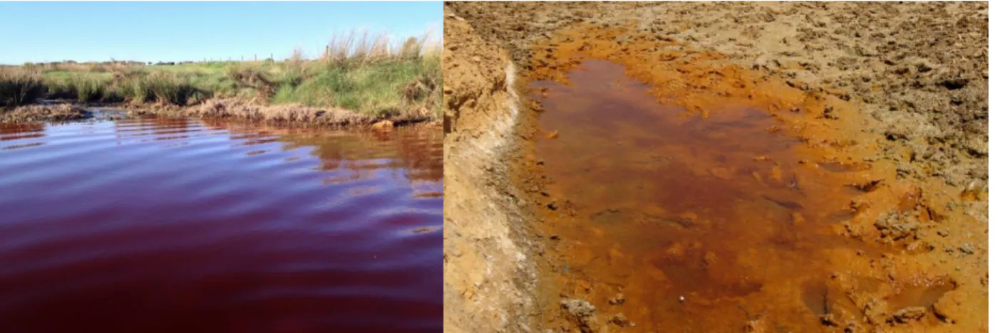
Agua Forte river, impacted by AMD coming from the Aljustrel mining site.
Fig.1.2
The river running through Sao Domingos mining site.
Fig.1.3
Minas de Almaden, and Arroyo de Valdefuentes in the Alcudia area.
Fig.1.4
A Perez’s frog adult with its eggs and tadpoles in the laboratory.
Fig.3.1
Expected theoretical frequencies (in %) of eggs (F1 generation), of Perez's frog (Pelophylax perezi) egg masses.
Fig.3.2
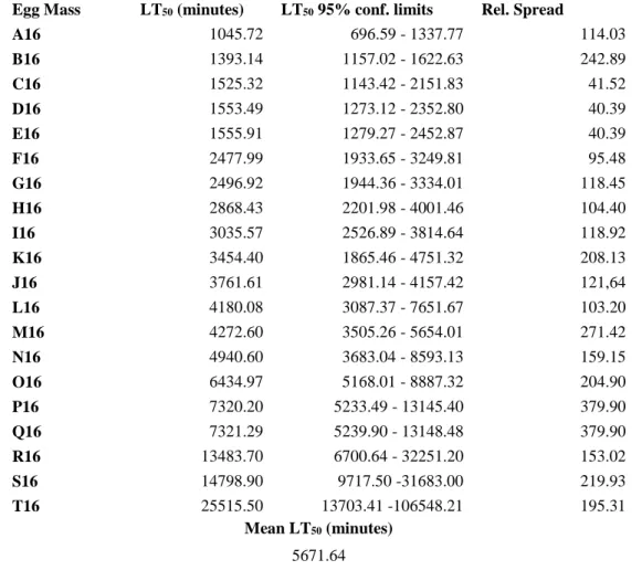
Box plots representing the median, the lower and the upper quartiles of lethal time values (exposure times after which 50, 25 and 75% of eggs died) of eggs within each of 21 Perez's frog (Pelophylax perezi) egg masses (A to U), collected in a reference pond, exposed to a 60% dilution of metal-rich acid mine drainage.
Fig.3.3
Frequencies (in %) of eggs belonging to eight classes of lethal tolerance to a 60% dilution of acid mine drainage, in each of 21 Perez's frog (Pelophylax perezi) egg masses (A to U), collected in a reference pond.
Fig.3.4
Relative spread (difference between the upper and lower quartiles relatively to the median) of egg tolerance to a 60% dilution of metal-rich acid mine drainage of 21 Perez's frog (Pelophylax perezi) egg masses, collected in a reference pond, versus the respective median lethal time values (LT50 – exposure time after which 50% of eggs died).
Fig.4.1
Expected theoretical frequencies (in %) of eggs (F1 generation), of Perez's frog (Pelophylax perezi) egg masses.
Fig.4.2
Box plots (2014 sampling above, 2016 sampling below) representing the median, the lower and the upper quartiles of lethal time values (exposure times after which 50, 25 and 75% of eggs died) of eggs within each of 20 Perez's frog (Pelophylax perezi) egg masses (A to T, 2014 and a to t, 2016), collected in a reference pond, exposed to a 60% dilution of metal-rich acid mine drainage.
Fig.4.3
Frequencies (in %) of eggs belonging to eight classes of lethal tolerance to a 60% dilution of acid mine drainage, in each of 40 Perez's frog (Pelophylax perezi) egg masses (A to U), collected in a reference pond. 24 24 24 26 78 81 83 85 96 100 101
12 Fig.4.4
Relative spread (difference between the upper and lower quartiles relatively to the median) of egg tolerance to copper of 40 Perez's frog (Pelophylax perezi) egg masses (2014 egg masses are represented by diamonds and 2016 by circles), collected in a reference pond, versus the respective median lethal time values (LT50 – exposure time after which 50% of eggs died).
Fig.4.5
Comparison of egg tolerance (median lethal time values, LT50) of Perez's frog (Pelophylax perezi) twenty one egg masses exposed to a dilution of metal-rich acid mine drainage and to copper.
Fig.5.1
Map of the sampling sites; Picon “Arroyo del Raso” (PAR – R1), Picon “abrevadero” (PAB – R2), Rio Bullaque (RB – R3), Arroyo del Tamujar (AT – HG1), Rio Guadalmez (RG – HG2), Arroyo de Valdefuentes (AV – PB1) and Arroyo de la Ribera (AR – PB2).
Fig.5.2
Levels of glutathione peroxidade (GPx), superoxide dismutase (SOD) and malondialdehyde (MDA) measured in tadpoles of Pelophylax perezi collected in the field.
Fig.5.3
Levels of metallothionein (MTs) and metal body burden (Hg and Pb) measured in tadpoles of Pelophylax
perezi collected in the field.
Fig.5.4
Cumulative mortality percentages of Pelophylax perezi tadpoles, per observation, exposed for 24h to lethal levels of mercury.
Fig.5.5
Levels of glutathione peroxidade (GPx), superoxide dismutase (SOD) and malondialdehyde (MDA) measured in tadpoles of Pelophylax perezi exposed to lead in the laboratory in comparison with the controls.
Fig.5.6
Levels of metallothionein (MTs) and metal body burden (Hg and Pb) measured in tadpoles of Pelophylax
perezi exposed to lead in the laboratory in comparison with the controls.
Fig.6.1
Map with the location of the sampling sites: Barragem de Reguengos 1 (BR1), Barragem de Reguengos 2 (BR2), Lagoa do Cao (LC) and Ribeira da Agua Forte (AF).
Fig.6.2
Similarity coefficient (UPGMA) between DGGE lanes deriving from Pelophylax perezi frog’s skin bacterial samples.
Fig.6.3
Rarefaction analysis curves of Pelophylax perezi skin bacterial communities.
Fig.6.4
Percentage composition of the whole Pelophylax perezi skin microbial community at family level as resulting from the DNA barcoding analysis.
Fig.6.5
PCoA for the total microbiome of the analyzed Pelophylax perezi skin bacterial samplesl.
107 109 124 128 130 131 133 134 150 156 157 158 159
13 Fig.6.6
Composition, in percentage, of the cultivable fraction of the microbial community (170 isolates), sampled in Pelophylax perezi skin, at family level.
Fig.6.7
Venn diagram of the whole Pelophylax perezi skin microbial community.
Fig.6.8
RDA analysis of Pelophylax perezi skin bacterial samples, based in the differences between metal contaminated and reference sites and between male and female frogs’ microbial community.
Fig.6.9
Average growth of Pelophylax perezi skin microbial community after being exposed to serial dilutions of acid mine drainage.
Fig.6.10
NMDS (Non-Metric-Multi-Dimensional-Scaling) analysis discriminating Pelophylax perezi skin samples’ microbial community’s tolerance to acid mine drainage (AMD) by site.
160
161
162
162
14
CHAPTER 1.
15
General introduction.
Brief overview on biodiversity
Biodiversity has been perceived and investigated in many different ways, as its main components have many times been questioned (Mace et al. 2012). The concept has been firstly defined in the United Nations Conference on Environment and Development, held in Rio de Janeiro in 1992 (Rio Convention, 1992). Biodiversity is largely interpreted as a measure of species richness, with species as its core unit, both for research and conservation purposes (Purvis and Hector, 2000). The concept also comprehends spatial-geographic components, thus considering the variation in habitats and ecosystems, among climatic geographical regions (Fischer and Young, 2007; Fleishman et al. 2006). This facilitated the designation of hotspot biodiversity areas as important for conservation, and the recognition of particular sites deserving to be promoted as protected areas (Olson et al., 2001). Indeed, the importance of species conservation is deeply connected with the preservation of the ecosystems in which they live (Mace et al. 2012). The concept established in the Rio convention was later officially accepted by the Parties of the Convention on Biological Diversity (CBD); the definition of biodiversity is thus given as: ‘the variability among living organisms from all sources including, inter alia, terrestrial, marine and other aquatic ecosystems and the ecological complexes of which they are part; this includes diversity within species, between species and of ecosystems’ (CBD, 2013; Rio Convention, 1992). Biodiversity, as agreed at the Rio convention, has three main components: a) diversity of landscapes and ecosystems inside them, b) species diversity, and c) genetic variability between and within populations of the same species (Agenda 21 : programme of action for sustainable development ; Rio Declaration on Environment and Development ; Statement of Forest Principles: The final text of agreements negotiated by governments at the United Nations conference on environment and devel, 1993). These components have been regarded as the three pillars of biodiversity, each one deserving to be taken into account when planning conservation policies (CBD, 2013). That is because biodiversity supports life on planet Earth, and all around the world is possible to spot a plethora of variation in nature (Odum and Barrett, 2004; Raven and Johnson, 2014; Sala et al. 2000). One of the important components included in the definition of biodiversity, as one of its three pillars, is the genetic variability, also because it can ensure population capability to adapt to future environmental changes (Boyce, 1992; Van Straalen and Timmermans, 2002). Scholars categorized organisms inhabiting Earth into taxa, creating different taxa-diversity levels. Although being an artificial classification, it is useful to understand reciprocal relatedness into group of living
16
beings (Wiley and Leiberman, 2011). Most important, taxa divisions were fundamental for research and for conservation. A lot of studies and research projects came out with a flag-species (charismatic and symbolic species with the purpose to stimulate conservation awareness) on the spot, because it was convenient and appealing (Dietz et al. 1994; Entwistle and Bowen-Jones, 2002). Indeed, protecting some noticeable or conspicuous species can end up protecting all the food-web-network around it, or even all ecosystem that harbors it (Simberloff, 1998). Even if species level has been at the core of conservation, none of the biodiversity pillars is inherently more important than the others, they interact with each other and with many factors that can impact them (Via and Lande, 1985). All together, concurring in shaping biodiversity (Agenda 21 : programme of action for sustainable development ; Rio Declaration on Environment and Development ; Statement of Forest Principles: The final text of agreements negotiated by governments at the United Nations conference on environment and devel, 1993; Odum and Barrett, 2004). Once again, impacts on one level of biodiversity affect the others two levels with upward and downward cascades. For example, habitat fragmentation can enable speciation (Odum and Barrett, 2004). But, if the isolated population is too small, it can experience a bottleneck, lowering its genetic diversity and precipitating its possible extinction. This process is more effective, in small populations, if the gene flow is interrupted or if the level of inbreeding is high (Hedrick and Kalinowski, 2000; Keller and Waller, 2002). On the other hand, the loss of a species may impact the ecosystem functions, altering its capabilities to provide services (Mace et al. 2012; Medina et al. 2007; Vinebrooke et al., 2004; Worm et al., 2006). Scholars investigated the mechanism and the causes behind biodiversity loss, to understand why it occurs and how to prevent it (Miller, 2005; Myers et al., 2000). Indeed, human activities produce a wide variety of adverse impacts on biodiversity. For example, urbanization enhances habitat fragmentation (Carr and Fahrig, 2001; Fahrig, 1997; McKinney, 2002; Wiegand et al. 2005), while agriculture and industry generate pollution by various kind of toxic compounds (Abler and Shortle, 2001; Bridges, 2000; Brühl et al., 2013; Nadal et al. 2004). Pollution is a concerning issue since it can lower biodiversity regarding all its three fundamental components. For example, marine ecosystem structure and services may be hindered by fertilizers, pesticides, sewage sludge, and metals; these pollutants can cause direct mortality of the primary and secondary consumers (edible crustaceans and fish), impairing the fishery industry. Furthermore, these waters would not be available for recreational use anymore (Islam and Tanaka, 2004). Chemical contamination is among the main causes of species decline nowadays (Fisker et al., 2011; Rouse et al., 1999; Wilcove et al., 1998). Moreover, contamination can impact genetic diversity directly; for example, through mutagenicity induced in plants by metal contaminated soil (Knasmüller et al., 1998). Metal contamination can also have drastic effects on microbial communities; the strains in this communities can develop tolerance to
17
metal contamination (Gadd and Griffiths 1977; Díaz-Raviña et al., 1994). Moreover, toxic compounds may cause genetic erosion (Fasola et al. 2015; Ribeiro and Lopes, 2013; Van Straalen and Timmermans, 2002). Therefore, it is important to enhance ecotoxicology research, because all human-generated impacts can alter nature’s processes in many more subtle ways than that which is conceivable at a single biodiversity level (Relyea and Diecks, 2008).
Impact of anthropogenic contamination on genetic diversity
Genetic erosion is the loss of genetic variation: the loss of genotypes determining a specific trait or set of traits (Fasola et al. 2015; Van Straalen and Timmermans, 2002). When genetic erosion is due to chemical contamination, it can be a serious threat to the viability of populations, especially small ones (Coutellec and Barata, 2011; Lopes et al., 2009; Luquet et al., 2011; Medina et al. 2007; Ribeiro and Lopes, 2013). This is because the loss of genetic variation, due to contamination, may impact: 1) average fitness of the population, 2) environmental plasticity, 3) co-tolerance mechanisms, 4) trade-off mechanisms, and 5) tolerance to pathogens or diseases (Fasola et al. 2015). When a population is exposed to a contaminant, the loss of some of its individuals (sensitive to this particular compound), either by death or emigration, causes a reduction of its genetic variability. Genetic erosion can be caused by contaminant-driven directional selection or contaminant-driven genetic drift, in practice inducing bottleneck effects (for example through mechanism of habitat fragmentation), leading to inbreeding (Fasola et al. 2015; Ribeiro and Lopes, 2013). The effects of contaminant-driven genetic erosion can be worsened if this process acts on already fragmented populations, that often show high levels of inbreeding (Channell and Lomolino, 2000; Chen et al., 2012; Hedrick and Kalinowski, 2000; Keller and Waller, 2002; Luquet et al., 2013). Those populations may be hardly impacted by contaminant-driven genetic variability loss, and deserve special attention and dedicated conservation practices (Channell and Lomolino, 2000; Fasola et al. 2015). In this context, vertebrates’ populations, especially those of endangered or vulnerable species, may be at higher risk of extinction (IUCN, 2013). Among them, amphibians arise major concerns about their conservation, as, at present, they are facing a global decline.
Global amphibian decline
Of the know species of amphibians in the world, 40% (about 6000 species; Pyron and Wiens, 2011), are considered to be declining, with 30% having a concerning conservation status according to the International Union for Conservation of Nature (IUCN, 2013). Most studies that have investigated
18
the amphibian decline point out that many populations are moving toward extinction (Alford and Richards, 2007; Blaustein and Bancroft, 2007; Blaustein et al. 1994; Collins and Storfer, 2003; Houlahan et al., 2000; Kiesecker et al. 2001; Stuart et al. 2004). Emblematic is the case of the golden toad (Incilius periglenes) and the harlequin frog (Atelopus varius), which have been linked to pollutants or diseases (Pounds and Crump, 1994). Amphibian populations are known to be fragmented and with low gene flow, given their relatively low dispersal capabilities (Beebee, 2005; Bielby et al., 2013; Smith and Green, 2005). Furthermore, those populations often present low effective sizes, increasing the probability of inbreeding and genetic erosion (Beebee, 2005; Beebee and Griffiths, 2005; Laan and Verboom, 1990). The majority of amphibians, mainly of the order Anura, go through metamorphosis, living in aquatic habitat as larvae and in the terrestrial compartment as adults (Pough and Kamel, 1984; Sparling et al., 2010; Wilbur, 1980; Wilbur and Collins, 1973). For this reason, amphibians enter in contact with contaminants characteristics of both aquatic and terrestrial ecosystems (Blaustein and Bancroft, 2007), again increasing the probability for contaminant-driven genetic erosion to act on their populations (Fasola et al. 2015; Medina et al. 2007). It has already been reported that many compounds, like pesticides, and pathogens can induce lethal effects, ultimately reducing populations and possibly leading to bottlenecks and decrease of the genetic pool of such populations (Baud and Beck, 2005; Bridges, 2000; Brühl et al., 2013; Corn and Vertucci, 1992; Hall and Henry, 1992; Hatch and Blaustein, 2003; Hua et al. 2013; Ireland, 1991; Macìas et al. 2007). Also, the gene flow among amphibian populations and genetic drift rates can be modified by humans’ activities, which often fragment or destroy habitats suitable for amphibians (Cushman, 2006). In addition, toxic compounds, deriving from human activities, may promote diseases in amphibians, because some pollutants have the capability to impair amphibian immunity (Jobling et al., 2013). Moreover, if the physiological condition of an individual is already lowered by the effect of contamination (for example by slowing its metabolism), then immune responses would be reduced in magnitude and efficacy (Fedorenkova and Vonk, 2012; Folt et al., 1999; Hatch and Blaustein, 2003; Mazanti et al., 2003; Vinebrooke et al., 2004; Wah-Chu and Chow, 2002; Yue et al., 2009). Another important factor within the frame of amphibians’ decline is the spread of diseases and pathogens, which are decimating many populations. For example, chytrid fungus, Ranavirus and water mould infections are a big threat to amphibians (Baláž et al., 2013; Fernández-Benéitez et al., 2011; Pearman and Garner, 2005; Pounds et al., 2006; Stevenson et al., 2013).
19
Amphibian tolerance to contamination and pathogens
Given the many adverse impacts that amphibian populations have to face nowadays, the tolerance mechanisms they possess, to several environmental perturbations, are crucial for survival. These capabilities may be due to selected genetic traits, in the population, leading to micro-evolutionary shifts in alleles frequencies (Dieckmann and Doebeli, 2004). Tolerance can also derive from phenotypic plasticity (Ghalambor et al., 2007; Via et al. 1995; Weitere et al., 2004). In either case, amphibians can face contamination and pathogens induced stress with many mechanisms. For example, Moor frogs’ (Rana arvalis) Swedish populations rapidly evolved tolerance to water acidification (Rasanen et al, 2003). The authors hypothesized that such increased tolerance was due to a physiologic mechanism likely related to the osmotic regulation or to a more efficient way for egg’s jelly coat to block hydrogen ions. This grants embryos and tadpoles the ability to tolerate low pH (Rasanen et al. 2003). Another study showed the origin, by natural selection, of osmotic stress tolerance in tadpoles of the natterjack toad (Bufo calamita) inhabiting brackish ponds, possibly resulting from drought tolerance by exaptation (Gomez-Mestre et al., 2004). Another study pointed out wood frogs’ (Lithobates sylvaticus) tadpoles capabilities for plastic responses toward pesticide contamination, by mean of inducible tolerance caused by previous exposure to sublethal concentrations of carbaryl (Hua et al. 2013). Wood frog was also an interesting case of maternal effects combined with genetically determined tolerance to acidification; the maternal effects determined embryo’s tolerance, while genetic variation determined tolerance of larvae, and thus embryo’s tolerance was not related with larval tolerance (Pierce and Sikand, 1985). Amphibians also harbor a rich and diverse microbial community onto their skin. This can be an effective defensive mechanism, which is not characteristic of the amphibian itself but of its skin associated microbial community. Some studies referred how their skin microbiome can contain bacteria strains tolerant to some pathogens or even pollutants (Becker et al., 2009; Choi et al., 2015; Díaz-Raviña et al., 1994; Dobson et al., 2012; Gadd and Griffiths 1977; Harris, Brucker e Walke, 2009; Woodhams et al., 2016). In this light, the microbial community residing into the amphibians’ skin mucus can possibly act as a primary barrier against environmental agents; for example, there is some evidence that microbial produced compounds can act to protect their hosts from pathogens like the chytrid fungus (Becker et al., 2009, 2011; Bletz et al., 2013; Harris et al., 2009; Pask, 2012). Therefore, it is important to investigate the diversity of this skin microbiome to understand if it can be impacted by pollution. If this is the case, amphibians living in polluted environments could come to lose a key component of their tolerance to environmental perturbations.
20
Metal contamination and mechanisms of toxicity
Metal contamination is recognized as having severe impacts on biodiversity all over the world. It can originate from many sources, namely industrial processes, water acidification (inducing metal ions leeching) or mining activities (Knasmüller et al., 1998; Linder and Grillitsch, 2000; Nadal et al., 2004; Roark and Brown, 1996; Santoro et al., 2008; Young and Harvey, 1991). The latter has a great impact on the soil surrounding the mine, in the water originating from it and in the surrounding water bodies contaminated by runoff and lixiviation (Knasmüller et al., 1998; Macnair, 1997; Nordstrom, 2011; Shaw, 1999). Metal -enriched waters, coming from mining activities, are characterized by a diverse composition and assortment of ions, which depends on the minerals present in the bedrock of the mine (Nordstrom and Alpers, 1999; US-EPA, 1989). These waters are acidic; their pH being often 5 or lower (Bowell and Bruce, 1995; Nordstrom, 2011; Nordstrom et al., 2000; Pereira et al. 2004; Sobral et al. 2013). Their effects on the surrounding environment also depend upon the degree of dilution the acid mine drainage suffers, due to rainfall or nearby rivers, exerting more severe effects near the extraction site (Nordstrom, 2011; Nordstrom and Alpers, 1999).
When interacting with living beings, metal ions usually bind to specific receptors in cell’s membranes (Di Toro et al., 2001; Wright, 1995). Most of the time, this process occur at the surface of respiratory organs in animals or in plant roots (but even leaves). Here, the binding ions can be pumped into the cells by active mechanisms or simply passing through by osmosis (Linder and Grillitsch, 2000; Di Toro et al., 2001). In microorganisms, the mechanism of tolerance to metal can involve: (1) hydrogen sulfide production to bind metal ions resulting in insoluble sulphates; (2) chelation of metal ions by active binding to organic substances; (3) sequestration of metal ions by binding to cell surfaces or by intracellular uptake sequestration; (4) change in valence and/or active conversion into organometallic compounds; (Branco et al., 2008a; Branco et al., 2008b; Francisco et al., 2002; Gadd and Griffiths, 1977; Santo et al., 2010; Sousa et al., 2013). Organisms purposely take in metal ions that are necessary for their physiological processes, like copper, zinc, iron, and other, which are essential for life (Di Toro et al., 2001; Rainbow, 2007). But even essential ions will become toxic when present at high concentrations, surpassing predetermined (and species specific) physiological thresholds (Rainbow, 2007; US-EPA, 1989; Wright, 1995). Their toxicity can be exerted when the ions bind to cellular receptors, which were meant to bind with other ions with equal charge, molecular weight or ionic valence (Linder and Grillitsch, 2000). In that case, especially when the concentration of the interfering ions fairly exceeds those of the proper one, the receptors became almost fully unavailable for the latter. Mutagenic metal ions can also bind to DNA altering its functions. Both mechanisms inhibit whichever correlated physiological reaction (Goyer and Clarkson, 2001; Linder and Grillitsch,
21
2000). Those mechanisms can block or lessen the production of particular compounds, enzymes or others proteins needed in the target organism, possibly leading to severe consequences (Goyer and Clarkson, 2001). If lethal concentrations are not reached the target organisms could experience a variety of sublethal effects: growth inhibition and delay, malformations, metabolic deficiencies, hormonal disequilibrium, sterility and unnatural behavior (Goyer and Clarkson, 2001; Linder and Grillitsch, 2000). The degree and kind of effect depends on many different variables, as those inherent to metal speciation and those concerning the organism on which it acts (Goyer e Clarkson, 2001; Linder and Grillitsch, 2000). Indeed, affected organisms can have a lot of different strategies to better cope with metal contamination. A simple choice, for animals able to do so, is to flee the polluted areas, being spatial avoidance one of the primary responses (Lopes et al. 2004; Lukkari and Haimi, 2005). Indeed, an option that plants and sessile or slow moving animals cannot afford, even if plants can make their roots grow toward deeper unpolluted soil (Dickinson et al. 1991). When spatial escaping is not possible, populations can adapt, with physiological plastic responses (Adlassnig et al., 2013; Peña-Castro et al., 2004; Via et al. 1995), trans-generational effects (Muyssen and Janssen, 2001; Pölkki et al. 2012) and/or genetic inheritable factors (Dieckmann and Doebeli, 2004; Lopes et al. 2006; Shaw, 1999). Either way, those mechanisms often involve metabolic pathways able to discriminate toxic ions or excrete them, when they are at a higher than desired concentration (Marsden and Rainbow, 2004; Rainbow, 2002, 2007). Such kind of responses came in a variety of ways; for example, plants can expel metal ions through their roots while crustaceans can accumulate them as tiny crystals in their carapaces, ready to be disposed with the subsequent mould (Marsden and Rainbow, 2004; Rainbow and Scott, 1979). Bacteria can either accumulate metal ions in their cells or at the membrane’s surface or bind them to other organic or inorganic compounds to make them not bioavailable (Gadd and Griffiths, 1979). Amphibians mainly enter in contact with metal pollutants in the aquatic medium; in which they live during embryonic and larval stages. Even when they are adult, most amphibians inhabit humid habitats, thus a constant layer of moisture surround their skin, even when they are not in the water. As a result, the diffusion of metal ions in the water is a key process to exert the toxicity of these compounds (Todd et al., 2011). The jelly coat surrounding the eggs is the first barrier that ions have to surpass to get to the embryo, while in later stages, after the development of the gills, these will likely be the target organ. As adults, the skin of the amphibians is the primary barrier against external agents, although it is highly permeable and unlikely to prevent the entrance of metal ions. The barrier effect of the skin is primarily exerted by its mucus and the bacterial community living in it. The complexity of this scenario is very high; the degree of the toxic effects on amphibians is not only related to their life stage, neither only with the kind and concentration of the metal contaminating the ponds in which they live. The characteristics of the
22
water body impact greatly this outcome, as pH, hardness and dissolved organic matter can interact with metal ions and alter their speciation in a variety of complex ways (Adlassnig et al., 2013; Freda, 1986; Freda 1991; Horne and Dunson, 1995; Rodríguez et al., 2009). Nevertheless, there are examples in which the toxicity caused by metals is mainly due to the content of metal ions present in the sediment; specifically when tadpoles feed near the bottom of a contaminated pond, they can uptake a quantity of metal by ingestion and end up showing toxic effects and/or bioaccumulation (Karasov et al. 2009; Loumbourdis et al., 2007; Sparling, 1996). The effect of metal contamination on amphibians range from death to malformations, to physiological reproductive or metamorphosis impairment, depending on the intensity of the contamination and its duration in time also. For example, chronic exposure can result in upregulation of some genes (Marques et al., 2013). The most likely effects, at low metal concentrations, are the ones lowering the individuals’ fitness; like reduced growth and fertility (Adlassnig et al., 2013; Todd et al., 2011; Zocche et al., 2014); these effects can be related to mechanisms of oxidative stress too (Borković-Mitić et al., 2016; Marques et al., 2011).
Objectives
Within the described context, the main objective of this work was to understand the tolerance to metals of amphibian aquatic stages and of their skin associated microbiomes. To attain this major goal the following specific objectives were delineated:
a) Resume a coherent and structured understanding of the genetic erosion mechanisms, focusing on amphibians as a model;
b) Contribute to the understanding of the heritable tolerance to metal contamination in amphibians;
c) Investigate if amphibians’ populations historically exposed to metals exhibit higher tolerance to this kind of contamination;
d) Assess if different environmental conditions (metal contamination) influence the bacterial diversity in amphibian´s skin.
e) Determine if bacteria strains, collected on frogs from a metal contaminated site, are more tolerant to such contamination than those from reference populations.
Acid mine drainage (AMD) was chosen as the pollutant factor and amphibians as the target group, being Perez’s frog (Pelophylax perezi) the model species.
23
The study sites
Mining was an important activity in the Iberian Peninsula since the Roman Empire ages, when the Romans already extracted various metals. Among those, precious metals like silver or gold, and others like iron, copper, lead, and tin (Edmondson, 1989). The geological conformation of the region gave origin to the Iberian pyrite belt in the Devonian period (Paleozoic era, 419,2 Mya – 358,9 Mya), due to massive volcanic activity, leading to huge sulfide deposits. It is a vast mining region, about 250 km long and 30 to 50 km wide. It lays approximately from Alcácer do Sal, in Portugal, to Sevilla, in Spain, running northwest to southeast (Gibbons and Teresa, 2002). This area saw his ancient mine exploitation diminishing during the middle ages but, returning at the economy’s leading activities with the industrial revolution. When industrial companies from northern Europe came to this region, they made the extraction of pyrite the pillar of this region for a century and a half. More than 250 extraction points were active in the pyrite belt at that time. The extraction continued in many areas until the end of the 19th century, declining since then (Gibbons and Teresa, 2002). Regarding the Portuguese portion of this region, the most important mines were in Aljustrel and São Domingos, both located in the Alentejo region, at both mines, iron was the main extracted metal. While the Aljustrel mine is still active, São Domingos was abandoned for more than 50 years, but still highly impacts the surroundings (Pereira et al. 2004; Sobral et al. 2013). Nevertheless, even if the impact of mining activities is more evident at São Domingos, its effect is present also in the Aljustrel district (Fig.1.1 and 1.2). More than a century of mining exposed sulfur rich minerals and led to huge deposits of slag accumulating around the mine pits. Over the years, the weather acted on those rocks and the rain pouring through it was enriched with metal and hydrogen ions. At present, these waters, coming from the mining spots, constitute acid drainage (AMD) that reaches pH values near 2 (Costa et al., 2016; Pereira et al. 2004; Sobral et al. 2013). The drainage from the Aljustrel mine results in a yellow/red colored water river (Ribeira de Água Forte, Fig. 1.1), while in São Domingos the same sort of water flows through a 10 km valley (Fig. 1.2). The Spanish portion of the Iberian pyrite belt was equally exploited, for example, in the mines of the Alcudia valley, Almaden and Horcajo; all located in the Province of Ciudad Real (Castilla-La Macha region) (Fig. 1.3). This mining area was the major lead producer in Spain during the second half of the 19th century and the Alcudia Valley was one of the most important districts (Rodríguez et al., 2009).The soils analyzed at the Almadén mine, now converted into a museum since 2000, showed increasing mercury levels, with a gradient in direction to the mine, caused by the erosion of the mineral deposits; mercury extracted from Almadén mine produced the largest amount of liquid mercury ever processed in our planet (Higueras et al., 2006; Lindberg et al., 1978). The Horcajo lead mine had a history similar to that of the Alcudia
24
valley (Baranda, 1994). A study reported analysis of high lead contamination in the soils, which was transferred into plants growing on it (for example grass of the Gramineae family or trees of the genus Quercus) and animals feeding on them (Cervus elaphus) (Reglero et al., 2008). In this context, six metal polluted locations, and seven unpolluted locations were chosen as sampling sites for our studies about Perez’s frog (eggs, and skin microbiome in Portugal; tadpoles in Spain) tolerance to AMD.
Fig.1.1: Água Forte river, impacted by AMD coming from the Aljustrel mining site.
Fig.1.2: The effluent running through São Domingos mining site.
25
The model organism
To perform the present work, the Perez’s frog (Pelophylax perezi, López-Seoane, 1885) is an advantageous model species, because it is easily encountered in the field and its larval stages can be maintained in the laboratory with no great effort. The Perez’s frog may inhabit eutrophic or even polluted habitats (Marques et al., 2013; Sillero and Ribeiro, 2010). Its ability to bear environmental stressors drove our choice of this species, for it is ideal to compare responses between pristine and polluted sites. Furthermore, there is no or little information about the microbiome composition of this species of frog. This frog is endemic and diffused in the Iberian Peninsula (Portugal and Spain) and southern France, though its northern range limit is still open to questions. The species was introduced in the Madeira, Balearic, Canary, and Azores islands and in the United Kingdom (Arnold and Ovenden, 2002; Loureiro et al., 2008). Its conservation status is of no concern and, more importantly, it can inhabit even highly polluted areas (Arnold and Ovenden, 2002; Bosch et al., 2009; Sillero and Ribeiro, 2010). The species was first described and attributed at the genus Rana, only subsequently being relocated to the newborn genus Pelophilax, which comprise all others former “Rana species”, like Pelophylax lessonae, Pelophylax ridibundus and Pelophylax kl. esculentus. The Perez’s frog Pelophylax perezi is a rather slender frog, agile and fast swimmer. It can measure up 10 cm snout to vent, but it is usually smaller and grows as large only after 2-3 years of life, also the males are 1-2 cm (snout to vent) smaller than females. This frog is usually green, with various darker spots on the dorsal side, but its color variability is huge and it is possible to encounter light green individuals as well as dark brown ones. A distinctive feature is the presence of 3 dorsal lines. The central line runs between the eyes to the rear in the middle of the dorsal region, while the others two go from behind the eye to the attachment of the hind-legs. The former is always present and better marked, while the remaining are often not quite marked or even absent. The lines use to be lighter than the dominant color of the frog but, once again, their color variability is very high (Arnold and Ovenden, 2002) (Fig. 1.4). The Perez’s frog hibernates during autumn and winter, in the riverbanks or lakes shores. It becomes active at early spring, as soon as the days lengthen, and males start calling for mates soon after. Their reproductive season typically spans from March to June in the Iberian Peninsula but can be longer or shorter at other latitudes (Arnold and Ovenden, 2002). The spawns typically present 50 to 200 eggs, grouped together, enveloped in gelatinous coats and secured under aquatic plants leaves (Arnold and Ovenden, 2002; Bosch et al., 2009). The eggs develop into swimming larvae in about a week, in the field (Arnold and Ovenden, 2002; Bosch et al., 2009). The tadpoles develop into herbivore grazers and occasional omnivores, starting their metamorphosis about two months after emerging from their egg (Arnold and Ovenden, 2002; Bosch et al., 2009). Pelophylax perezi thrives
26
in stream, rivers, lakes, ponds, and agricultural ditches, usually selecting slow current zones. Its temperatures limits go up to 35°C and down to 3°C and this frog can live at altitudes up to 2300 meters, in Sierra Nevada (Spain) (Arnold and Ovenden, 2002; Loureiro et al., 2008).
Fig.1.4: A Perez’s frog adult with its eggs (left) and tadpoles (Gosner stage 27) in the laboratory (right).
Detailed description of each chapter
Chapter 1 – Corresponds to the general introduction of this document and provides an overall view of the basic knowledge underlying the purpose of the research performed during this work, as well as the explanation of its intended objectives.
Chapter 2 – This chapter tackles the first specific objective (a). It joins the previous contributions about genetic erosion theory and looked at them both with a broad view, open to various applications, and with a special focus on amphibians. It is pointed out that genetic erosion is the loss of populations’ genetic variation: the loss of genotypes determining a specific trait or set of traits. It is reported, in a coherent way, studies focused on genetic variability altered by microevolutionary processes. As much as a third of known amphibian species is reported as endangered. A decreasing genetic variability can lower their future adaptability, ultimately leading to local extinction. This chapter highlights how amphibians’ populations are naturally fragmented, often highly inbred, or bottlenecked; and how those populations already harbor lowered genetic diversity (in the light of the studies available in literature). Amphibians populations could so be a model to assess how contaminant-driven effects can decrease their genetic variability. This chapter specify that contaminant-driven genetic erosion impacts: 1) fitness, 2) environmental plasticity, 3) co-tolerance mechanisms, 4) trade-off mechanisms, and 5) tolerance to pathogens; in amphibian populations.
27
Chapter 3 - This third chapter was aimed to understand how inheritance, of genetically determined, tolerance to metal contamination stands behind individuals’ sensitivity. Each individual expression, of tolerance to metal, would be due to the specific conditions of the trait’s dominance status. This chapter investigated the recessive tolerance inheritance (working-) hypothesis, which is verified if tolerance is due to the action of a recessive or incompletely dominant trait. In this case, contaminant-driven genetic erosion could be able to completely remove a large portion of the genetic variability, in a population impacted by metal contamination. The results seem to support this hypothesis, as we found the genetically determined tolerance to AMD, tested in Pelophylax perezi eggs, to be determined by incomplete dominance mechanism.
Chapter 4 - The fourth section is a further investigation of the mechanisms of genetically determined tolerance. Carried out with the same methodologies but in respect of the toxicity of a single metal ion: copper. Indeed, copper is one of the main components of AMD coming from Aljustrel and São Domingos mines. Focusing on a single metal is also useful to exclude the confounding factors deriving from the study of a highly complex mixture, with such a low pH, like AMD is. Using a copper solution, it is possible to perform the toxicity assays characterized only by an excess of copper ions. The pH of such solution, being almost neutral (near pH 7) thus excluding the potential effect of a low pH. To further corroborate the observed patterns this work was repeated, with the same procedures and in the same frogs’ population, in two different years: 2014 and 2016. The genetically determined tolerance to copper, was found again to be likely determined by incomplete dominance. This chapter also includes a short review about the various tolerance mechanisms to metals. Being able to insert our own findings into a broader context.
Chapter 5 - The fifth chapter studies the tolerance to metal contamination in historically-exposed populations of Perez’s frog tadpoles. These were sampled in sites known to be impacted by AMD coming from Minas de Alcudia, Minas de Almadén and Minas de Horcajo (Castilla-La-Mancha region, south-western Spain). This work intended to explore if, exposure to low levels of mercury and lead through the aquatic developmental stages could enable the tadpoles to better tolerate a subsequent stronger (but short lasting) metal contamination pulse. No evidence on that was found. Tadpoles, already living in contact with low Hg and Pb levels, did not cope better than the controls to controls to an exposure to high concentrations of these two metals.
Chapter 6 - This chapter investigated the diversity of Pelophylax perezi skin microbiome. Researchers increasingly understand that the microbial community, being hosted by a larger
28
organism, is an integrating factor in the host’s physiology and immune system’s responses. Some bacterial strains have been found to provide their hosts with substances able to inhibit infectious agents or pathogens. For example, violacein, found in north American salamanders’ skin microbiome, can lower the growth of Bathracochytrium dendrobatidis (Becker et al., 2009). In this context, some studies followed, characterizing skin microbial communities for various amphibian species, sampled in natural parks or pristine sites. The research group of the university of Aveiro started to publish in 2016, to address if such microbial communities could be impacted or altered (as much as their hosts) by pollution. Indeed, this study confirms and extends the previous findings. The obtained results suggest that acid mine drainage can alter the composition of the bacteria community in amphibians’ skin, both lowering their diversity and shifting the composition of such a community; the results being corroborated both by cultivable approach and microbiome next generation sequencing (NGS; 16S rRNA gene-based metagenomics). This is not only a loss of biodiversity related to bacterial species, but also a loss of a potentially precious physiological symbiosis relationship. Which in turn is part of the ecological diversity of the Perez’s frog populations. This study reported, for the studied populations and for the first time, different microbial communities’ compositions between male and female individuals. Thus, it is important to consider both unpolluted and contaminated sites, with matching sex-ratio, samples to deepen our understanding of the above-mentioned dynamics. Furthermore, the growth inhibition assay, conducted on isolated bacterial strains exposed to AMD, showed that there is no difference in the level of tolerance to AMD of the strains isolated from reference or metal polluted sites. However, a 75% AMD concentration succeeded in inhibit the growth of all the isolated strains (except for four genera: Erwinia, Serratia, Stenotrophomonas and Phyllobacterium).
Chapter 7 – This chapter corresponds to the general conclusion that resumes all the evidences obtained during this work and connects them into a broader framework.
References
ABLER, David Gerrard; SHORTLE, J. S. - Environmental policies for agricultural pollution control. [S.l.] : CABI
Publishing (2001). ISBN 00851993990.
ADLASSNIG, Wolfram; SASSMAN, Stefan; GRAWUNDER, Anja; PUSCHENREITER, Markus; HORVATH, Amadea; KOLLER-PEROUTKA, Marianne - Amphibians in metal-contaminated habitats. Salamandra. 49:3 (2013) 149–158.
Agenda 21 : programme of action for sustainable development ; Rio Declaration on Environment and Development ; Statement of Forest Principles: The final text of agreements negotiated by governments at the United Nations conference
29
on environment and devel - Em . Rio de Janeiro, Brazil. : New York, NY: United Nations Dept. of Public Information (1993).
ALFORD, Ross A.; RICHARDS, Stephen J. - Global amphibian declines: a problem in applied ecology. Annual Review
of Ecology and Systematics. 30:1999 (2007) 133–165.
ARNOLD, Nicolas; OVENDEN, Denys - Reptiles and Amphibians of Britain and Europe. [S.l.] : Collins; New edition (2002).
BALÁŽ, Vojtech; VOROS, Judit; IVIS, Petr; VOJAR, Jiri; HETTYEY, Attila; SOS, Endre; DANKOVICS, Róbert; JEHLE, Robert; CHRISTIANSEN, Ditte G; CLARE, Frances; FISHER, Matthew C; GARNER, Trenton W J; BIELBY, Jon. - Assessing Risk and Guidance on Monitoring of Batrachochytrium dendrobatidis in Europe through Identification of Taxonomic Selectivity of Infection. Conservation biology : the journal of the Society for Conservation Biology. ISSN 1523-1739. (2013) 1–11. doi: 10.1111/cobi.12128.
BARANDA, Borja Sainz - The Horcajo mines. Mineralogical Record. 25:1 (1994) 1–21.
BAUD, D. R.; BECK, M. L. - Interactive effects of UV-B and copper on spring peeper tadpoles (Pseudacris crucifer).
Southeastern Naturalist. 4 (2005) 15–22.
BECKER, Matthew H.; BRUCKER, Robert M; CHRSTIAN, R; HARRIS, Reid N; MIMBIOLE, Kevin P C; SCHWANTES, Christian R - The Bacterially Produced Metabolite Violacein Is Associated with Survival of Amphibians Infected with a Lethal Fungus The Bacterially Produced Metabolite Violacein Is Associated with Survival of Amphibians Infected with a Lethal Fungus. Applied and Environmental Microbiology. 75:21 (2009) 6635–6638. doi: 10.1128/AEM.01294-09.
BECKER, Matthew; HARRIS, Reid N; MINBIOLE, Kevin P C; GRATWICKE, Brian - Towards a better understanding of the use of probiotics for preventing chytridiomycosis in Panamanian golden frogs. EcoHealth. ISSN 16129210. 8:4 (2011) 501–506. doi: 10.1007/s10393-012-0743-0.
BEEBEE, T. J. C. - Conservation genetics of amphibians. Heredity. . ISSN 0018-067X. 95:6 (2005) 423–7. doi: 10.1038/sj.hdy.6800736.
BEEBEE, Trevor J. C.; GRIFFITHS, Richard A. - The amphibian decline crisis: A watershed for conservation biology?
Biological Conservation. . ISSN 00063207. 125:3 (2005) 271–285. doi: 10.1016/j.biocon.2005.04.009.
BIELBY, J.; BOVERO, S.; AGELINI, C.; FAVELLI, M.; GAZZANIGA, E.; PERKINS, M.; SOTGIU, G.; TESSA, G.; GARNER, T. W. J. - Geographic and taxonomic variation in Batrachochytrium dendrobatidis infection and transmission within a highly endemic amphibian community. Diversity and Distributions. ISSN 13669516. 19:9 (2013) 1153–1163. doi: 10.1111/ddi.12085.
BLAUSTEIN, AR; BANCROFT, BA - Amphibian population declines: evolutionary considerations. BioScience. 57:5 (2007).
BLAUSTEIN, AR; WAKE, DB; SOUSA, WP - Amphibian declines: judging stability, persistence, and susceptibility of populations to local and global extinctions. Conservation Biology (1994).
BLETZ, Molly C.; LOUNDON, Andrew H.; HARRIS, Reid N. - Mitigating amphibian chytridiomycosis with bioaugmentation: Characteristics of effective probiotics and strategies for their selection and use. Ecology Letters. ISSN 1461023X. 16:6 (2013) 807–820. doi: 10.1111/ele.12099.
BOSCH, J.; TEJEDO, M.; BEJA, P.; MARTINEZ-SOLANO, I.; SALVADOR, A.; GARCIA-PARIS, M.; RECUERO Gil, E.; BEEBEE, T.J.C. - Pelophylax perezi. The IUCN Red List of Threatened Species. [On line], available at: http://dx.doi.org/10.2305/IUCN.UK.2009.RLTS.T58692A11812894.en.
30
Geochemistry. ISSN 08832927. 10:2 (1995) 237–250. doi: 10.1016/0883-2927(94)00036-6.
BOYCE, M. S. - Population viability analysis. Ann. Rev. Ecol Syst. 23 (1992) 481–506.
BORKOVIĆ-MITIĆ, Slavica S.; PROKIC, M.; SAICIC, Zorica S. - Biomarkers of oxidative stress and metal accumulation in marsh frog (Pelophylax ridibundus). Environmental Science and Pollution Research. ISSN 16147499. 23:10 (2016) 9649–9659. doi: 10.1007/s11356-016-6194-3.
BRANCO, Rita; CHUNG, Ana Paula; VASCONCELO MORAIS, Paula; ZHITCOVIC, Anatoly - The chromate-inducible chrBACF operon from the transposable element TnOtChr confers resistance to chromium(VI) and superoxide.
Journal of bacteriology. ISSN 1098-5530. 190:21 (2008) 6996–7003. doi: 10.1128/JB.00289-08.
BRANCO, Rita; CHUNG, Ana-Paula; VASCONCELO MORAIS, Paula V - Sequencing and expression of two arsenic resistance operons with different functions in the highly arsenic-resistant strain Ochrobactrum tritici SCII24T. BMC
microbiology . ISSN 1471-2180. 8:1 (2008) 95. doi: 10.1186/1471-2180-8-95.
BRIDGES, C. M. - Long-term effects of pesticide exposure at various life stages of the southern leopard frog (Rana
sphenocephala). Archives of Environmental Contamination and Toxicology. 39 (2000) 91–96.
BRÜHL, C; SCHMIDT, T; PIEPER, S; ALSCHER, A - Terrestrial pesticide exposure of amphibians: An underestimated cause of global decline? Scientific reports (2013) 1–4. doi: 10.1038/srep01135.
CARR, L. W.; FAHRIG, L. - Impact of road traffic on two amphibian species of differing vagility. Conservation Biology. 15 (2001) 1071–1078.
CBD - Convention on Biological Diversity [On line], available at: http://www.cbd.int/.
CHANNELL, R.; LOMOLINO, M. V. - Dynamic biogeography and conservation of endangered species. Nature. 403 (2000) 84–86.
CHEN, Shao-Yu; ZHANG, Yi-Jun; WANG, Xiu-Ling; QU, Liang-Hu; ZHOU, Hui; Sun, JIAN-YUN; Xue, YAN; Zhang, Peng - Extremely Low Genetic Diversity Indicating the Endangered Status of Ranodon sibiricus (Amphibia: Caudata) and Implications for Phylogeography. PLoS ONE. ISSN 1932-6203. 7:3 (2012) e33378. doi: 10.1371/journal.pone.0033378.
CHOI, Seong Yeol; YOON, Kyoung Hye; LEE, Jin Il; MITCHELL, Robert J. - Violacein: Properties and production of a versatile bacterial pigment. BioMed Research International. ISSN 23146141. (2015). doi: 10.1155/2015/465056. COLLINS, James P.; STORFER, Andrew - Global amphibian declines: sorting the hypotheses. Diversity and
Distributions. 9 (2003) 89–98.
CORN, P. S.; VERTUCCI, F. A. - Descriptive risk assessment of the effects of acidic deposition on Rocky Mountain amphibians. Journal of Erpethology. 26 (1992) 361–369.
COSTA, S.; NEVES PROENÇA, D.; RIBEIRO, R; LOPES, I; VASCONCELO MORAIS, P - Diversity of cutaneous bacterial community of Pelophylax perezi populations inhabiting different environments. Science of the Total
Environment. ISSN 1098-6596. in press (2016). doi: 10.1016/j.scitotenv.2016.07.230.
COUTELLEC, Marie-Agnès; BARATA, Carlos - An introduction to evolutionary processes in ecotoxicology.
Ecotoxicology (London, England). ISSN 1573-3017. 20:3 (2011) 493–6. doi: 10.1007/s10646-011-0637-x.
CUSHMAN, Samuel A. - Effects of habitat loss and fragmentation on amphibians: A review and prospectus. Biological
Conservation. ISSN 00063207. 128:2 (2006) 231–240. doi: 10.1016/j.biocon.2005.09.031.
DÍAZ-RAVIÑA, M.; BÅÅTH, E.; FROSTEGÅRD, A. - Multiple heavy metal tolerance of soil bacterial communities and its measurement by a thymidine incorporation technique. Applied and environmental microbiology. ISSN 0099-2240. 60:7 (1994) 2238–47.