Article
Printed in Brazil - ©2017 Sociedade Brasileira de Química0103 - 5053 $6.00+0.00*e-mail: [email protected]
Effect of Endophytic Fungal Associations on the Chemical Profile of
in vitro
Vochysia divergens
Seedlings
Bruna A. S. Parpinelli,a Katia A. Siqueira,a Luis C. Kellner Filho,b Letícia P. Pimenta,b Ricardo M. da Costa,b Renato L. T. Parreira,b Rodrigo C. S. Veneziani,b
Marcio L. Andrade e Silva,b Wilson R. Cunha,b Patrícia M. Pauletti,b Marcos A. Soaresa and Ana H. Januario*,b
aDepartamento de Botânica e Ecologia, Instituto de Biociências, Universidade Federal de Mato Grosso,
Av. Fernando Corrêa da Costa, 2367, Boa Esperança, 78060-900 Cuiabá-MT, Brazil
bNúcleo de Pesquisa em Ciências Exatas e Tecnológicas, Universidade de Franca,
Av. Dr. Armando Salles de Oliveira, 201, 14404-600 Franca-SP, Brazil
Vochysia divergens (Vochysiaceae) is considered an invasive species in the wetlands of the Brazilian Pantanal, which hinders the cultivation of agricultural species. In this study, we evaluated the chemical profile by HPLC-DAD (high-performance liquid chromatography-diode
array detector) of leaves extracts from V. divergens seedlingsinoculated with endophytic fungi
isolated from V. divergens roots. These fungi were collected on dry (D) and wet (W) seasons
in the Pantanal. The presence of tannin hexahydroxydiphenoyl (HHDP)-galloyl-glucose and flavone 3’,5’-dimethoxy-luteolin were predominant in the seedlings inoculated with endophytic fungi W experiments at 100 and 80%, respectively. Likewise, flavone
3’,5-dimethoxy-luteolin-7-O-β-glucoside showed a similar representation in the two evaluated periods, compared with
5-methoxy-luteolin, which was detected only in seedlings inoculated with W endophytic fungi.
This approach is new to V. divergens, which has no scientific data on its in vitro elicitation, in the
search for a better understanding of the ecological relationships of this species.
Keywords:Vochysia divergens, Pantanal, endophytic fungi, 5-methoxy-flavones
Introduction
The Pantanal of Mato Grosso State, Brazil (16-20°S, 55-58°W) is a large wetland in the center of South America; it covers approximately 160,000 km2, of which
approximately 140,000 km2 belong to Brazil. Seasonal
flooding is the most important ecological phenomenon in the Pantanal.1-3 This large continental savanna wetland is
strongly affected by its hydrology and is characterized by wet (October to April) and dry (May to September) seasons.
Vochysia divergens Pohl (Vochysiaceae), also named
Cambará, is a native species from the Amazon Basin and the Cerrado (Brazilian savanna) biomes, and it is considered an invasive species in the wetlands of the Brazilian Pantanal.
V. divergens has a curious ability to quickly and extensively spread under the extreme water stress of the Pantanal, both in prolonged flooding or dryness, which results in extensive monospecific forests known as Cambarazal.4 Several
researchers studied the physiological aspects, phenology, vegetative structure, soil nutrient content and energetic balance correlated with climate variation of V. divergens.5-9
Machado et al.9 studied 14 species in the Pantanal flooding
season and found an absolute predominance of V. divergens
at 73%. However, the reason for the invasion of this flood-adapted species and how this species survives and persists in habitats with broadly differing hydrology remains poorly understood.9 The invasion and predominance
of V. divergens become a serious ecological problem
because it replaces areas of natural pastures, damaging the livestock sector in the region and hinder the cultivation of agricultural species.10 The competitive abilities of weedy
plants may be increased when mutualistic associations are established with symbiotic microbes.11,12 Endophytes are
non-pathogenic bacteria and fungi that colonize and grow within the interior spaces or cells of healthy plants.13 These
microorganisms benefit plants through auxin production,14
N2 fixation or increased mineralization of soil nutrients,15,16
plants increase their tolerance to stresses, including soils contaminated by heavy metals.17 Natural products
synthesized by endophytic bacteria can induce resistance to plant pathogens18 or biocontrol phytopathogens by
lipopeptides that result in plant growth.19 Previous
studies by our research group20 found that the endophytes
colonization of Hyptis marrubioides seedlings Epling results in a qualitative and quantitative modification of the phytochemical profile of the host. In this study, we decided to investigate the relationship of V. divergens and their endophytic fungi collected in dry and wet seasons in Pantanal. With this purpose, initially, in vitro V. divergens
seedlings were obtained and theninoculated with different endophytic fungi collected from this species in both seasons in the Pantanal. The chemical profile by HPLC-DAD (high-performance liquid chromatography-diode array detector) of methanol extracts of the seedlings was compared to control samples. This approach is new to studying V. divergens, which characterizes the importance of this work in the search for a better understanding of the ecological relationships of this species.
Experimental
Chemicals and reagents
The MeOH used in the experiments was of HPLC grade and was obtained from J. T. Baker. Ultrapure water was obtained by passing redistilled water through a Direct-Q UV3 system from Millipore.
Plant materials and sample preparations
Vochysia divergens seeds were collected in the
Pantanal region (S16°35’22.90’’ and W56°47’83.40’’). A voucher specimen was deposited in the Herbarium of Federal University of Mato Grosso, Brazil (UFMT 39559). Surface disinfected seeds (disinfected with 2.5% sodium hypochlorite for 5 min and then rinsed 5 times with autoclaved distilled water) were germinated on plates that contained mineral medium (MM: 0.68 g (NH4)2SO4; 0.95 g KNO3;
0.22 g (CaCl2)2H2O; 0.18 g MgSO4.7H2O; 0.08 g KH2PO4;
9 g agar). Plates were incubated for 15 days at ambient laboratory temperature in 12 h alternating light/dark cycle. Seedlings from these plates without presence of visible microbe growth were considered endophyte-free (E-)21,22
and were used for inoculation experiments. It was used 14 strains of endophytic fungi (Table 1) that were previously isolated from the Cambará roots.21 DNA was extracted from
a representative strain of each morphological group with an Axygen Biosciences (Union City, USA) kit according
to the manufacturer’s recommendations. The ITS5 and ITS4 primers were used for the amplification of the ITS region.23 PCR (polymerase chain reaction) products were
purified and bidirectionally sequenced using the Sanger method. Sequences were compared to sequences deposited in the UNITE database (https://unite.ut.ee/analysis.php) and GenBank using the BLASTn tool (http://www.ncbi. nlm.nih.gov). The strains were activated on PDA (potato dextrose agar) for seven days at 28 °C. Seedlings were transplanted to new plates, and after four days, fragments of mycelium were inoculated close to the roots’ seedlings. The plates were incubated at ambient laboratory temperature in the 12 h alternating light/dark cycle. Root colonization of the host was evaluated, seedlings were collected after 30 days, sufficient time for the host’s root system to be superficially colonized by fungal lineages. The material was kept in a drying oven at 60 °C until dry. Dry powder samples from the leaves of each seedling (20 mg) were dissolved in 3 mL methanol HPLC grade (J. T. Baker), sonicated in an ultrasonic bath (Unique®, Ultra Cleaner 1400A, Brazil) for
30 min and filtered through a 0.45 nylon membrane prior to the HPLC analysis. The same procedure was repeated for V. divergens leaves in nature (Vd). The experiment was performed only once.
HPLC analysis conditions
The analytical HPLC analyses were carried out on a Shimadzu Prominence LC-20AD binary system equipped with a DGU-20A5 degasser, an SPD-20A series diode array detector, a CBM-20A communication bus module, an SIL-20A HT autosampler, and a CTO-20A column oven. The chromatographic separations of the microplants extracts were performed on a Phenomenex Gemini C18 (particle diameter 5 µm, 250 × 4.60 mm) column equipped with a pre-column with the same material. The mobile phase used was a linear gradient CH3OH/H2O/CH3COOH
(5:94.9:0.1 v/v/v) to 100% methanol for 30 min, followed by elution with 100% methanol for 10 min, oven at 40 °C, flow 1.0 mL min-1, and a 10 µL injection volume. The total
analysis time was 60 min, including returning to the initial condition and equilibration. The detector wavelength was set at 254 nm. Data were analyzed using LC solution, 1.25 version software (Shimadzu, Japan).
Nuclear magnetic resonance (NMR) and MS analysis
1H and 13C NMR (nuclear magnetic resonance) spectra
were recorded in methanol-d4 for compounds 2 (methyl
gallate), 3 (3’,5-dimethoxy-luteolin-7-O-β-glucoside),
dimethyl sulphoxide-d6 for 1 (HHDP-galloyl-glucose)
and in pyridine-d5 for compound 5
(3’,5’-dimethoxy-luteolin) on a Bruker® DRX-500 spectrometer using
TMS as the internal standard. The electrospray ionization mass spectrometry (ESI-MS) mass spectrometry analyses were performed in a micrOTOF-Q II ESI-TOF Mass Spectrometer (Bruker Daltonics, Billerica, MA, USA) by direct infusion. Experimental conditions: nitrogen was used as dry gas (temperature of 180 °C, flow of 4 L min-1) and
as nebulizer gas (pressure of 0.4 bar). The capillary voltage was set up to 3500 volts. Internal calibration was performed with sodium trifluoroacetate solution 10 mg mL-1.
Standard compounds
The compounds 1-6 were previously isolated from leaves in ethanol extract of in naturaV. divergens by our research group and their spectral data24 are in agreement
with published data.25-30
Structural identification of the compounds
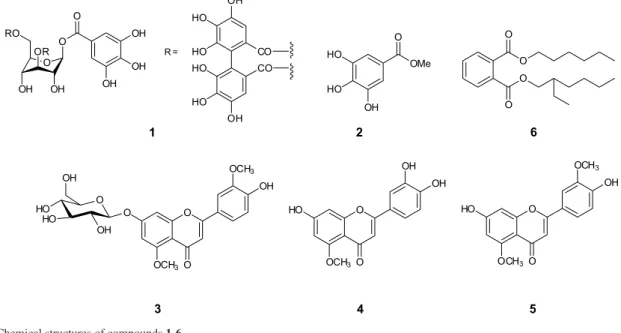
According to the ESI-MS data presented in Table 2, compound 1 was identified as an isomer of HHDP-galloyl-glucose [M − H]− at m/z 633.0748. The identification was
corroborated by the presence of the fragment m/z 481, confirming the loss of a galloyl moiety from this precursor ion, the fragment m/z 463 associated with the loss of a gallic acid unit and the fragment m/z 301 corresponding to the hexahydroxydiphenoyl (HHDP) unit after lactonization to ellagic acid.31
Data analysis
The software R version 3.2.1 (The R Foundation for Statistical Computing) was used to perform hierarchical clustering analysis (HCA) (details can be found in the Supplementary Information). The calculation of the degree of similarity between the elements of the Cartesian space was done based on the Euclidean distance equation:
(1)
where dii’ is the Euclidean distance between the pair of
individuals i and i’, Xi’j and Xij the numerical values of the jth
coordinates i’ and i, respectively. The calculation of distance was applied until all elements of a group were more similar to each other and dissimilar to the elements of different groups. Ward’s method32 allowed the rearrangement of the
formed clusters. The results were presented as a hierarchical
tree, a two-dimensional graph also known as a dendrogram, in which the lengths of the branches represent the degree of similarity between the objects.33
Results and Discussion
The HPLC-DAD chromatographic analyses of crude extracts from V. divergens seedlings inoculated with endophytic fungi isolated from V. divergens roots collected on dry and wet seasons in the Pantanal allowed the detection of at least thirteen chromatographic bands at 254 nm (Table 1); six of these bands were identified as the tannin HHDP-galloyl-glucose (1, retention time (RT) 12.67 min); methyl gallate (2, RT 17.63 min), the flavones 3’,5-dimethoxy-luteolin-7-O-β-glucoside (3, RT 19.210 min); 5-methoxy-luteolin (4, RT 22.14 min), 3’,5’-dimethoxy-luteolin (5, RT 23.85 min) and bis (2-ethylhexyl) phthalate (6, RT 36.69 min) by comparison of retention time, UV spectra with authentic standards obtained from in natura V. divergens by our research group in previous studies24 and are in accordance
with literature data (Figure 1, Table 2).
Based on the cluster analysis using Ward’s method, the seedlings inoculated with endophytic fungi from the dry period (D) and wet period (W) could be ranked according to their HPLC chemical profile in four and three groups, respectively (Figures 2 and 3).
Tannin 1 occurred in all the samples, including the control. In contrast, the methyl gallate 2 (C6-C1 phenolic compound) was observed only in V. divergens in nature. Comparing the flavones occurrence in seedlings inoculated with fungi collected from both seasons, the data revealed that flavone 3 had the same representation in both fungi groups (20%). However, flavone 4 occurred only in seedlings inoculated with fungi isolated on the wet period with 20% of occurrence. In contrast, flavone 5 had a predominance of 80% of the V. divergens seedlingsinoculated with endophytic fungi collected on the wet season compared with 30% of occurrence of strains from the dry period.
Regarding the HPLC analysis, the chromatographic band with RT 36.69 min was observed in all samples except for the 2D strain and was associated with the compound bis (2-ethylhexyl) phthalate (6). This phthalate have been isolated previously from tubers of
Humirianthera ampla;34 bis (2-ethylhexyl) phthalate was
also isolated from Nauclea officinalis leaves.30 Although
several phthalates were found in the Burkholderia cepacia
bacterium, including dibutyl and dioctyl phthalate,35 and
bis (2-ethylhexyl) phthalate was isolated from fungal strain No. 7088, associated with the plant Erica arborea;36 in this
Figure 1. Chemical structures of compounds 1-6.
Table 1. Chromatographic bands observed in HPLC analysis of in vitroV. divergens seedlings inoculated with endophytic fungi from the dry and wet periods compared with the control
Code
Endophytic fungi identification (GenBank access number,
Similarity)
Chromatographic bands RT / min
12.67 (1) 15.64 17.63 (2) 19.21 (3) 21.41 22.14 (4) 23.85 (5) 29.21 33.80 36.14 36.69 (6) 38.02 39.77
Dry period
2D Eutypella scoparia
(KY083415, 98%)
+ + + + +
12D Paraconiothyrium brasiliense
(KY083416, 12%)
+ + + +
14D Paraconiothyrium cyclothyrioides
(KY083417, 99%)
+ + + + + +
15D Fungal sp. ASR-179 (KY083418, 99%)
+ +
18D Leptosphaeriasenegalensis
(KY083419, 99%)
+ + +
21D Melanconiella elegans
(KJ173701, 96%)
+ +
35D Uncultured Pleosporales
(KY083420, 99%)
+ + + + +
57D Fungal sp. ARIZ L80 (KY083421, 92%)
+ + + + + + + + + +
66D Wuestneiamolokaiensis
(KY083422, 99%)
+ + + + +
Wet period 1W Neosartoryafischeri
(KY083410, 99%)
+ + + + +
51W Fungal sp. ARIZ L80 (KY083411, 99%)
+ + + + +
49W Eupenicillium sp. (KY083412, 98%)
+ + + + + + +
54W Gongronellabutleri
(KY083413, 99%)
+ + + + +
66W Chaetomium sp.
(KY083414, 99%)
+ + + + + +
Controla + + + + + + + +
Vdb + + + + + + + + + + + + +
Table 2. Identified compounds in V. divergens microplants infected with D and W strains
Compound Identification RT / min UV λmax / nm Molecular formula MW ESI-MS (m/z)
125 HHDP-galloyl-glucosea,b 12.67 194, 225, 270 C
27H22O18 634 657.0461 [M + Na]+
633.0748 [M − H]
-unknown 15.64 195, 228, 278
226 methyl gallateb,c 17.63 241, 264, 339 C
8H8O5 184
327 3’,5-dimethoxy luteolin-
7-O-β-glucoside a,b,c,d
19.21 245, 265, 339 C23H24O11 476 477.14115 [M + H]+
unknown 21.41 196, 252, 243
428 5-methoxy luteolin a,b,c,d 22.14 243, 284, 339 C
16H12O6 300 301.07404 [M + H]+
529 3’,5-dimethoxy luteolin a,b,c,d 23.85 244, 264, 338 C
17H14O6 314 315.08903 [M + H]+
unknown 29.20 197, 243, 279
unknown 33.80 198, 251
unknown 36.14
630 bis (2-ethylhexyl) phthalate a,b,c,d 36.69 267, 330 C
24H38O4 390
unknown 38.02 268, 330
unknown 39.77 257, 316
aIdentification by MS; bidentification by literature data; cidentification by NMR spectra; didentification by comparison with reference standards. HHDP:
hexahydroxydiphenoyl; RT: retention time; MW: molecular weight; ESI-MS: electrospray ionization mass spectrometry.
Figure 3. Cluster analysis using Ward’s method of endophytic fungal strains collected in (a) dry (D) and (b) wet (W) seasons from the Pantanal Biome. The codes are described in Table 1.
Figure 2. Cluster analysis using Ward’s method of endophytic fungal strains collected in the dry (D) and wet (W) seasons from the Pantanal Biome associated with in vitro V. divergens seedlings. The codes are described in Table 1.
product or a contaminant; therefore, additional studies are necessary to gain a better understanding of these results.
We observed that the effect of inoculation with endophytic fungi in in vitro microplants varied according to the season and the strain used. For example, the production of flavones 3 and 4 seems to have been favored by inoculation with strains acquired in the wet period.
Studies regarding the interactions of V. divergens and their endophytic microbiota are rare; however, Savi et al.38
describe the isolation of an endophytic actinomycete strain from V. divergens.
The influence of V. divergens leaves extracts on the germination and growth of lettuce and tomato were investigated by Oliveira et al.39 and they suggest a
possible allelopathic effect of this species associated with the presence of phenolic compounds, which include flavonoids. Several authors reported that the presence and increase of flavonoids in plants may be associated with several abiotic factors including UVB, temperature and drought among others, such as environmental stress.40,41
In line with this, Guidi et al.42 studied the interactions
of water stress and solar irradiance on the physiology and biochemistry with special emphasis on flavonoid production of Ligustrum vulgare. They observed thatthe content of quercetin and luteolin derivatives increased in response to full sunlight irrespective of the water treatment; however, the phenylpropanoid concentrations increased in response to water stress only in shaded leaves.
Ahuja et al.43 mention that environmental stress factors
such as drought, elevated temperature, salinity and rising CO2 or multiple environmental stress in combination affect
plant growth, and they reprogram the plant to survive in a changing climate. Responses to perturbations are usually accompanied by major changes in the plant transcriptome, proteome and metabolome.
In this work, the re-isolation of the endophytic fungi from the in vitro seedlings was not carried out to effectively confirm the endophyte-host interaction.
Comparing the flavone structures reveals that all of them have a methoxy group at C-5 position, and 3 and 5 have one additional methoxy group at C-3’ carbon. In addition, 5-methoxy flavones and flavones with no substitution in this position are shown active as growth inhibitors of navel orange worm (NOW), a citrus pest that also attacks other cultures, especially walnuts and almonds. These flavones appear to be associated with the resistance of citrus to this pathogen, whereas flavones with hydroxyl-substitution at the 5-position are inactive. Since a 5-hydroxy substituent is strongly hydrogen bonded to the 4-carbonyl oxygen, the growth inhibition is correlated with the availability of this carbonyl.44
In contrast, polymethoxy flavones are related to the defense mechanisms of the plant itself, and they have an antiviral and antimicrobial capacity that confers resistance against microbial infections in citrus.45,46
The 5-methoxy flavonoids 5-methoxy-luteolin and 3’,5-dimethoxy luteolin have been reported as active nod gene inducers in Rhizobium meliloti together with the
flavonoids luteolin, luteolin-7-O-glucoside and 3 methoxy-luteolin (chrysoeriol). This gene is responsible for the nitrogen fixation in alfalfa.47
Conclusions
Therefore, the presence of 5-methoxy flavones in
V. divergens leaves suggests the possible correlation of this class of substances with the V. divergens resistance to flooding, pathogens attack and allelopathic action. This approach is new to V. divergens, with no scientific data on in vitro fungi elicitation; however, further studies are necessary to gain a better understanding of the ecological relationships of this species.
Supplementary Information
Supplementary data, including 1H NMR, 13C NMR,
mass spectra and HPLC chromatogram, are available free of charge at http://jbcs.sbq.org.br as PDF file.
Acknowledgments
The authors are grateful to Jessica Potomatti Batista for her technical support and to José Carlos Tomaz for his help with the EM-AR analysis (Faculdade de Ciências Farmacêuticas de Ribeirão Preto, Universidade de São Paulo).
The authors are grateful to the Mato Grosso Research Foundation (FAPEMAT; grant number 331950/2012); the São Paulo Research Foundation (FAPESP; grant number 2011/00631-5); INAU; Coordenadoria de Aperfeiçoamento de Pessoal do Ensino Superior (CAPES); and Conselho Nacional de Desenvolvimento Científico e Tecnológico (CNPq) for their fellowships.
References
1. Junk, W. J.; Brown, M.; Campbell, I. C.; Finlayson, M.; Gopal, B.; Ramberg, L.; Warner, B. G.; Aquat. Sci. 2006, 68, 400. 2. Junk, W. J.; Cunha, C. N.; Wantzen, K. M.; Petermann, P.;
Strussmann, C.; Marques, M. I.; Adis, J.; Aquat. Sci.2006, 68, 278.
3. Alho, C. J. R.; Braz. J. Biol. 2008, 68,957.
4. da Cunha, C. N.; Junk, W. J.; Appl. Veg. Sci.2004, 7, 103. 5. Dalmolin, A. C.; Dalmagro, H. J.; Lobo, F. A.; Antunes Jr., M.
Z.; Ortíz, C. E. R.; Vourlitis, G. L.; Photosynthetica2013, 51, 379.
6. Vourlitis, G. L.; Nogueira, J. S.; Lobo, F. A.; Sendall, K. M.; Paulo, S. R.; Dias, C. A. A.; Pinto Jr., O. B.; Andrade, N. L. R.;
7. Vourlitis, G. L.; Lobo, F. A.; Lawrence, S.; Holt, K.; Zappia, A.; Pinto Jr, O. B.; Nogueira, J. S.; Plant Ecol.2014, 215, 963. 8. Dalmolin, A. C.; Lobo, F. A.; Vourlitis, G.; Silva, P. R.;
Dalmagro, H. J.; Antunes Jr., M. Z.; Ortíz, C. E. R.; Plant Ecol.
2015, 216, 407.
9. Machado, N. G.; Sanches, L.; Silva, L. B.; Novais, J. W. Z.; Aquino, A. M.; Biudes, M. S.; Pinto-Junior, O. B.; Nogueira, J. S.; Appl. Ecol. Environ. Res.2015, 13, 289.
10. Pott, A.; Pott, V. J.; Plantas do Pantanal, 1a ed.; Embrapa: Brasilia, DF, Brazil, 1994.
11. Andonian, K.; Hierro, J. L.; Biol. Invasions2011, 13, 2957. 12. Aschehoug, E. T.; Callaway, R. M.; Newcombe, G.; Tharayil,
N.; Chen, S.; Oecologia2014, 175, 285.
13. Bacon, C. W.; White, J. F.; Microbial Endophytes; Marcel Dekker Inc.: New York, 2000.
14. Perez-Garcia, O.; Escalante, F. M.; de-Bashan, L. E.; Bashan, Y.; Water Res. 2011, 45, 11.
15. Zhang, Y. F.; He, L. Y.; Chen, Z. J.; Wang, Q. Y.; Qian, M.; Sheng, X. F.; Chemosphere2011, 83, 57.
16. Lima, J. V. L.; Weber, O. B.; Correia, D.; Soares, M. A.; Senabio, J. A.; Plant Soil2015, 389, 25.
17. Li, H. Y.; Li, D. W.; He, C. M.; Zhou, Z. P.; Mei, T.; Xu, H. M.;
Fungal Ecol. 2012, 5, 309.
18. Berg, G.; Müller, H.; Zachow, C.; Opelt, K.; Scherwinski, K.; Tilcher, R.; Ullrich, A.; Hallmann, J.; Grosch, R.; Sessitsch, A.;
Simbiogenetics2008, 6, 17.
19. Qiao, J. Q.; Wu, H. J.; Huo, R.; Gao, X. W.; Borriss, R.; Chem. Biol. Technol. Agric.2014, 1, 12.
20. Vitorino, L. C.; Silva, F. G.; Lima, W. C.; Soares, M. A.; Pedroso, R. C. N.; Silva, M. R.; Dias Junior, H.; Crotti, A. E. M.; Silva, M. L. A.; Cunha, W. R.; Pauletti, P. M.; Januario, A. H.; Quim. Nova2013, 36, 1014.
21. Soares, M. A.; Li, H.-Y.; Kowalski, K. P.; Bergen, M.; Torres, M. S.; White, J. F.; Biol. Invasions2016, 6, 1.
22. de Siqueira, K. A.; Brissow, E. R.; Santos, J. L.; White, J. F.; Santos, F. R.; de Almeida, E. G.; Soares, M. A.; Symbiosis2016,
71, 211.
23. White, T. J.; Bruns, T.; Lee, S. J. W. T.; Taylor, J. W. In PCR Protocols: a Guide to Methods and Applications; Innis, M.;
Gelfand, D.; Sninsky, J.; White, T., eds.; Academic Press: Orlando, Florida, 1990, p. 315.
24. Pimenta, L. P.; Kellner Filho, L. C.; Liotti, R. G.; Soares, M. A.; Aguiar, D. P.; Magalhães, L. G.; Oliveira, P. F.; Tavares, D. C.; Andrade e Silva, M. L.; Cunha, W. R.; Pauletti, P. M.; Januario, A. H.; Adv. Pharmacoepidemiol. Drug Saf.2015, 4, 182. 25. Santos, S. A.; Freire, C. S.; Domingues, M. R.; Silvestre, A. J.;
Pascoal Neto, C.; J. Agric. Food Chem.2011, 59, 9386. 26. Hayat, S.; Atta-ur-Rahman; Choudhary, M. I.; Khan, K. M.;
Abbaskhan, A.; Chem. Pharm. Bull. 2002, 50, 1297.
27. Osawa, T.; Sakuta, H.; Negishi, O.; Kajiura, I.; Biosci., Biotechnol., Biochem.1995, 59, 2244.
28. Ueli, A. H.; Carl, A. M.; Cecillia, M. J.; Donald, A. P.; Plant Physiol.1990, 92, 116.
29. Monache, G. D.; de Rosa, M. C.; Scurria, R.; Monacelli, B.; Pasqua, G.; Dall’Olio, G.; Botta, B.; Phytochemistry1991, 30, 1849.
30. Su, K.; Gong, M.; Zhou, J.; Deng, S.; Int. J. Chem. 2009, 1, 77. 31. Boulekbache-Makhlouf, L.; Meudec, E.; Chibane, M.;
Mazauric, J. P.; Slimani, S.; Henry, M.; Cheynier, V.; Madani, K.; J. Agric. Food Chem.2010, 58, 12615.
32. Mingoti, S. A.; Análise de Dados através de Métodos de
Estatística Multivariada: Uma Abordagem Aplicada, 1a ed.;
Universidade Federal de Minas Gerais: Belo Horizonte, MG, Brazil, 2013.
33. Ferreira, M. M. C.; Quimiometria: Conceitos, Métodos e Aplicações, 1a ed.; Unicamp: Campinas, São Paulo, Brazil,
2015.
34. Graebner, I. B.; Morel, A. F.; Burrow, R. A.; Mostardeiro, M. A.; Ethur, E. M.; Dessoy, E. C. M.; Scher, A.; Rev. Bras. Farmacogn.
2002, 12, 80.
35. Sultan, M. Z.; Moon, S.-S.; Park, K.; J. Sci. Res.2010, 2, 191. 36. Hussain, H.; Krohn, K.; Ullah, Z.; Draeger, S.; Schulz, B.;
Biochem. Syst. Ecol. 2007, 35, 898.
37. Paiva, N. L.; J. Plant Growth Regul.2000, 19, 131.
38. Savi, D. C.; Shaaban, K. A.; Vargas, N.; Ponomareva, L. V.; Possiede, Y. M.; Thorson, J. S.; Glienke, C.; Rohr, J.; Curr. Microbiol.2015, 70, 345.
39. Oliveira, A. K. M.; Ribeiro, J. W. F.; Fontoura, F. M.; Matias, R.; Allelopathy J.2013, 31, 129.
40. Chalker-Scott, L.; Photochem. Photobiol.1999, 70, 1. 41. Yaginuma, S.; Shiraishi, T.; Ohya, H.; Igarashi, K.; Biosci.,
Biotechnol., Biochem.2002, 66, 65.
42. Guidi, L.; Degl’Innocenti, E.; Remorini, D.; Massai, R.; Tattini, M.; Tree Physiol. 2008, 28, 873.
43. Ahuja, I.; de Vos, R. C.; Bones, A. M.; Hall, R. D.; Trends Plant Sci. 2010, 15, 664.
44. Mahoney, N. E.; Roitman, J. N.; Chan, B. C.; J. Chem. Ecol.
1989, 15, 285.
45. Ortuno, A. M.; Arcas, M. C.; Benavente-Garcia, O.; Del Rio, J. A.; Food Chem.1999, 66, 217.
46. Li, S.; Pan, M. H.; Lo, C. Y.; Tan, D.; Wang, Y.; Shahidi, F.; Ho, C. T.; J. Funct. Foods2009, 1, 2.
47. Hartwig, U. A.; Maxwell, C. A.; Joseph, C. M.; Phillips, D. A.;
Plant Physiol.1990,92, 116.
Submitted: January 18, 2017