Article
Printed in Brazil - ©2017 Sociedade Brasileira de Química0103 - 5053 $6.00+0.00*e-mail: [email protected]
Rare Earth-Indomethacinate Complexes with Heterocyclic Ligands:
Synthesis and Photoluminescence Properties
João Batista M. Resende Filho,a Paulo R. Santos,a Juliana A. Vale,a
Wagner M. Faustino,a Danyelle S. Farias,b Hermi F. Brito,c Maria C. F. C. Felintod and Ercules E. S. Teotonio*,a
aDepartamento de Química, Universidade Federal da Paraíba (UFPB),
58051-970 João Pessoa-PB, Brazil
bInstituto Federal de Educação, Ciência e Tecnologia da Paraíba (IFPB),
Av. Primeiro de Maio, 720, 58015-435 João Pessoa-PB, Brazil
cDepartamento de Química Fundamental, Instituto de Química, Universidade de São Paulo (USP),
Av. Prof. Lineu Prestes, 748, 05508-900 São Paulo-SP, Brazil
dInstituto de Pesquisas Energéticas e Nucleares (IPEN-CQMA), Av. Prof. Lineu Prestes, 2242,
05508-000 São Paulo-SP, Brazil
In this work, synthesis, characterization and photophysical properties of trivalent rare earth complexes with a nonsteroidal anti-inflammatory drug [the indomethacinate (indo), presenting formulas RE(indo)3(H2O)x (x = 3, for Eu3+ and Gd3+, and x = 4 for Tb3+), RE(indo)3(bipy) and
RE(indo)3(phen) (bipy: 2,2’-bipyridine, and phen: 1,10-phenanthroline)] were investigated. Based
on photoluminescent results, the intramolecular energy transfer process from T1 triplet states of
indo, phen and bipy ligands to the 5D
0 emitting level of the Eu3+ ion in the coordination compounds
is discussed. Accordingly, it is proposed two possible intramolecular energy transfer mechanisms between indomethacinate ligand and rare earth ions, which involve the participation of excited electronic states of the heterocyclic ligands as intermediate ones.
Keywords: indomethacin, rare earth, heterocyclics, photoluminescence, energy transfer
Introduction
In recent decades, metallotherapeutic drugs based on transition metal and trivalent rare earth ions have attracted considerable attention owing to their potential
applications in medicine as anti-inflammatory,1
antitumor,2 antiproliferative,3 antifungal,4,5 antiviral6,7 and
antimicrobial.8 Many of these compounds present enhanced
biomedical activity, reduced toxicity1 and diminished
collateral effects as compared with non-coordinated parent drugs. Among various classes of metallotherapeutic drugs, those based on the nonsteroidal anti-inflammatory drugs (NSAIDs) from the carboxylate or oxicam families have received particular attention because of their great ability to coordinate to different metal ions, resulting thermodynamically and kinetically stable complexes.
The molecular structures of NSAIDs are characterized by the presence of chromophore groups and low-lying
triplet state with energy in the range of 20,000-25,000 cm-1,
which may act as luminescent sensitizers for trivalent rare
earth ions.9 Additionally, molecules of NSAIDs may act
as multidentate ligands for RE3+ ion through their hard
N– and O– sites. It is well-known that RE3+ ions play a
wide range of roles in fluoroimmunoassay methods,10
therapy11 and as luminescent probe for biomolecules12 due
to their unique spectroscopic properties such as narrow emission bands, high luminescence intensities and the long
lifetimes of the 2S+1L
J emitting levels. Furthermore, the rare
earth ions may be indirectly excited via coordinated ligand (antenna effect) in a spectral region far from those where emission is generally monitored.
Recently, Zhou et al.13 have investigated the structural and
sensitizer for the Tb3+ ion, but not for the Eu3+ ion.
Luminescence properties of the Tb3+ complexes with
ibuprofen and orthofen containing additional organic bases
were also studied by Teslyuk et al.9 in order to propose
an analytical method to determine NSAID drugs. The
chemiluminescent method, based on Eu3+ emission for the
determination of naproxen in pharmaceutical and urine, has
been developed by Kaczmarek.14
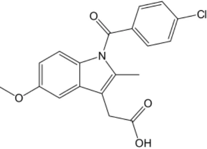
A m o n g N S A I D d r u g s , i n d o m e t h a c i n
(1-(4-chlorobenzoyl)-5-methoxy-2-methyl-1H
-indole-3-acetic acid (Hindo)) (Figure 1) is considered one of the
most clinically investigated.1 Its coordination complexes
with transition metals have been extensively investigated
in terms of structural and spectroscopic properties.15
Generally, these complexes present other donor ligands because indomethacinate (indo) cannot saturate the coordination sphere of the metal ions. In the case of the rare earth complexes, neutral ligands such as heteroaromatic and phosphine oxide ligands have been not only used for this purpose, but have been shown that they may also play an essential role in the luminescence sensitization that involves either a direct intramolecular energy transfer to the metal ion or an indirect mechanism via energy transfer to other ligands. The main evidences for this latter mechanism were
reported for Eu3+-tris(diketonate) complexes,16-18 however,
the interligand energy transfer in rare earth complexes is still not clear.
In particular, excited triplet states of some heteroaromatic ligands are almost resonant with that for indomethacinate (indo) ligand, consequently, these states may contribute to an operative interligand energy transfer in which the heteroaromatic ligands act as energy acceptor from ionic ligands and, subsequently, transfer their energy to the trivalent rare earth ion increasing the luminescence intensity. Thus, the investigation of this intramolecular energy process may contribute to design of new highly luminescent complexes based on carboxylate ligands for different practical applications. Besides, up to now, to the best of our knowledge, no investigation has been carried
out on the quantitative photophysical properties of the
RE3+ coordination compounds in the solid state containing
indomethacin.
Herein, it is reported the synthesis, characterization, intramolecular ligand-to-metal energy transfer and
photophysical properties study of the RE3+ complexes
(where RE3+: Eu3+, Gd3+ or Tb3+) with indomethacinate
(indo) and ancillary heteroaromatic ligands (2,2’-bipyridine (bipy), and 1,10-phenanthroline (phen)), in the solid
state. Photoluminescent properties of the Eu3+ complexes
have been thoroughly investigated taking into account
the radiative (Arad) and non-radiative (Anrad) emission
coefficients, experimental intensity parameters (Ω2 and
Ω4) and emission quantum efficiency (η). Additionally,
the synthesized complexes may be used to perform new methods for luminescence determination of NSAID drugs
and potential application as target medicine.9
Experimental
Materials
The indomethacin ligand was extracted from the
INDOCID® drug using ethyl acetate as solvent and
purified in a flash column chromatography on silica
gel (Tedia, SiliaFlash® F60 40-63 µm, 230-400 mesh),
using a mixture of hexane and ethyl acetate (1:1) as eluent. The commercially available 2,2’-bipyridine (bipy) 99% and 1,10-phenanthroline (phen) monohydrated 99% heteroaromatic ligands were purchased from Sigma-Aldrich and were used without previous purification.
The rare earth chlorides (RECl3·6H2O) were prepared by
the reaction between their corresponding rare earth oxides,
RE2O3 (99.999%) (RE: Eu3+ and Gd3+) and Tb4O7 (99.999%)
from Sigma-Aldrich, and concentrated hydrochloric acid
(37%, Merck) as reported in the literature.19,20
Instrumentation
C, H and N analyses were performed on a microanalytical analyzer model 2400 (PerkinElmer). Infrared absorption spectra were recorded in a Shimadzu FTIR spectrophotometer model IRPrestige-21 by using the technique of KBr pellets in
the interval of 400-4000 cm-1. Thermogravimetric analyses
(TGA) of the RE3+-indomethacinate complexes from room
temperature to 900 oC were conducted with a thermobalance
model DTG-60H Shimadzu by using platinum crucibles with approximately 2 mg of complexes. A dynamic synthetic air
atmosphere (50 mL min-1) and a heating rate of 10 °C min-1
were used as instrumental conditions. Steady-state luminescent spectra were acquired by using a Horiba
JobinYvon Fluorolog-3 spectrofluorimeter presenting double grating 0.22 m monochromator (SPEX 1680) and a 450 W Xenon lamp as excitation source. Luminescence decay data were also collected by using a phosphorimeter SPEX 1934D accessory coupled to the Fluorolog-3 spectrofluorimeter.
Synthetic methodology
Synthesis of the RE(indo)3(H2O)x complexes
A NaOH aqueous solution (1 mol L-1) was added slowly
(dropwise) to a suspension of indomethacin ligand (400.0 mg, 1.12 mmol) in 50 mL of water until the apparent pH value of
ca. 7, followed by the dropwise addition of a RECl3·6H2O
aqueous solution (0.37 mmol: 135.6 mg (EuCl3·6H2O),
137.4 mg (GdCl3·6H2O) and 138.2 mg (TbCl3·6H2O)),
yielding immediately a yellow solid precipitate. The powder samples were filtered, washed thoroughly with cooled water
and dried under reduced pressure (358.9 mg, 76% for Eu3+;
312.8 mg, 66% for Gd3+; and 308.6 mg, 65% for Tb3+); CHN
anal. calcd. for C57H51Cl3EuN3O15 (1): C, 53.64; H, 4.03;
N, 3.29; found: C, 53.46; H, 4.17; N, 3.17; FTIR (KBr)
ν / cm-1 3390m, 3088w, 2992w, 2926m, 2849w, 2833w,
1682s, 1591s, 1545s, 1477s, 1416s, 1402s, 1323s, 1229s, 1090m, 1014w, 924w, 835w, 754w; CHN anal. calcd. for
C57H51Cl3GdN3O15 (2): C, 53.42; H, 4.01; N, 3.28; found:
C, 53.49; H, 4.15; N, 3.35; FTIR (KBr) ν / cm-1 3418m,
3088w, 3069w, 2994w, 2955w, 2928m, 2833w, 1682s, 1591s, 1553s, 1477s, 1435s, 1400s, 1369s, 1323s, 1230s, 1148m, 1090m, 1070m, 1036m, 1014w, 926w, 835w, 754w; CHN
anal. calcd. for C57H53Cl3TbN3O16 (3): C, 52.61; H, 4.11;
N, 3.23; found: C, 52.26; H, 4.08; N, 3.03; FTIR (KBr)
ν / cm-1 3424m, 3088w, 3067w, 2994w, 2928m, 2849m,
2833w, 1682s, 1591s, 1553s, 1477s, 1437s, 1400s, 1321s, 1229s, 1148m, 1090m, 924m, 835m, 754m.
Synthesis of the RE(indo)3(bipy) complexes
To a solution of bipy (37.4 mg, 0.24 mmol) in ethyl acetate (10 mL) was added dropwise a solution
of RE(indo)3(H2O)x complexes (0.24 mmol: 300.0 mg
for Eu3+, 307.5 mg for Gd3+, and 308.0 mg for Tb3+
complex) in ethyl acetate (40 mL). A precipitate (yellow powder) was obtained after the solvent evaporation. The product was thoroughly washed with cooled ethyl acetate (ca. 10 mL), filtered and dried under reduced pressure resulting in yellow solid (281.5 mg, 84% for
Eu3+; 289.2 mg, 86% for Gd3+; and 276.2 mg, 82%
for Tb3+). Although the complexes are very soluble in
several solvents (dichloromethane, chloroform, ethyl acetate, acetone, toluene, etc.), and all attempts to obtain single crystals were unsuccessful; CHN anal. calcd. for
C67H53Cl3EuN5O12 (4): C, 58.38; H, 3.88; N, 5.08; found
(%): C, 57.76; H, 3.84; N, 5.07; FTIR (KBr) ν / cm-1
3428w, 3069w, 3042w, 2992w, 2926w, 2832w, 1682s, 1601s, 1566s, 1477s, 1458s, 1437s, 1398s, 1321s, 1227s, 1148m, 1088m, 1036m, 924w, 835m, 756m; CHN anal.
calcd. for C67H53Cl3GdN5O12 (5): C, 58.15; H, 3.86; N,
5.06; found: C, 57.26; H, 4.24; N, 4.93; FTIR (KBr)
ν / cm-1 3428w, 3071w, 2990w, 2928w, 2849w, 2832w,
1682s, 1601s, 1566m, 1477s, 1458m, 1437s, 1321s, 1227s, 1148m, 1088m, 1036m, 1014m, 924w, 835m,
756m; CHN anal. calcd. for C67H53Cl3TbN5O12 (6): C,
58.08; H, 3.86; N, 5.05; found: C, 57.26; H, 4.36; N,
4.96; FTIR (KBr) ν / cm-1 3428w, 3084w, 2990w, 2926w,
2849w, 2833w, 1682s, 1603s, 1564s, 1477s, 1437s, 1317s, 1227s, 1148m, 1090m, 1036m, 926m, 835m, 756m.
Synthesis of the RE(indo)3(phen) complexes
To a solution of phen monohydrated (47.5 mg, 0.24 mmol) in ethyl acetate (10 mL) was added dropwise
a solution of RE(indo)3(H2O)x complexes (0.24 mmol:
300.0 mg (Eu3+), 307.5 mg (Gd3+) and 308.0 mg (Tb3+)) in
ethyl acetate (40 mL). A precipitate (yellow powder) was obtained after the solvent evaporation. The product was thoroughly washed with cooled ethyl acetate (ca. 10 mL), filtered and dried under reduced pressure resulting in yellow
solid (279.5 mg, 82% for Eu3+; 280.5 mg, 82% for Gd3+; and
267.2 mg, 78% for Tb3+). Although the complexes are very
soluble in several solvents (dichloromethane, chloroform, ethyl acetate, acetone, toluene, etc.), and all attempts to obtain single crystals were unsuccessful; CHN anal. calcd.
for C69H53Cl3EuN5O12 (7): C, 59.09; H, 3.81; N, 4.99; found:
C, 58.73; H, 3.98; N, 5.55; FTIR (KBr) ν / cm-1 3437w,
3076w, 2992w, 2926w, 2832w, 1680s, 1607s, 1553m, 1477s, 1427s, 1356s, 1325s, 1227s, 1148m, 1088m, 1068m, 920m,
833m, 754m; CHN anal. calcd. for C69H53Cl3GdN5O12 (8):
C, 58.87; H, 3.79; N, 4.97; found: C, 57.63; H, 4.24; N,
4.86; FTIR (KBr) ν / cm-1 3447m, 3078w, 2992w, 2953w,
2953w, 2832w, 1678s, 1607s, 1589s, 1556m, 1477s, 1427s, 1398s, 1356s, 1229s, 1144m, 1090m, 1069m, 922w, 837m,
754m. CHN anal. calcd. for C69H53Cl3TbN5O12 (9): C, 58.80;
H, 3.79; N, 4.97; found: C, 57.36; H, 3.91; N, 4.85; FTIR
(KBr) ν / cm-1 3597w, 3428w, 3078w, 2992w, 2953w, 2928w,
2832w, 1678s, 1609s, 1589s, 1557m, 1477s, 1427s, 1356s, 1325s, 1229s, 1148m, 1090m, 922m, 835m, 754m.
Results and Discussion
Synthesis and characterization
complexes (1 and 2), 1:3:4 for hydrated complex (3) and
1:3:1 for rare earth complexes (4-9) which contain bipy
and phen as ancillary ligands.
FTIR spectrum for indomethacin ligand extracted from
the INDOCID® drug is in agreement (Figure S1 in the
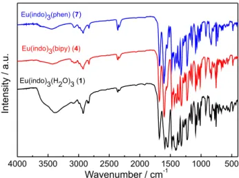
Supplementary Information (SI) section). Figure 2 shows the
FTIR spectra for the Eu3+-indomethacinate complexes, while
FTIR spectra for Gd3+ and Tb3+ are presented in Figures S2
and S3 in the SI section. In general, the absorption spectra for
the RE3+ complexes (1-3) exhibit a broad band in the range
of 2600-3400 cm-1 that is assigned to the O–H stretching of
the water molecules. As can be seen, this band is not present in the vibrational spectra for the complexes containing the heteroaromatic ancillary ligands, indicating that water molecules have been substituted by neutral ligands. The characteristic absorption bands assigned to the carboxylic
group (COOH) of the Hindo free ligand, e.g., ν(O–H) at
3300 cm-1 and ν(C=O) at 1734 cm-1, have been substituted
by the intense absorption bands assigned to νas(COO) (at
1613 cm-1) and ν
s(COO) (at 1416 cm-1) of the carboxylate
group of the RE3+ complexes ( 2). Furthermore, the values for
∆[∆as(COO) – ∆s(COO)] are falling in the range of 197 cm-1
for all the complexes (1-9), indicating that indo ligand is
coordinated to the RE3+ as chelating or bridging mode.21-23
These results strongly suggest that those complexes are either binuclear or coordination polymers. Moreover, the
absorption bands around 1468 and 1423 cm-1 indicate that
bipy and phen heteroaromatic ligands are coordinated to the
metal ions through their nitrogen atoms.16,24
The presence of water molecules in the complexes (1-3)
was also characterized by thermogravimetric analysis. TGA curves (Figure 3) present a weight loss around 3.5%
in the temperature interval between 60-150 oC that is
attributed to the release of three water molecules for the
complex (1) (similar to complex (2), see Figure S4 in the
SI file). This result is similar to the thermal decomposition
for the Ce(indo)3(H2O)3 complex reported in reference.15
Figure 3 also displays the TGA curves for the Eu3+
complexes (4 and 7) containing bipy and phen ligands.
TGA curves for other coordination compounds are shown in Figures S4 and S5 in the SI section. For the
complex (3), TGA curve shows a weight loss around 5%
in the same temperature interval that is attributed to the release of four water molecules which corroborate with the CHN elemental analysis data. The resulting anhydrous compounds undergo thermal decomposition in successive
steps that occur from 230 to 670 oC. In contrast to the
hydrated complexes, TGA curves for the complexes (4) to
(9) do not exhibit any weight loss until 150 oC, confirming
the anhydrous form of these coordination compounds.
The coordination compounds (3, 6 and 9) present
successive steps that take place in the range of 300-750 oC,
starting with a weight loss that corresponds to the release of the phen ligand. The TGA curves show that complexes
with bipy ligand (2, 5 and 8) are less stable than those ones
containing phen ligand (3, 6 and 9). The first weight loss of
these complexes (around 170 oC) may be assigned to the
release of the bipy ligand. However, no well-defined level is observed in the TGA curves owing to the subsequent
decomposition of the RE(indo)3 products. The final products
remain stable until 900 oC for the thermal decomposition of
all complexes, which can be assigned to the corresponding
rare earth oxide, RE2O3 (RE: Eu and Gd) and Tb4O7.
Phosphorescent behavior
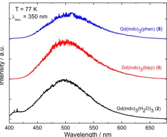
The emission spectra of the Gd3+ complexes (2, 5
and 8) were recorded with excitation on the S0 → S1
Figure 2. FTIR absorption spectra of the Eu3+ complexes (1, 4 and 7)
recorded in the range of 4000-400 cm-1 in KBr pellets.
Figure 3. TGA curves of the complexes (1, 4 and 7) recorded in the interval of 25-900 oC, using dynamic synthetic air atmosphere (50 mL min-1) and
indomethacinate centered transition at 350 nm (Figure 4), showing two overlapped broad bands with maxima around 420 (blue region) and 500 nm (green region) assigned to
the fluorescence (S1→ S0) and phosphorescence (T → S1)
intraligand transitions, respectively.
Since the highest intensity band is that due to the
phosphorescence, the Gd3+-indomethacinate complexes
present green emission color under UV radiation excitation. Interestingly, no obvious difference between the emission
spectra profiles of the Gd3+ complexes (5 and 8) and that
for the precursor complex, 2, is observed, indicating that
photoluminescence properties of all the Gd3+ complexes
are mainly due to the indomethacinate ligand coordinated
to the RE3+ ion. For these coordination compounds, the
indomethacinate triplet state (T1) was determined taking
into account the 0-0 phonon transition from the time-resolved phosphorescence spectra recorded with time delay of 0.04 ms (Figure S6 in the SI section).
The absence of the short-lived fluorescence emission band around 420 nm in those spectra (Figure 4) allowed for a more
precise determination of the T1 state energies, as following:
22,124, 21,882 and 21,552 cm-1 for the Gd(indo)
3(H2O)3 (2),
Gd(indo)3(bipy) (5) and Gd(indo)3(phen) (8) complexes,
respectively. These optical data indicate that triplet states of indo ligand should be in a good resonance condition of the
emitting levels of the Eu3+ ion, acting as an efficient antenna
for europium complexes.16
Luminescence spectra of the Eu3+
As to the emission monitored on the intraconfigurational
5D
0→7F2 hypersensitive transition centered on the Eu3+
ion (at approximately 612 nm), all excitation spectra for
solid-state of the complexes (1, 4 and 7) exhibit similar
spectral profiles. These spectra (Figure 5) present a strong broad band in the range of 250-450 nm assigned
to the S0→ S1 transition belong to indo ligand, indicating
that even in the complexes with bipy and phen, the
luminescence sensitization mechanism of the Eu3+ ion
started with radiation absorption by indomethacinate ligand. However, minor contributions due to the absorption from heterocyclic ligands are observed. It is worth noting that this band exhibits higher intensity than those ones from
the intraconfigurational 7F
0→2S+1LJ transitions at around
468 and 525 nm, confirming the potential ability of the
ligand to transfer energy to the Eu3+ ion.
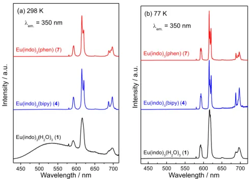
The emission spectra of all the RE3+ coordination
compounds were recorded at 298 and 77 K, upon
excitation on the ligand S0 → S1 transition (at 350 nm)
of the indomethacinate ligand (Figure 6). Under this
condition, all the Eu3+-indomethacinate coordination
compounds (1, 4 and 7) display high red luminescence at
77 K. A qualitative analysis shows that the Eu3+ complexes
with ancillary heteroaromatic ligands (4 and 7) exhibit
significantly higher luminescent intensities than the
hydrated complex (1).
The emission spectra of the complex (1) in the range
of 430-720 nm recorded at 298 and 77 K exhibit different spectral profiles (Figure 6). When recorded at 298 K, a broad band arising from intraligand transition is observed, that is only partially quenched in the emission spectrum recorded at 77 K. This band overlaps the standard set of well-resolved narrow emission bands owing to the
Eu3+ centered transitions: 5D
0→7F0 (579 nm), 5D0→7F1
(589 nm), 5D
0→7F2 (613 nm), 5D0 →7F3 (635 nm) and
5D
0 → 7F4 (717 nm).24 On the other hand, one of the
Figure 5. Excitation spectra of the Eu3+ complexes (1, 4 and 7), in solid
state, recorded at 77 K with emission monitored at 613 nm.
Figure 4. Phosphorescence spectra of the Gd3+ complexes (2, 5 and 8),
main features to be outlined from the photoluminescent
spectra for complexes 4 and 7, both recorded at 298 and
77 K, is the absence of the broad emission bands due to the indomethacinate fluorescence or phosphorescence. All of these photoluminescence spectra display similar
spectral profile, in which only the intraconfigurational-4f6
transitions are observed, being dominated by the
well-characterized hypersensitive 5D
0 →7F2 transition
allowed by forced electric dipole mechanism. It is also important to note the lower relative intensity ratio between the 5D
0→7F2 and 5D0→7F1 transitions in comparison with
those for low symmetry Eu3+tris-β-diketonate coordination
compounds containing the bipy and phen, or their derivative
ligands.25,26 These results altogether suggest that a chemical
environment around the rare earth ion is close to Cn or Cnv
symmetry.
Although indomethacinate ligand dominates the antenna effect, emission data give evidences that excited states centered on the heterocyclic ligands (bipy and phen) can also play an important role as intermediate states in the intramolecular energy transfer from indomethacinate ligand
to Eu3+ ion. A possible explanation for this behavior lies
in the fact of heterocyclic ligands present their low-lying
excited triplet states close in energy to the T1 triplet excited
state of the indomethacinate ligand. It is also close in energy to 5D
0 (ca. 17293 cm-1) and 5D1 (ca. 19027 cm-1) emitting
levels of the Eu3+ ion. According to the literature, the
intramolecular energy transfer in the Eu3+-indomethacinate
complexes can be divided into two mechanisms, which is
shown in Figure 7.27,28
For the first mechanism, indomethacinate ligand absorbs
energy being excited to its S1 states, and subsequently
transfers this energy to the T1 excited state belonging to
heterocyclic ligand (Figure 7). Therefore, this latter ligand undergoes energy transfer from the intraligand triplet state to 5D
1 or 5D0 levels of the Eu3+ ion (T1→5D1→5D0),
followed by radiative and non-radiative deactivation
centered on the central metal ion (5D
0→5FJ). By means
of the second mechanism, indomethacinate ligand absorbs
energy being excited to its singlet state S1, which undergoes
intersystem crossing energy to the own T1 state and transfers
to the triplet excited level (T1) of the heterocyclic ligand,
followed by sensitization of the excited levels of the Eu3+
(Figure 7). In order to give an insight into the proposed
Figure 6. Emission spectra of the Eu3+ complexes (1, 4 and 7), in solid state, measured at (a) 298 K and (b) 77 K, under excitation on the S
1 state of the
indomethacinate ligand at 350 nm.
Figure 7. Partial energy level diagrams of the intramolecular energy transfer mechanisms for Eu3+-indomethacinate complexes containing
heterocyclic ligands. S1 and T1 represent excited singlet and triplet states,
mechanisms above, it is listed the energies corresponding
to the S1 and T1 excited states for all the coordinated
ligands: S1(indo) at 29,411 cm-1, T1(indo) at 21,790 cm-1,
S1(phen) at 32,894 cm-1, T1(phen) at 22,100 cm-1, S1(bipy)
at 30,300 cm-1 and T
1(bipy) at 22,100 cm-1.
Following the standard methodology described
by Sá et al.,28 the photophysical properties of the
Eu3+ ion into the novel complexes were quantitatively
investigated. The spectroscopic parameters (Arad),
Judd-Ofelt intensity parameters (Ωλ, λ = 2, 4 and 6) and emission
quantum efficiency (η) were directly determined from the
photophysical data.
The radiative rates (Arad) of the Eu3+-indomethacinate
complexes were determined taking into account the
following equation:27
(1)
where, A01 = 0.31 × 10⁻¹¹η3σ3 and A0J is the radiative
rates assigned to the intraconfigurational 5D
0 →7F1 and
5D
0→7FJ (J = 2, 4 and 6) transitions, respectively. S0J and
σ0J correspond, respectively, to the integrated intensities
and barycenters of the intraconfigurational transitions.
Each Judd-Ofelt intensity parameters (Ωλ, λ = 2, 4 and 6)
of the Eu3+ ion presents a direct correlation with the A
0J
for a particular 5D
0→7FJ transition, which is expressed by
the well-known equation reported in our previous work.29
Importantly, luminescence quantum efficiency (η) for the
Eu3+-indomethacinate complexes was determined from the
product η = Aradτ, where τ is the luminescence decay lifetime
of the 5D
0 emitting level. The values of τ were determined
by the well-adjusted single-exponential luminescence decay
curves of the Eu3+ complexes upon excitation and emission
monitored at 350 and 613 nm, respectively. The non-radiative
rate, Anrad, was estimated from the difference between the
value of Atotal (1/τ) and Arad value.
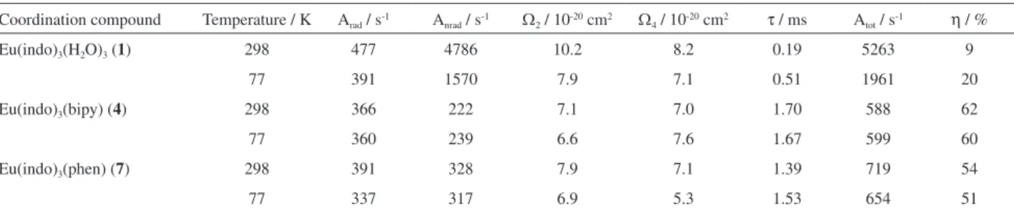
Table 1 presents the spectroscopic parameters obtained
for all three Eu3+-indomethacinate compounds at 298 and
77 K. As can be seen, the values of radiative rates for the complexes are comparable, indicating that no marked change occurs in the symmetry around the central metal ion when water molecules are substituted by the heterocyclic ligands or when the temperature is changed. On the other
hand, a significant decrease in non-radiative rates (Anrad)
is observed when water molecules are replaced by those ligands, which is expected due to the ability of water molecules act as luminescence quenchers via multiphonon
decay mechanism.30
Eu3+ complex (1) presents a considerably high dependence
of Anrad on the temperature in comparison with all of the
other complexes investigated in this work (Table 1). This result suggests that there is the multi-phonon luminescent quenching effect, promoted by water molecules, and the additional luminescence quenching channel, probably a ligand-to-metal charge transfer state (LMCT). As a result, the luminescence quantum efficiency for the hydrated
Eu3+-indomethacinate complex at low temperature (η = 20%)
is the double of the value at room temperature (η = 9%). The
low values of η reflect the luminescence quenching of the
Eu3+ ion. Such behavior agrees with what is observed for
some Eu3+-aromatic carboxylate coordination compounds
reported in the literature.31
The Anrad values of the complexes (4) and (7) are
very low and practically independent of temperature.
Although the low values of Arad as compared with the
Eu3+-β-diketoante complexes reported in the literature,30
which present values of Arad close to 1000 s-1, suggest
that the low values of Anrad contribute for the augment of
emission quantum efficiency (η) in more than six times as
compared with hydrated complex (1) (Table 1).
The Judd-Ofelt intensity parameters (Ω2 and Ω4) present
very similar values and reflect the small ratio between
intensities of the 5D
0 → 7F2 and 5D0 → 7F4 transitions
(Table 1). Moreover, the Ω2 values change only slowly
when water molecules are substituted by neutral ligands
in the first coordination sphere of the Eu3+ ion, indicating
that the parameter is mostly influenced by small angular
Table 1. Experimental intensity parameters (Ωλ = 2, 4), radiative rate (Arad), non-radiative rate (Anrad), lifetime (τ) and emission quantum efficiency (η) of the 5D
0 emitting level determined for the Eu3+ complexes (1, 4 and 7) recorded at 298 and 77 K
Coordination compound Temperature / K Arad / s-1 A
nrad / s-1 Ω2 / 10-20 cm2 Ω4 / 10-20 cm2 τ / ms Atot / s-1 η / %
Eu(indo)3(H2O)3 (1) 298 477 4786 10.2 8.2 0.19 5263 9
77 391 1570 7.9 7.1 0.51 1961 20
Eu(indo)3(bipy) (4) 298 366 222 7.1 7.0 1.70 588 62
77 360 239 6.6 7.6 1.67 599 60
Eu(indo)3(phen) (7) 298 391 328 7.9 7.1 1.39 719 54
changes in the local coordination geometry.32-35 However,
they are quite similar for Eu3+-indomethacinate complexes
containing heterocyclic ligands.
Luminescence spectra of the Tb3+
F i g u r e 8 s h ow s t h e ex c i t a t i o n s p e c t r a f o r
Tb3+-indomethacinate complexes recorded at 77 K in the
spectral range of 250-550 nm. These spectra exhibit broad absorption bands with maxima at around 350 nm and a sharp absorption band located at 480 nm that reflect the indirect
(via ligand) and direct excitation of the Tb3+ ion, respectively.
The emission spectra of Tb3+-indomethacinate
complexes were recorded at 298 and 77 K in the spectral range of 450-700 nm, under excitation on the intraligand
S0→ S1 transition (Figure 9). As shown in Figure 9a, all
emission spectra measured at 298 K present a broad emission band due to the indomethacinate ligand that overlaps the
sharp emission bands assigned to the intraconfiguration-4f8
of the Tb3+ ion: 5D
4→7F6 (480 nm), 5D4→7F5 (545 nm),
5D
4→7F4 (584 nm), 5D4→7F3 (621 nm), and 5D4→7F2
(650 nm). The energy gap ∆E(Tindomethacinate − 5D4) is only
approximately 1000 cm-1. These optical data indicate
that the indomethacinate ligand is a better luminescence
sensitizer for the Eu3+ than for the Tb3+ ion. This result is
in according to Latva’s empirical rule, which indicates
that optimal sensitizing for Tb3+ and Eu3+ luminescence
requires an energy gap between donor and acceptor states
in gate 2100-4500 cm-1. This fact indicates that energy
back-transfer process from the Tb3+ ion to indo ligand is
operative, acting as a luminescence quenching channel. The CIE (International Comission on Illumination)
color coordinates of the Eu3+- and Tb3+-indomethacinate
complexes measured from emission spectra35 recorded
at 298 and 77 K are presented in Figure 10. According to
these results, the emission color of the Eu3+ complex (1)
is tunable from pink to red when temperature is lowed. This behavior reflects the complete quenching of the phosphorescence band from indomethacinate coordinated ligand for the emission spectrum recorded at 77 K. Besides, taking into consideration that the emission spectra of the
Eu3+ coordination compounds (4) and (7) recorded at both
temperatures are dominated by the intraconfigurational-4f6
narrow emission bands centered on the Eu3+ ion, the CIE
color coordinates are located in the red region of CIE
Figure 8. Excitation spectra of the Tb3+-complexes (3, 6 and 9), in solid
state, recorded at 77 K with emission monitored at 545 nm.
Figure 9. Emission spectra of the Tb3+ complexes (3, 6 and 9), in solid state, recorded at (a) 298 and (b) 77 K, under excitation on the singlet state S 1 of
chromaticity diagram, as expected. Thus, the europium complexes exhibit pure red emission color.
As mentioned above, all emission spectra of the
Tb3+-indomethacinate compounds recorded at 298 K
exhibit the high intense phosphorescence band assigned
to T1→ S0 transition from indomethacinate ligand (Figure
9a). Therefore, they present CIE coordinates color in the green-yellow region. On the other hand, the green emission colors displayed by these complexes at 77 K reflect the dominating intensities of the narrow emission bands arising
from the intraconfigurational-4f8 transitions centered on the
Tb3+ ion (Figure 9b).
E x c i t a t i o n s p e c t r a o f t h e s o l i d s o l u t i o n s
Eu0.5Tb0.5(indo)3(H2O)3 and Eu0.5Tb0.5(indo)3(L), where
L: phen or bipy, have been also recorded at 77 K in order to investigate the monomeric or polymeric nature of the synthesized systems (Figure S7 in the SI section). These systems were prepared based on the experimental procedures for pure complexes, using equimolar mixtures
of Eu3+ and Tb3+ ions. All excitation spectra (Figure S7 in the
SI section) of the solid solutions recorded under emissions
monitored at 679 nm (5D
0→7F4 transition of the Eu3+ ion
exhibit a narrow band at 487 nm, which corresponds to the
intraconfigurational 5F
6→5D4 Tb3+-centered transition. This
result suggests that RE-indomethacinate complexes are dinuclear or polymeric systems. Similar results were also observed for RE-ibuprofen complexes containing donor
nitrogen ligands36,37 and for RE-dipivaloyl complexes with
tppo as neutral ligand.38
Conclusions
Herein, nine novel rare earth indomethacinate complexes were successfully prepared using simple synthetic routes. The coordination compounds presenting formulas
RE(indo)3(H2O)x, RE(indo)3(bipy) and RE(indo)3(phen)
(RE: Eu3+, Gd3+ and Tb3+, indo: indomethacinate ligand,
bipy: 2,2’-bipyridine, and phen: 1,10-phenanthroline, x = 3
for Eu3+ and Gd3+ complexes, and x = 4 for Tb3+ complex)
were characterized by elemental analysis of C, N and H, thermogravimetric analysis and infrared spectroscopic data. Based on photoluminescent parameters, non-radiative rates
(Anrad) were drastically decreased when water molecules
were substituted by heterocyclic ligands, which improved significantly the emission quantum efficiencies of the
complexes. On the other hand, the radiative rates (Arad) and
the Judd-Ofelt Ωλ = 2, 4 parameters were almost insensitive
to the coordination changes.
Referring to the coordination of heterocyclic ligands, the intramolecular energy transfer processes in the
RE3+ coordination compounds were also improved. The
spectroscopic results indicated that T1 excited states of
the heterocyclic ligands play an important role as an intermediate state on the luminescence sensitization of
the Eu3+ ion by the indomethacinate ligand. Finally, the
properties of RE3+-indomethacinate complexes in the
solid state described in this work are highly appropriate for light conversion molecular devices, such as those used as luminescent markers for bioassays and bioimage fields.
Supplementary Information
FTIR spectra, thermogravimetric data and luminescence
spectra for compounds 2, 5, 8, 3, 6 and 9 are available free
of charge at http://jbcs.sbq.org.br as PDF file.
Acknowledgments
The authors are grateful for the financial support from the CNPq (Conselho Nacional de Desenvolvimento Científico e Tecnológico), INCT-INAMI (CNPq), CNPq-FACEPE-PRONEX, CAPES, FAPESP (Fundação de Amparo à Pesquisa do Estado de São Paulo), Financiadora de Estudos e Projetos (FINEP), PET/IFPB (Programa de Educação Tutorial / Instituto Federal de Educação, Ciência e Tecnologia da Paraíba).
References
1. Weder, J. E.; Dillon, C. T.; Hambley, T. W.; Kennedy, B. J.; Lay, P. A.; Biffin, J. R.; Regtop, H. L.; Davies, N. M.; Coord. Chem. Rev. 2002, 232, 95.
2. Kostova, I.; Curr. Med. Chem. Anti-Cancer Agents2005, 5, 591.
3. Lopez-Sandoval, H.; Londono-Lemos, M. E.; Garza-Velasco, R.; Poblano-Melendez, I.; Granada-Macias, P.; Gracia-Mora, I.; Barba-Behrens, N.; J. Inorg. Biochem. 2008, 102, 1267.
Figure 10. CIE chromaticity diagram presenting (x, y) color coordinates for Eu3+ and Tb3+ complexes at (a) room temperature and (b) liquid
nitrogen temperature, under excitation at 350 nm. Photographs of the RE3+ complexes (insert) taken with a digital camera displaying the green
4. Miodragovic, D. U.; Bogdanovic, G. A.; Miodragovic, Z. M.; Radulovic, M. D.; Novakovic, S. B.; Kaludjerovic, G. N.; Kozlowski, H.; J. Inorg. Biochem. 2006, 100, 1568.
5. Devereux, M.; McCann, M.; Shea, D. O.; Kelly, R.; Egan, D.; Deegan, C.; Kavanagh, K.; McKee, V.; Finn, G.; J. Inorg. Biochem. 2004, 98, 1023.
6. Takeuchi, T.; Bottcher, A.; Quezada, C. M.; Meade, T. J.; Bioorg. Med. Chem. 1999, 7, 815.
7. Epstein, S. P.; Wallace, J. A.; Cornea1998, 17, 550.
8. Mothilal, K. K.; Karunakaran, C.; Rajendran, A.; Murugesan, R.; J. Inorg. Biochem. 2004, 98, 322.
9. Teslyuk, O. I.; Bel’tyukova, S. V.; Yegorova, A. V.; Yagodkin, B. N.; J. Anal. Chem. 2007, 629, 330.
10. Terreno, E.; Castelli, D. D.; Viale, A.; Aime, S.; Chem. Rev.
2010, 110, 3019.
11. Tweedle, M. F.; Acc. Chem. Res.2009, 42, 958. 12. Horrocks, W. D.; Acc. Chem. Res. 1981, 14, 384.
13. Zhou, X. J.; Zhao, X. Q.; Wang, Y. J.; Wu, B.; Shen, J.; Li, L.; Li, Q. X.; Inorg. Chem. 2014, 53, 12275.
14. Kaczmarek, M.; J. Fluoresc. 2011, 2201, 21.
15. Refat, M. S.; Mohamed, G. G.; Ibrahim, M. Y. S.; Killa, H. M. A.; Fetooh, H.; Russ. J. Gen. Chem.2013, 83, 2479. 16. Nolasco, M. M.; Vaz, P. D.; Carlos, L. D.; New J. Chem.2011,
35, 2435.
17. Xu, H.; Huang, W.; J. Photochem. Photobiol., A2011, 217, 213. 18. Li, X. N.; Si, Z. J.; Zhou, L.; Liu, X. J.; Zhang, H. J.; Phys.
Chem. Chem. Phys.2011, 11, 9687.
19. Bemquerer, M. P.; Bloch, C.; Brito, H. F.; Teotonio, E. E. S.; Miranda, M. T. M.; J. Inorg. Biochem. 2002, 91, 363. 20. Deacon, G. B.; Phillips, R. J.; Coord. Chem. Rev.1980, 33, 227. 21. Nakamoto, K.; Infrared and Raman Spectra of Inorganic and
Coordination Compounds, Applications in Coordination,
Organometallic, and Bioinorganic, 6th ed.; Wiley: New Jersey,
USA, 2009.
22. Gerasimova, T. P.; Katsyuba, S. A.; Dalton Trans. 2013, 42, 1787.
23. Chen, Y. J.; Xing, Z. F.; Cao, S.; Wang, Y.; J. Rare Earths2016,
34, 240.
24. Lin, M. J.; Wang, X. P.; Tang, Q.; Ling, Q.; J. Rare Earths2013,
31, 950.
25. Holz, R. C.; Thompson, L. C.; Inorg. Chem. 1993, 32, 5251.
26. Resende-Filho, J. B. M.; Silva, J. C.; Vale, J. A.; Brito, H. F.; Faustino, W. M.; Espínola, J. G. P.; Felinto, M. C. F. C.; Teotonio, E. E. S.; J. Braz. Chem. Soc. 2014, 25, 2080. 27. Kisel, K. S.; Linti, G.; Starova, G. L.; Sizov, V. V.; Melnikov,
A. S.; Pushkarev, A. P.; Bochkarev, M. N.; Grachova, E. V.; Tunik, S. P.; Eur. J. Inorg. Chem. 2015, 2015, 1734.
28. de Sá, G. F.; Malta, O. L.; Mello-Donegá, C.; Simas, A. M.; Longo, R. L.; Santa-Cruz, P. A.; Silva-Jr, E. F.; Coord. Chem. Rev. 2000, 196, 165.
29. Niyama, E.; Teotonio, E. E. S.; Brito, H. F.; Brito, G. E. S.; Cremona, M.; Reyes, R.; Felinto, M. C. F. C.; Spectrochim. Acta, Part A2005, 61, 2643.
30. Horrocks-Jr, W. W.; Sudnick, D. R.; Acc. Chem. Res. 1981, 14, 384.
31. Teotonio, E. E. S.; Brito, H. F.; Viertler, H.; Faustino, W. M.; Malta, O. L.; de Sá, G. F.; Felinto, M. C. F. C.; Santos, R. H. A.;
Polyhedron2006, 25, 3488.
32. Brito, H. F.; Malta, O. L.; Felinto, M. C. F. C.; Teotonio, E. E. S. In Luminescence Phenomena Involving Metal Enolate, vol. 1; Zabicky, J., ed.; John Wiley & Sons: Chichester, UK, 2009. 33. Silva, H. R. M.; Fonseca, M. G.; Espínola, J. G. P.; Brito, H. F.;
Faustino, W. M.; Teotonio, E. E. S.; Eur. J. Inorg. Chem. 2014,
11, 1914.
34. Teotonio, E. E. S.; Brito, H. F.; de Sá, G. F.; Felinto, M. C. F. C.; Santos, R. H. A.; Fuquen, R. M.; Costa, I. F.; Kennedy, A. R.; Gilmore, D.; Faustino, W. M.; Polyhedron2012, 38, 58. 35. Santa-Cruz, P. A.; Teles, F. S.; SpectraLux Software, version 2.0;
Ponto Quântico Nanodispositivos/RENAMI: Recife, PE, Brasil, 2003.
36. Marques, L. F.; Cuin, A.; Ribeiro, S. J. L.; Machado, F. C.;
Inorg. Chim. Acta2016, 441, 67.
37. Marques, L. F.; Correa, C. C.; Garcia, H. C.; Ribeiro, S. J. L.; Dutra, J. D. L.; Freire, R. O.; Machado, F. C.; J. Lumin.2014,
148, 307.
38. Miranda, Y. C.; Pereira, L. L. A. L.; Barbosa, J. H. P.; Brito, H. F.; Felinto, M. C. F. C.; Malta, O. L.; Faustino, W. M.; Teotonio, E. E. S.; Eur. J. Inorg. Chem. 2015, 2015, 3019.
Submitted: February 1, 2017