Original article (short paper)
Acute effects of resistance exercise performed on
ladder on energy metabolism, stress, and muscle
damage in rats
João Guilherme Silvestre
Universidade de São Paulo, São Paulo, SP, Brasil
Guilherme Fleury Fina Speretta
Universidade Federal de Santa Catarina, Florianópolis, SC, Brasil
Fernando Fabrizzi Gilberto Moraes
Ana Claudia Garcia de Oliveira Duarte
Universidade Federal de São Carlos, São Carlos, SP, Brasil
Abstract — Aim: To evaluate the acute effects of a resistance exercise session performed on ladder on energy metabolism, stress, and muscle damage in rats. Methods: Male Wistar rats were randomly distributed in Exercise (E) (n=30) and Control (C) (n = 20) groups. The E group performed a resistance exercise session on a vertical ladder with weights on their tails. Blood samples were collected at rest and after each climb to analyze lactate levels and ten minutes after the last climb to analyze lactate dehydrogenase (LDH), creatine kinase (CK), and corticosterone levels. Results: Blood lactate levels remained stable during exercise. Serum corticosterone, blood glucose, LDH and CK levels increased and glycogen content decreased in the E group, when compared to the C group. Conclusion: These results suggest that resistance exercise performed on ladder is a model of high-intensity exercise. However, the stabilization of lactate during the session suggests that the aerobic metabolism is an important factor during the intervals between climbs.
Keywords: resistance exercise; metabolism; stress; muscle damage
Introduction
Over the last decade, several studies have described the acute and chronic physiological effects of resistance exercise1,2. These indings have established the importance of this modality of exercise to improve physical functioning and quality of life in healthy people2 or in patients with chronic diseases such as cancer3 or diabetes4. Thus, the American College of Sports Medicine recommends resistance exercise as a primary intervention for health maintenance in young and old people1 and for prevention and treatment of obesity5.
Regarding the acute physiological responses of the resistance exercise, it is well known that, during high-intensity activities, the phosphagen system (ATP-PCr) and anaerobic glycolysis, respectively, are crucial to ATP synthesis and maintenance of exercise intensity. Glycolysis generates lactic acid, which breaks down into lactate and hydrogen ions as co-products, and lactate is removed from the active iber muscles to the blood6. The enzymes creatine kinase (CK) andlactate dehydrogenase (LDH) are required in the ATP-PCr and in lactate production, respectively, and both are released into the bloodstream when
tissue damage occurs7,8. The aerobic system predominates dur-ing the interval between sets and exercises, usdur-ing lipids as main substrate and metabolizing the increased lactate in the heart, liver, and non-working muscles6.
The glucocorticoids secreted from the adrenal cortex have an important role in the metabolic responses in high-intensity exercises9. Corticosterone acts along with the sympathetic ner-vous system and catecholamines, increasing gluconeogenesis and glycogenolysis10,11. Thus, corticosterone blood levels are considered a reliable physiological index of the stress imposed by exercise10,12.
Different animal models have been used experimentally to understand the mechanisms behind the resistance exercise-induced beneits13,15. Among them, stands out the resistance exercise performed on ladder, described by Hornberger and Farrar16. The authors showed this speciic model mimics many of the chronic physiological adaptations observed in human progressive resistance exercise, increasing the total strength and enhancing myoibrillar protein16 and muscular glycogen17. Additionally, a growing body of evidence has shown that the resistance exercise performed on ladder is also interesting to understand the effects of this exercise modality in the preven-tion and treatment of chronic diseases, such as obesity and cardiovascular diseases15,18,19.
exercise performed on ladder on lactate, corticosterone blood levels, CK and LDH activity, and glucose and lipid metabolism.
Materials and methods
Animals
A total number of 50 male Wistar rats weighting 340-360g were used. The animals were maintained in collective polypropylene cages (5 animals per cage) with food (MP-77; Primor, São Paulo, Brazil) and water provided ad libitum, in a room with controlled temperature (23 ± 2 ºC) and humidity (55 ± 10%). Lights were on from 7:00 am to 7:00 pm. The Ethics Committee for Animal Care and Use of the Federal University of São Carlos approved the experimental protocols used in this study (protocol number CEUA 009/2011), and the latter have been carried out in ac-cordance with the Animal Experimentation Ethical Principles adopted by the Brazilian College of Animal Experimentation (COBEA).
Experimental design
The rats were randomly assigned to two experimental groups: Control (C) (n = 10) and Exercise (E) (n = 20). All animals were familiarized with climbing a ladder for two non-consecutive days. After 72 hours, E and C groups performed the maximum voluntary carrying capacity (MVCC). After two days, the E group performed a single session of resistance exercise and the C group did not perform any type of exercise. The resistance exercise session consisted of four to nine ladder climbs. During the irst four ladder climbs, the rats carried 50%, 75%, 90%, and 100% of their previous maximum voluntary carrying capacity (MVCC). During subsequent ladder climbs, an additional 30g load was progressively added until the rat could not climb the entire length of the ladder or complete nine climbs. The rest between sets was 120 seconds. Blood samples (25 µl) were collected from an incision at the tail tip (20) at rest (res); after each climb on the ladder; and at recovery (rec), ive minutes after the last climb. Ten minutes after the last climb, another blood sample (200 µl) was collected for analyses of LDH and CK. The blood sample from the C group was collected immediately after collecting the samples from the E group.
Another set of animals was randomly allocated in the C group (n = 10) or E group (n = 10) and performed the same experimental protocol. The animals were euthanized by decapitation at rest (C group) or shortly after acute exercise session (E group) for trunk blood collection in plastic tubes with heparin, to assess blood glucose, glycogen, free fatty acids (FFA), and corticosterone hormone levels. Importantly, a subgroup of animals was used for these analyses to avoid the inluence of tail cut.
Resistance exercise
The resistance exercise protocol was adapted from Hornberger and Farrar16, according to the requirements of this study. The rats
were adapted to the resistance exercise (RT) protocol by climbing a vertical ladder (1.1 m; 0.18 m, 2-cm grid, 80° inclination) with a load apparatus without additional weight. The load apparatus was ixed to the tail by wrapping its proximal portion with a self-adhesive foam strip. With the load apparatus ixed to the tail, each rat was placed at the bottom of the ladder and familiarized with the climbing procedure. If necessary, a stimulus with tweezers was applied to the animal’s tail to initiate movement. When the rats reached the top of the ladder (house chamber), they were allowed to rest for 120 s. This procedure was repeated until they voluntarily climb the ladder for three consecutive turns without any stimuli.
Each animal performed a test for the evaluation of its MVCC, which consisted of climbs with progressive heavier loads. The initial climb was performed with 75% of the animal’s body mass and, after that, an additional 30g weight was added until reaching a load with which the rat could not climb the entire length of the ladder. The highest load that the animal successfully carried through the ladder was considered the MVCC for that training session. Failure was determined when the animal could not move up the ladder after three consecutive stimuli to the tail21. Three days after MVCC test, the resistance exercise protocol was performed as described above in the Experimental design.
Lactate analysis
Samples were immediately deposited in tubes containing 50 µl sodium luoride (1%). To avoid dilution of blood lactate with residual water at the tail of the animal, the rats were quickly dried with a towel immediately before blood collection. The lactate concentrations were determined in a lactate analyzer (YSI model 1500 SPORT; Yellow Springs Instruments Co, Yellow Springs, Ohio, USA), as previously described20.
Corticosterone, glucose, and free fat acids
Corticosterone was analyzed using a Corticosterone EIA Kit (Cayman Chemical cat. n° 500655, Ann Arbor, MI, USA)22. This kit is based on the competition between corticosterone and a corticosterone-acetylcholinesterase conjugate (Corticosterone Tracer) for a restricted number of corticosterone-speciic sheep antiserum binding sites. The plates were pre-coated with rabbit anti-sheep IgG and blocked with a proprietary formulation of proteins. The samples were incubated with tracer and antiserum in the wells of the plate for two hours at room temperature on an orbital shaker. The plate was washed to remove any unbound reagents and then Ellman’s Reagent was added to the wells. The intensity of the product was determined spectrophotometrically at 405 nm on a Dynex MRX TC Revelation Microplate Reader (Dynex Technologies, Chantilly, VA). Corticosterone analyses were performed in triplicate.
Trunk blood was collected shortly after euthanasia and serum glucose was analyzed using a glucometer device (One Touch UltraMini/Johnson&Johnson, Milpitas, CA, USA).
α-nitroso-β-naftol, following the method described by Novák23. Optical densities at 500 nm were measured after ifteen minutes using a spectrophotometer and compared with a standard consisting of 0.4 mM palmitic acid. These analyses were performed in duplicate.
Hepatic and muscular glycogen
To determine glycogen, tissue samples from the liver and soleus and gastrocnemius muscles were dissolved in 10 volumes of 6N KOH in a boiling-water bath for ive minutes15. The extract was combined with an equal volume of 10% ethanol and one volume of 10% K2SO4 was added to precipitate the glycogen. The pellet was resuspended in 3.0 ml of distilled water and glucose was determined using phenol-sulfuric acid24. The absorbance was measured at 480nm using a spectrophotometer. These analyses were performed in duplicate.
LDH and CK activity assays
LDH activity was assayed in plasma samples by a spectropho-tometric method based on the reduction of pyruvate to lactic acid coupled to NADH oxidation, as previously described25. The decrease in absorbance at 340 nm was monitored at 37°C. CK activity was assayed in plasma samples by a spectropho-tometric method based on the phosphorylation of ADP to ATP coupled to the reduction of NADP to NADPH, catalyzed by glucose-6-phosphate dehydrogenase. The increase in absorbance at 340 nm was monitored at 30°C, as previously described26. All analyses were performed in duplicate.
Statistical analysis
Values were reported as means ± standard error of the mean (SEM). Data were initially examined for normality using Kolmogorov-Smirnov test and for homoscedasticity using Bartlett criterion. The unpaired Student’s t-test was used to compare groups E and C. Lactate data were analyzed using paired Student’s t-test. The software package GraphPad Prism 5 (San Diego, CA, USA) was used and values of P < 0.05 were considered statistically signiicant.
Results
Weights, maximal workload, and time to climb
Table 1 shows the body weight and maximal workload in the maximal voluntary carrying capacity (MVCC) test for Control and Exercise groups. No differences were found in these parameters between the groups (P = 0.8979 and P = 0.8283, respectively). Table 1 also shows the time to climb during the exercise session for the Exercise group.
No differences were found between the groups in liver (P =
0.2793), soleus (P = 0.9432), and gastrocnemius weights (P =
0.9999), as shown in Table 2.
Table 1. Body weight andmaximal workload.
Body Weight (g) Maximal Workload (g)
Time to climb Exercise session (seconds) C 351.62 ± 7.75 440.85 ± 12.09 12.39 ± 0.66
E 350.10 ± 8.10 436.95 ± 12.16
-All values are presented as means ± standard error of the mean; C: control group (n=20); E: exercise group (n=30).
Lactate
The E group showed an increase (P < 0.05) in blood lactate from the second climb (75%) up to recovery, when compared with resting values (RES), as shown in Figure 1.
Table 2. Tissue weights from control (C) and exercised (E) groups.
Liver Weight (g) Soleus Weight (g) Gastrocnemius Weight (g) C 13.62 ± 0.49 0.169 ± 0.07 2.09 ± 0.09
E 12.75 ± 0.56 0.163 ± 0.05 2.09 ± 0.04 All values are presented as means ± standard error of the mean; C: control group (n=20); E: exercise group (n=30).
Corticosterone, serum glucose, and FFA
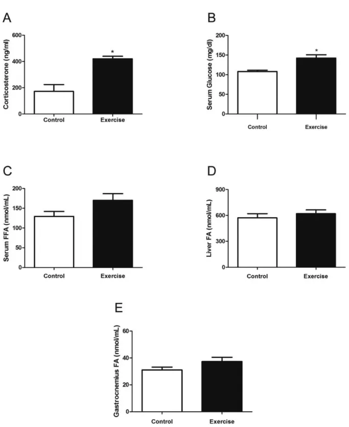
The acute exercise increased serum corticosterone concentra-tions (Figure 2A) in the E group, when compared with the C group (81.43 ± 29.74 ng/ml vs 420.30 ± 19.50 ng/ml; P < 0.05). Regarding serum glucose (Figure 2B), we found an increase when comparing C and E groups (107.9 ± 3.33 vs 142.3 ± 8.43 mg/dL; P < 0.05). On the other hand, we did not detect changes in serum FFA (129.46 ± 11.77 vs 170.12 ±15.93 nmol/mL; P = 0.109), liver (572.75 ± 47.35 vs 620.50 ± 43.52 nmol/mL; P = 0.575), and gastrocnemius (31.08 ± 1.98 vs 37.38 ± 2.90 nmol/ mL; P = 0.219) FA content.
Figure 2. (A) Serum corticosterone concentrations after a single exhaustive resistance exercise session. (B) Serum glucose concentrations after an exercise session. (C) Serum free fatty acid (FFA) concentrations after an exercise session. (D) Liver fatty acid (FA) content after an exercise session.
(E) Gastrocnemius FA content after an exercise session. Each bar represents the mean ± SEM of rats. * different from Control group; P < 0.05.
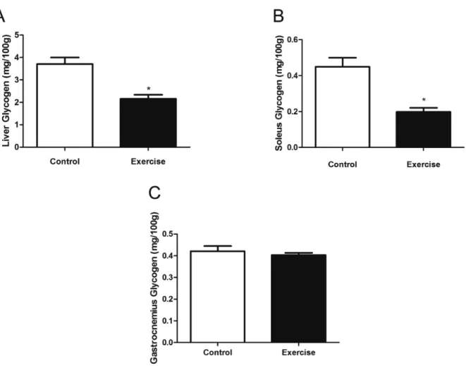
Hepatic and muscle glycogen
We also assessed the glycogen content in the liver, observing a decrease after acute exercise in the E group, when compared with the C group (2.15 ± 0.18 vs 3.71 ± 0.29 mg/100g; P < 0.05) (Figure
Serum LDH and CK
Muscle damage was indirectly estimated by changes in serum LDH and CK activities, as shown in Figure 4A. LDH activity was higher in the E group, when compared with the C group
(1,216.3 ± 67.54 vs 810.6 ± 79.07 U/ml; P < 0.05), after a single session of resistance exercise.
CK activity was also higher in the E group than in the C group (1,472.2 ± 91.85 vs 1,097.0 ± 133.71 U/ml; P < 0.05) (Figure 4B).
Figure 4. (A) Mean serum lactate dehydrogenase (LDH) activities after a single exhaustive resistance exercise session. (B) Mean serum creatine kinase (CK) activities after a single exhaustive exercise session. Each bar represents the mean ± SEM of rats. * different from Control group; P < 0.05.
Discussion
To our knowledge, this study is the irst to characterize the acute physiological responses of a resistance exercise performed on ladder. Our main indings are that an acute session of this exercise model increases biomarkers of muscle damage and circulatory stress biomarkers as well as reduces hepatic and muscle glyco-gen content. Additionally, blood lactate levels remained stable during the resistance exercise session in this particular model.
The resistance exercise increased blood lactate concentra-tions after the irst climb. However, they tended to stabilize during the exercise session, despite the progressive increase in load after each climb. These results suggest a balance between lactate production and lactate elimination20. The average time to climb was approximately 12 seconds, which does not allow a signiicant lactate production, and 120 seconds of recovery between climbs seems to be suficient for the removal of lactate through oxidation in the heart and skeletal muscles27. It is well established that this process is responsible for 70-75% of lactate removal and that the remaining lactate (30-25%) is metabolized through gluconeogenesis (i.e., the Cori cycle) by the liver28-30. The present data contrast with a previous study that has shown an exponential rise in blood lactate concentrations in the MVCC test performed on ladder in ovariectomized female rats31. Differences by sex in the enzyme activities and energy metabolism during exercise32,33 might explain these differences.
It was previously demonstrated that a single session of in-termittent jump exercise34 or exhaustive exercise performed on treadmill35 promote increases in corticosterone plasma levels in rats. These data are consistent with our indings, which showed higher serum corticosterone concentration (~2.4 fold) after a single session of resistance exercise performed on ladder by the E group. Corticosterone secretion is a physiological response to stress, suggesting that the exercise induced changes in the HPA axis36,37. Of note, this particular exercise model seems to promote lower relative change in corticosterone levels than a single exercise session of swimming, as previously observed11. This difference may be due to the necessity for the animals to keep exercising in the water to avoid drowning11.
Glucocorticoids have important metabolic functions, includ-ing their effects on the metabolism of glucose, lipids, and proteins, inducing increases in blood glucose, mobilization of fatty acids from fat reserves to active tissues11,36, and rapid mobilization of fat and amino acids from stores for using in both the synthesis of compounds (such as glucose) and as energetic sources38. This is in line with our results, which have shown increases in blood glucose concentrations and reduction in liver and soleus glycogen content in the exercise group, when compared with the control group. However, we found no differences in the gastrocnemius contents between the two groups. These results were expected, since it has been shown that the soleus muscle is more recruited during the movement of climb, when com-pared with the gastrocnemius muscle16. This decrease seems to be linked with the glycogenolysis resulting from the sharp increase of sympathetic activity and glucocorticoid hormones induced by exercise39,40.
There were no differences between groups in serum, liver,
and gastrocnemius fatty acid content. These results suggest a predominant use of glycogen in comparison to lipids, consistent with the concept that high-intensity exercises are associated with the depletion of muscle glycogen40.
In this study, animals that performed a single session of re-sistance exercise showed higher serum LDH and CK activities. Both enzymes have been used as indirect biomarkers for muscle damage8,41-43. Our indings are in line with a previous study that showed increases in LDH and CK activity after a single session of high-intensity exercise performed on treadmill until exhaus-tion in rats13. It is possible that the alterations in these enzymes relect muscle damage, which could explain in part the skeletal muscle hypertrophy observed in previous studies that used the same exercise protocol16,44. On the other hand, another study showed that a single session of high-intensity resistance exer-cise, which required full range of motion at the ankle, did not promote increases in LDH and CK in rats45. Differently from our study, the measurements of LDH and CK were performed after the ifth session and not after the irst, as we did in our study. It could explain these discrepancies, since it is well accepted that the adaptation of muscles to the exercise decreases muscle dam-age42,46. For example, it was demonstrated that eccentric resistance exercise in pre-trained individuals did not increase CK activity42. Further studies using direct markers for muscle damages, such as morphology, are necessary to conirm muscle damage after a single session of resistance exercise performed on ladder.
Conclusion
In conclusion, we showed that a single session of resistance exercise performed on a ladder increases indirect markers of muscle damage and corticosterone blood levels. Our results thus suggest an increased use of glycogen instead of lipid, consis-tent with the hypothesis that this is a high-intensity model of exercise. However, the tendency to stabilization of the blood lactate suggests that the aerobic metabolism has a key role during the intervals between climbs. Further studies are necessary to evaluate other variables, including the time-course of muscle damage after a single session and whether adaptation occurs after long-term of training. Furthermore, the impact of different rest intervals between sets on metabolic responses, particularly in lactate concentrations, should be examined.
References
1. Ratamess NA, Alvar BA, Evetoch TK, Housh TJ, Kibler WB, Kraemer WJ, et al. American College of Sports Medicine posi-tion stand. Progression models in resistance training for healthy adults. Med Sci Sports Exerc. 2009;41(3):687-708.
2. Faigenbaum AD, Kraemer WJ, Blimkie CJ, Jeffreys I, Micheli LJ, Nitka M, et al. Youth resistance training: updated position statement paper from the national strength and conditioning as-sociation. J Strength Cond Res. 2009;23(5 Suppl):S60-79. 3. Norris MK, Bell GJ, North S, Courneya KS. Effects of resistance
prostate cancer survivors: a pilot randomized controlled trial. Prostate Cancer Prostatic Dis. 2015;18(3):281-7.
4. Daly RM, Miller EG, Dunstan DW, Kerr DA, Solah V, Menzies D, et al. The effects of progressive resistance training combined with a whey-protein drink and vitamin D supplementation on glycaemic control, body composition and cardiometabolic risk factors in older adults with type 2 diabetes: study protocol for a randomized controlled trial. Trials. 2014;15:431.
5. Donnelly JE, Blair SN, Jakicic JM, Manore MM, Rankin JW, Smith BK. American College of Sports Medicine Position Stand. Appropriate physical activity intervention strategies for weight loss and prevention of weight regain for adults. Med Sci Sports Exerc. 2009;41(2):459-71.
6. Wells GD, Selvadurai H, Tein I. Bioenergetic provision of energy for muscular activity. Paediatr Respir Rev. 2009;10(3):83-90. 7. Brancaccio P, Lippi G, Maffulli N. Biochemical markers of
mus-cular damage. Clin Chem Lab Med. 2010;48(6):757-67. 8. Cooke MB, Rybalka E, Stathis CG, Cribb PJ, Hayes A. Whey
protein isolate attenuates strength decline after eccentrically-induced muscle damage in healthy individuals. J Int Soc Sports Nutr. 2010;7:30.
9. Hackney AC. Stress and the neuroendocrine system: the role of exercise as a stressor and modiier of stress. Expert review of endocrinology & metabolism. 2006;1(6):783-92.
10. França SC, Barros Neto TL, Agresta MC, Lotufo RF, Kater CE. [Divergent responses of serum testosterone and cortisol in ath-lete men after a marathon race]. Arq Bras Endocrinol Metabol. 2006;50(6):1082-7.
11. Contarteze RV, Manchado Fde B, Gobatto CA, De Mello MA. Stress biomarkers in rats submitted to swimming and treadmill running exercises. Comp Biochem Physiol A Mol Integr Physiol. 2008;151(3):415-22.
12. Vale RGdS, de Oliveira RD, Pernambuco CS, de Meneses YPdSF, Novaes JdS, de Andrade AdFD. Effects of muscle strength and aerobic training on basal serum levels of IGF-1 and cortisol in elderly women. Arch Gerontol Geriatr. 2009;49(3):343-7. 13. Malaguti M, Angeloni C, Garatachea N, Baldini M, Leoncini E,
Collado PS, et al. Sulforaphane treatment protects skeletal muscle against damage induced by exhaustive exercise in rats. J Appl Physiol. 2009;107(4):1028-36.
14. Marqueti RC, Prestes J, Stotzer US, Paschoal M, Leite RD, Perez SE, et al. MMP-2, jumping exercise and nandrolone in skeletal muscle. Int J Sports Med. 2008;29(7):559-63.
15. Speretta GF, Rosante MC, Duarte FO, Leite RD, Lino AD, Andre RA, et al. The effects of exercise modalities on adiposity in obese rats. Clinics (Sao Paulo). 2012;67(12):1469-77.
16. Hornberger TA, Jr., Farrar RP. Physiological hypertrophy of the FHL muscle following 8 weeks of progressive resistance exercise in the rat. Can J Appl Physiol. 2004;29(1):16-31.
17. Rodrigues MD, Borin SH, Silva CAd. Relações metabólicas em ratos sob o treinamento anaeróbio em escada. Rev. Bras. Ciênc. Esporte.
18. Prestes J, de Cassia Marqueti R, Shiguemoto GE, Leite RD, Pereira GB, Selistre-de-Araujo HS, et al. Effects of ovariectomy and resistance training on MMP-2 activity in skeletal muscle. Appl Physiol Nutr Metab. 2009;34(4):700-6.
19. Leite RD, Prestes J, Bernardes CF, Shiguemoto GE, Pereira GB, Duarte JO, et al. Effects of ovariectomy and resistance training on lipid content in skeletal muscle, liver, and heart; fat depots; and lipid proile. Appl Physiol Nutr Metab. 2009;34(6):1079-86. 20. Gobatto CA, de Mello MA, Sibuya CY, de Azevedo JR, dos Santos
LA, Kokubun E. Maximal lactate steady state in rats submitted to swimming exercise. Comp Biochem Physiol A Mol Integr Physiol. 2001;130(1):21-7.
21. Speretta GF, Silva AA, Vendramini RC, Zanesco A, Delbin MA, Menani JV, et al. Resistance training prevents the cardiovascular changes caused by high-fat diet. Life Sciences. 2016;146:154-62. 22. Wu LM, Hu MH, Tong XH, Han H, Shen N, Jin RT, et al. Chronic
unpredictable stress decreases expression of brain-derived neuro-trophic factor (BDNF) in mouse ovaries: relationship to oocytes developmental potential. PLoS One. 2012;7(12):e52331. 23. Novák M. Colorimetric ultramicro method for the determination
of free fatty acids. J. Lipid Res. 1965;6(3):431-3.
24. Dubois M, Gilles KA, Hamilton JK, Rebers PA, Smith F. Colorimetric Method for Determination of Sugars and Related Substances. Anal Chem. 1956;28:350-6.
25. Korzeniewski C, Callewaert DM. An enzyme-release assay for natural cytotoxicity. J Immunol Methods. 1983;64(3):313-20. 26. Noda L, Nihei T, Morales MF. The enzymatic activity and
inhibi-tion of adenosine 5'-triphosphate-creatine transphosphorylase. J Biol Chem. 1960;235:2830-4.
27. Faude O, Kindermann W, Meyer T. Lactate threshold concepts: how valid are they? Sports Med. 2009;39(6):469-90.
28. Brooks GA. Intra- and extra-cellular lactate shuttles. Med Sci Sports Exerc. 2000;32(4):790-9.
29. Brooks GA. Cell-cell and intracellular lactate shuttles. J Physiol. 2009;587(Pt 23):5591-600.
30. Gladden LB. The role of skeletal muscle in lactate exchange during exercise: introduction. Med Sci Sports Exerc. 2000;32(4):753-5. 31. Sanches IC, Conti FF, Sartori M, Irigoyen MC, De Angelis K.
Standardization of resistance exercise training: effects in diabetic ovariectomized rats. Int J Sports Med. 2014;35(4):323-9. 32. Green HJ, Fraser IG, Ranney DA. Male and female differences in
enzyme activities of energy metabolism in vastus lateralis muscle. J Neurol Sci. 1984;65(3):323-31.
33. Ruby BC, Coggan AR, Zderic TW. Gender differences in glucose kinetics and substrate oxidation during exercise near the lactate threshold. J Appl Physiol (1985). 2002;92(3):1125-32.
34. Rogatto GP, Oliveira CA, Faria MC, Luciano E. Acute metabolic responses of Wistar rats to intermittent jump exercise. Motriz. 2004;10:61-6.
35. de Araujo GG, Gobatto CA, Marcos-Pereira M, Dos Reis IG, Verlengia R. Interval versus continuous training with identical workload: physiological and aerobic capacity adaptations. Physiol Res. 2015;64(2):209-19.
36. Franca SC, Barros Neto TL, Agresta MC, Lotufo RF, Kater CE. [Divergent responses of serum testosterone and cortisol in ath-lete men after a marathon race]. Arq Bras Endocrinol Metabol. 2006;50(6):1082-7.
38. Andersen ML, Bignotto M, Machado RB, Tuik S. Different stress modalities result in distinct steroid hormone responses by male rats. Braz J Med Biol Res. 2004;37(6):791-7.
39. Ramel A, Wagner K-H, Elmadfa I. Correlations between plasma noradrenaline concentrations, antioxidants, and neutrophil counts after submaximal resistance exercise in men. Br J Sports Med. 2004;38(5):e22.
40. Cambri LT, de Araujo GG, Ghezzi AC, Botezelli JD, Mello MA. Metabolic responses to acute physical exercise in young rats re-covered from fetal protein malnutrition with a fructose-rich diet. Lipids Health Dis. 2011;10:164.
41. Cooke MB, Rybalka E, Williams AD, Cribb PJ, Hayes A. Creatine supplementation enhances muscle force recovery after eccentrically-induced muscle damage in healthy individuals. J Int Soc Sports Nutr. 2009;6:13.
42. Flann KL, LaStayo PC, McClain DA, Hazel M, Lindstedt SL. Muscle damage and muscle remodeling: no pain, no gain? J Exp Biol. 2011;214(Pt 4):674-9.
43. Totsuka M, Nakaji S, Suzuki K, Sugawara K, Sato K. Break point of serum creatine kinase release after endurance exercise. J Appl Physiol (1985). 2002;93(4):1280-6.
44. Leite RD, Durigan Rde C, de Souza Lino AD, de Souza Campos MV, Souza M, Selistre-de-Araujo HS, et al. Resistance training may concomitantly beneit body composition, blood pressure and muscle MMP-2 activity on the left ventricle of high-fat fed diet rats. Metabolism. 2013;62(10):1477-84.
45. Zanchi NE, Lira FS, Seelaender M, Lancha-Jr AH. Experimental chronic low-frequency resistance training produces skeletal mus-cle hypertrophy in the absence of musmus-cle damage and metabolic stress markers. Cell Biochem Funct. 2010;28(3):232-8.
46. Brown SJ, Child RB, Day SH, Donnelly AE. Exercise-induced skeletal muscle damage and adaptation following repeated bouts of eccentric muscle contractions. J Sports Sci. 1997;15(2):215-22.
Acknowledgments
The author thanks CAPEs and CNPq for the inancial support. The author also
thanks Dr. Orlando de Castro e Silva Junior and the Special Liver Transplantation Unity, Departments of Surgery and Anatomy, Ribeirão Preto School of Medicine, University of São Paulo, Brazil, and Dra. Fernanda de Freitas Anibal, Department of Morphology and Pathology, Center of Biological and Health Sciences, Federal University of São Carlos, Brazil.
Corresponding author
Guilherme Fleury Fina Speretta
Federal University of Santa Catarina, Department of Physiological Sciences,
Cam-pus Universitário Reitor João David Ferreira Lima, Trindade. Florianopolis, SC,
Brazil.
Email: [email protected]
Manuscript received on August 23, 2016 Manuscript accepted on October18, 2016