Introduction
High-intensity training sessions during a training program are widely utilized to maximize athletes’ performance in different sports1. However, these training sessions need to comprise an adequate recovery period for the achievement of better athletes’ performance. When the athlete is submitted
to an overtraining (OT) period (i.e., an intensiied training
period) and the time destined to recovery is not respected,
the nonfunctional overreaching (NFOR) – deined as a drop
of performance that can be reversed after weeks or months of recuperation2 – may occur.
Recently, da Rocha et al.3 veriied that mice in NFOR, in -duced by an OT protocol based on downhill running sessions, presented an improvement of the hepatic insulin signaling path-way when compared to other two OT protocols with the same external load (i.e., the product between intensity and volume of training), but performed in uphill and without inclination. Also, these mice displayed high phosphorylation status of the glycogen synthase kinase 3 beta (GSK-3beta), a direct target that is phosphorylated and inactivated by the protein kinase B (Akt). When GSK-3beta is not phosphorylated, the glycogen synthase activity is inhibited, and the glycogen storage is de-creased4. Interestingly, these mice showed increased levels of hepatic glycogen even with high contents of tribbles-like protein
3 (TRB3), an Akt inhibitor3. These contradictory results may be better elucidated with the investigation of other proteins that are involved in this pathway modulation, such as the adaptor proteins APPL1 and APPL2.
APPL1 and APPL2 are homologous proteins that bind to diverse groups of trans-membrane receptors or signaling proteins5. APPL1 acts on the adiponectin signaling cascade, communicating signals of the adiponectin receptors (AdipoR1 and AdipoR2) to downstream targets through direct interaction with the NH2-terminal region6. According to Ryu and cowork-ers7, APPL1 also interacts directly to the insulin receptor beta
(IRbeta). Also, these authors veriied that the high APPL1
expression increased the interaction between IRbeta and insu-lin receptor substrate-1 (IRS-1), as well as Akt activation. On the other hand, APPL1 suppression impaired Akt activation and glucose transporter type 4 (GLUT4) translocation to the membrane when stimulated by insulin in L6 myocytes and 3T3-L1 adipocytes6,8.
Although the physiological role of APPL2 is not fully understood it is interesting to note that only APPL1, and not APPL2, interacts directly with Akt9. Wang et al.10 demonstrated that under basal conditions, APPL2 interacts with AdipoR1 and competes with APPL1, negatively regulating adiponectin signaling in muscle cells. Therefore, APPL2 has an inhibitory role in the insulin sensitizing effects, blocking the adiponectin Original Article (short paper)
Nonfunctional overreaching and hepatic
adaptations of APPL1 and APPL2
Gustavo P. Morais Larissa Gaioto de Vicente
Luciana da C. Oliveira Ana P. Pinto Alisson L. da Rocha
Bruno C. Pereira
Universidade de São Paulo, Ribeirão Preto, SP, Brasil
José R. Pauli
Universidade Estadual de Campinas, Limeira, SP, Brasil
Adelino S. R. da Silva
Universidade de São Paulo, Ribeirão Preto, Brasil
Abstract — Aims: Previously, we veriied that overtrained mice upregulated the TRB3 levels, its association with Akt,
and the hepatic concentrations of glycogen. It is known that APPL1 can limit the interaction between TRB3 and Akt,
playing an important role in the glucose homeostasis. Thus, we veriied the effects of three overtraining protocols on
the hepatic levels of APPL1 and APPL2. Methods: Rodents were divided into control (CT), overtrained by downhill running (OTR/down), overtrained by uphill running (OTR/up) and overtrained by running without inclination (OTR).
The hepatic contents of APPL1 and APPl2 were measured by the immunoblotting technique. Results: Signiicant
elevation of APPL1 observed in the OTR/down and OTR/up groups, as well as the tendency of increase (p=0.071) observed in the OTR group. Conclusion: These results indicate that this particular protein is likely to participate in the glucose homeostasis previously observed in response to these OT protocols.
effect in Akt activation stimulated by insulin. Interestingly, APPL2 also seems to have an involvement in macrophages
inlammatory response, suppressing it via the toll-like recep -tor 4 (TLR4)11.
Regarding their physiological importance and capacity to
modulate the aforementioned molecular pathways, our irst
aim was to verify the effects of the OT protocol based on downhill running sessions on the hepatic contents of APPL1 and APPL2. Although the other two OT protocols performed in uphill and without inclination led to similar decrements in the analyzed performance parameters3,12,13, they induced particular modulations of the hepatic insulin signaling pathway3. Thus, we also evaluated their effects on the hepatic protein levels of APPL1 and APPL2.
Methods
Experimental Animals
Male C57BL/6 mice from the Central Animal Facility of the Ribeirão Preto campus of the University of Sao Paulo (USP) were maintained in individual cages with controlled temperature (22±2°C) on a 12:12-h light-dark inverted cycle (light: 6 PM to 6 AM, dark: 6 AM to 6 PM) with food (Purina chow) and water ad libitum. The experimental procedures were performed by the Brazilian College of Animal Experimentation and were approved by the Ethics Committee of the University of Sao Paulo (ID 14.1.873.53.0). Eight-week-old C57BL/6 mice were divided into four groups: control (CT; sedentary mice; n = 8), overtrained by downhill running (OTR/down; performed the OT protocol based on downhill running; n = 8), overtrained by uphill running (OTR/up; performed the OT protocol based on uphill running; n = 8) and overtrained by running without inclination (OTR; performed the OT protocol based on running without inclination; n = 8). The CT, OTR/down, OTR/up and OTR mice were manipulated and/or trained in a dark room between 6 to 8 am.
Overtraining protocols and performance evaluations
The eight-week OT protocols based on downhill running, uphill running and running without inclination were performed as previously described13, and each experimental week consisted of
ive days of training followed by two days of recovery. During the irst four weeks of the OT protocols (i.e. irst stage), the in -tensity was maintained at 60% of the exhaustion velocity (EV) that was obtained by the incremental load test (ILT), and the volume was gradually increased to 60min per day in the fourth
week. In this irst stage, rodents ran at a grade of 0%. In the ifth week of the OT protocols, the intensity and volume were
maintained, but the rodents ran at a grade of – 14% (OTR/ down), 14% (OTR/ up) or 0% (OTR).
These running grades were maintained until the end of the OT protocols. In the sixth week of the OT protocols, the inten-sity was increased to 70% of EV. In the seventh week of the OT protocols, the intensity and volume were increased to 75% of EV and 75min, respectively. In the eighth week of the OT protocols, the number of daily sessions was doubled. The resting interval between daily sessions during the eighth week was 4h. The performance evaluations were made on week 0 and 48 h after the last sessions of the OT protocols at the end of week 8 and consisted of a rotarod test12,14,15, the ILT3,12,14,16-18, an exhaus-tive test3,12,14,16-18 and a grip force test3,12,14,16-18.The description of these tests and their results were recently published3 using the same sample as the current study.
Liver Extraction
Mice were anesthetized at the end of week 8. After an overnight fast (12h), rodents were anesthetized with an intraperitoneal
(i.p.) injection of 2-2-2 tribromoethanol 2.5% (10–20μL.g−1). As soon as the effect of anesthesia was conirmed by the loss of pedal relexes, the abdominal cavity was opened and each
mouse liver was removed. Subsequently, the livers were quickly stored at – 80 ° C for later analysis of the contents of APPL1 and APPL2 by the immunoblotting technique.
Immunoblotting technique
The liver samples were homogenized in extraction buffer (1% de Triton X-100, 100 mM Tris, pH 7.4, containing 100 mM
sodium pyrophosphate, sodium luoride 100 mM, EDTA 10
mM, 10 mM sodium vanadate, 2 mM de PMSF and 0.1 mg.ml-1 aprotinin) at 4oC with a Polytron PTA 20S generator (Brinkmann Instruments model PT 10/35), operated at a maximum speed of 30s. The extracts were centrifuged (9900g) for 40 minutes at 4°C to remove insoluble material, and the supernatants of these homogenates were used for protein quantitation using the Bradford method20.
Proteins were denatured by boiling in Laemmli21 sample buffer containing 100mM DTT, run on SDS-PAGE gel and transferred to nitrocellulose membranes (GE Healthcare, Hybond
ECL, RPN303D). The eficiency of transfer to nitrocellulose membranes was veriied by the brief staining of bands with
Ponceau. These membranes were blocked with Tris-buffered saline (TBS) containing 5% BSA and 0.1% Tween-20 for 1 hour at 4°C. Subsequently, the membranes were incubated in
1:1000 dilution overnight with a speciic antibody anti-APPL1
(SC-67402) and anti-APPL2 (SC-67403) from Santa Cruz Biotechnology (Santa Cruz, CA, USA). Sixteen hours after the
incubation with the speciic primary antibody, the membranes
were washed 5 times (5min each time) with TBS containing 0.1% Tween-20. Then, all the membranes were incubated for 1 hour at 4°C with secondary antibody conjugated with a
horse-radish peroxidase. The speciic immunoreactive bands were
were acquired using the C-DiGitTM Blot Scanner (LI-CORR,
Lincoln, Nebraska, USA) and quantiied using the software
Image Studio for C-DiGit Blot Scanner.
Statistical analysis
The results are expressed as mean ± standard error (SE). According to the Shapiro-Wilk W-test, data were normally
distributed and homogeneity was conirmed by Levene’s test.
Hence, one-way analysis of variance (ANOVA) was used to verify the effects of the experimental groups on the hepatic contents of APPL1 and APPL2. When one-way ANOVA indicated
statisti-cal signiicance, a Bonferroni’s post hoc test was used. When
Bonferroni’s post hoc indicated tendency (P<0.1), Cohen’s d
test was used to measure the effect size. Statistical analysis was performed using STATISTICA 8.0 computer software (StatSoft1,
Tulsa, OK) and the signiicance level was set at p< 0.05.
Results
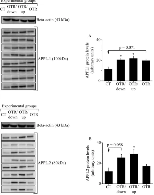
The protein levels of APPL1 were signiicantly higher in
the OTR/down and OTR/up groups compared to the CT group. Also the OTR group presented a tendency (p=0.071) to increase APPL1 compared to the CT group (Figure 1A). Regarding APPL2, OTR/up group increased its protein levels compared to the CT group, and OTR/down group presented a tendency (p=0.058) to increase APPL2 compared to the CT group (Figure 1B).
* *
p = 0.071 40
20
0
A
P
P
L
1 prot
ei
n l
eve
ls
(a
rbi
tra
ry uni
ts
)
A
CT OTR/ down
OTR/ up
OTR
* p = 0.058 40
20
0
A
P
P
L
2 prot
ei
n l
eve
ls
(a
rbi
tra
ry uni
ts
)
B
CT OTR/ down
OTR/ up
OTR CT OTR/
down OTR/
up OTR Experimental groups
Beta-actin (43 kDa)
APPL.1 (100kDa)
CT OTR/ down
OTR/ up OTR Experimental groups
Beta-actin (43 kDa)
APPL.2 (80kDa)
Regarding tendency values, we have calculated the effect size values by Cohen’s test. The result for APPL1 was Cohen’s d=1.469139 when OTR was compared to CT. The result for APPL2 was Cohen’s d=1.414457 for APPL2 when OTR/down
was compared to CT. Both Cohen’s d results were classiied as
a strong effect (i.e., larger than 0.8).
Discussion
The main indings of the present investigation were: a) In general,
the OT protocols increased the hepatic APPL1 protein levels; b) OTR/up group also presented high values of hepatic APPL2 protein levels. Taken together, these results indicated that APPL1 could be part of the molecular mechanism that contributes to the glucose homeostasis in the liver of overtrained mice. Da Rocha et al.3 using the same liver samples of the current investigation
veriied that mice from the OTR/down group improved some
initial proteins of the hepatic insulin signaling pathway, but upregulated the TRB3 and its association with Akt, implying a limitation on this downstream point of the insulin signaling pathway. The authors also observed high levels of hepatic glycogen3, which leads to the hypothesis that another molecule could be responsible for this result especially in the OTR/down and OTR/up groups.
It is known that APPL1 can limit the interaction between Akt and TRB3 in mouse liver tissue, which is accompanied by an increase in the membrane translocation of APPL1, Akt activation, and enhancement in response to insulin stimula-tion22,23. Also, adiponectin also stimulates APPL1 in mouse hepatocyte cells with activation of another class of proteins, the p38 mitogen-activated protein kinases (p38MAPK), which activates glucose transporters protein. The APPL1 overexpression
was suficient to enhance GLUT4 membrane translocation to a
level comparable with that induced by adiponectin6. Therefore, considering the increased values of liver glycogen concentration previously related for OTR/down and OTR/up groups3 and the
current signiicant elevation of APPL1 in these same groups, we
consider that APPL1 plays a major role in the hepatic glucose homeostasis of overtrained mice.
Studies using depletion of APPLs also revealed their
regu-lation of signaling for glucose metabolism and inlammatory
responses by the interaction with adiponectin receptors, phos-phorylating its downstream targets: AMP-activated protein kinase (AMPK), GLUTs, peroxisome proliferator-activated receptor-gamma coactivator 1 alpha (PGC1-alfa), and FoxO1 11,24. Da Rocha et al.3 demonstrated that the phosphorylation levels of FoxO1 and GSK-3beta are increased in the OT groups after insu-lin stimulation. Also, consistent evidence shows the potential role of insulin-mediated APPL125, resulting in a markedly enhanced GSK-3beta phosphorylation and increased glycogen storage in the mouse liver. APPL1 downregulation is associated with GSK-3beta phosphorylation decreased in liver of obese mice under fasting conditions. However, in response to endurance exercise training, these obese mice increased APPL1 expression, Akt/ APPL1 association, and GSK-3beta phosphorylation23, which corroborates our consideration above.
Recently, Pereira et al.26 demonstrated that the currently used OT protocols impaired the insulin signaling pathway in skeletal
muscles with different iber type speciicities, but did not induce signiicant differences in the insulin tolerance test (ITT). The
authors concluded that other tissues such as liver played a major role in the maintenance of glucose homeostasis3. It is known that APPL1 also plays a key role in the regulation of glucose metabolism by mediating the adiponectin signaling6,27. In fact, the adiponectin-stimulated activation of AMPK can reduce the glucose levels in vivo, being considered as an antidiabetic strat-egy28. Thus, we may hypothesize that the current high levels of hepatic APPL1 (i.e., in the OTR/down and OTR/up groups) and its interaction with adiponectin contributed to the maintenance of glucose homeostasis in these OT groups.
The interaction between APPL1 and adiponectin may also
be linked to an anti-inlammatory effect29. Previously, our research group showed that the NFOR induced by the current
OT protocols led to low-grade chronic inlammation state12. Also, the OTR/down group increased the hepatic levels of interleukin-10 (IL- 10)3, a classical anti-inlammatory cytokine.
Although the anti-inlammatory effects of adiponectin are not
well documented30, it is known that this adipocyte-speciic proteincan increase the expression of anti-inlammatory me -diators such as the IL-1031. Finally, APPL1 is necessary for the anti-inflammatory and cytoprotective effects induced by adiponectin6,32.
Regarding APPL2, it is known that this protein can negatively modulate the adiponectin signaling by direct connection with AdipoR1 and/or AdipoR2 via BAR domain, thereby prevent-ing the APPL1 interaction with the Adipo receptors9,25. On the other hand, adiponectin modulates the dissociation of the APPL1/APPL2 heterodimers, which can also be triggered by insulin. Interestingly, Cheng et al.22 demonstrated that APPL2 inhibits insulin-stimulated glucose uptake in skeletal muscle. In this study, only the OTR/up group increased the hepatic levels of APPL2, which may be linked to the increased phos-phorylation of the insulin receptor substrate 1 (IRS-1) at serine 307 3, a molecular marker directly related to insulin signaling pathway impairment33.
Conclusion
In summary, the OTR/down and OTR/up protocols increased the protein levels of APPL1, but only the OTR/up group increased the protein levels of APPL2 in liver. These results suggest a pos-sible molecular pathway that potentiates hepatic glucose uptake
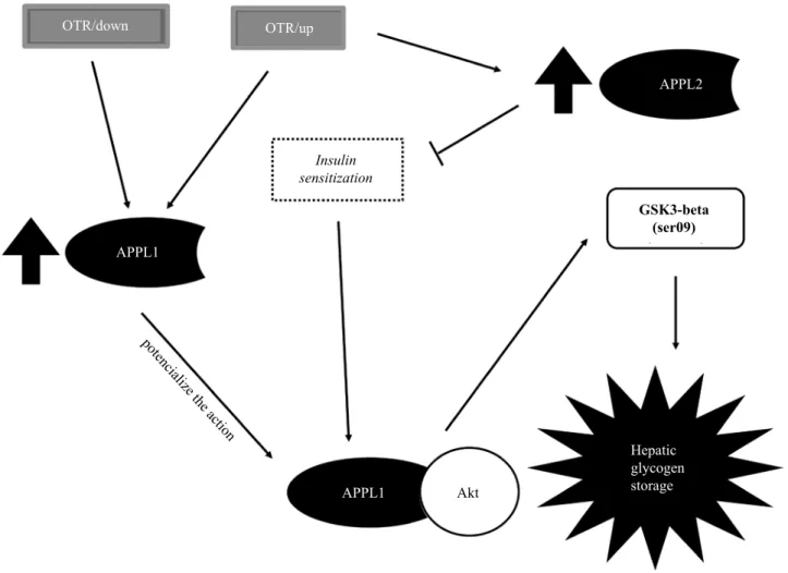
in overtrained mice. Finally, igure 2 summarizes the current indings. In future investigations, we will perform an experiment silencing APPL1 and APPL2 to conirm their contributions to
OTR/down OTR/up
APPL1
APPL1
Hepatic glycogen storage
APPL2
Akt Insulin
sensitization
GSK3-beta (ser09)
pot enc
ialize the
a ction
Figure 2. A squematic igure representing the possible mechanisms by which APPL1 and APPL2 act in the liver of overtrained mice. The over -training protocols based on downhill and uphill running upregulated the protein levels of APPL1, which can potentiate the action of Akt. The higher the activation of Akt, the higher is the phosphorylation and inactivation of GSK-3beta, activating glycogen synthase and increasing hepatic glycogen storage. The OTR/up group also upregulated APPL2, which may decrease insulin sensitization. It is important to highlight that the pro-tein levels of Akt and GSK-3beta, as well as the glycogen contents of the OTR/down, OTR/up and OTR groups, were recently published3 using the same liver samples as the current investigation.
References
1. Armstrong LE, VanHeest JL. The unknown mechanism of the overtraining syndrome – Clues from depression and psychoneu-roimmunology. Sports Med. 2002;32(3):185-209.
2. Meeusen R, Duclos M, Foster C, Fry A, Gleeson M, Nieman D, et al. Prevention, diagnosis and treatment of the overtraining syndrome: Joint consensus statement of the European College of Sport Science (ECSS) and the American College of Sports Medicine (ACSM). Med Sci Sports Exerc. 2013;13(1):1-24. 3. da Rocha AL, Pereira BC, Pauli JR, Cintra DE, de Souza CT, Ropelle
ER, et al. Downhill Running-Based Overtraining Protocol Improves Hepatic Insulin Signaling Pathway without Concomitant Decrease of Inlammatory Proteins. PloS one. 2015;10(10):e0140020. 4. Cross DA, Alessi DR, Cohen P, Andjelkovich M, Hemmings BA.
Inhibition of glycogen synthase kinase-3 by insulin mediated by protein kinase B. Nature. 1995;378(6559):785-9.
5. Chial HJ, Lenart P, Chen YQ. APPL proteins FRET at the BAR: direct observation of APPL1 and APPL2 BAR domain-mediated interactions on cell membranes using FRET microscopy. PloS one. 2010;5(8):e12471.
6. Mao X, Kikani CK, Riojas RA, Langlais P, Wang L, Ramos FJ, et al. APPL1 binds to adiponectin receptors and mediates adipo-nectin signalling and function. Nat Cell Biol. 2006;8(5):516-23. 7. Ryu J, Galan AK, Xin X, Dong F, Abdul-Ghani MA, Zhou L, et
al. APPL1 potentiates insulin sensitivity by facilitating the binding of IRS1/2 to the insulin receptor. Cell Rep. 2014;7(4):1227-38. 8. Saito T, Jones CC, Huang S, Czech MP, Pilch PF. The
interac-tion of Akt with APPL1 is required for insulin-stimulated Glut4 translocation. J Biol Chem. 2007;282(44):32280-7.
10. Wang C, Xin X, Xiang R, Ramos FJ, Liu M, Lee HJ, et al. Yin-Yang regulation of adiponectin signaling by APPL isoforms in muscle cells. J Biol Chem. 2009;284(46):31608-15.
11. Yeo JC, Wall AA, Luo L, Condon ND, Stow JL. Distinct roles for APPL1 and APPL2 in regulating Toll‐like receptor 4 signaling in macrophages. Trafic. 2016; 17(9): 1014-26
12. Pereira BC, da Rocha AL, Pauli JR, Ropelle ER, de Souza CT, Cintra DE, et al. Excessive eccentric exercise leads to transitory hypothalamic inlammation, which may contribute to the low body weight gain and food intake in overtrained mice. Neuroscience. 2015;311:231-42.
13. Pereira B, Lucas G, Da Rocha A, Pauli J, Ropelle E, Cintra D, et al. Eccentric exercise leads to glial activation but not apoptosis in mice spinal cords. Int J Sports Med. 2015;36(05):378-85. 14. Pereira BC, Pauli JR, de Souza CT, Ropelle ER, Cintra DE,
Rocha EM, et al. Nonfunctional overreaching leads to inlam-mation and myostatin upregulation in swiss mice. Int J Sports Med. 2014;35(2):139-46.
15. Turgeman T, Hagai Y, Huebner K, Jassal DS, Anderson JE, Genin O, et al. Prevention of muscle ibrosis and improvement in muscle performance in the mdx mouse by halofuginone. Neuromuscul Disord: NMD. 2008;18(11):857-68.
16. da Rocha AL, Pereira BC, Pauli JR, de Souza CT, Teixeira GR, Lira FS, et al. Downhill Running Excessive Training Inhibits Hypertrophy in Mice Skeletal Muscles with Different Fiber Type Composition. J Cell Physiol. 2016;231(5):1045-56.
17. Pereira BC, Pauli JR, De Souza CT, Ropelle ER, Cintra DE, Freitas EC, et al. Eccentric exercise leads to performance decrease and insu-lin signainsu-ling impairment. Med Sci Sports Exerc. 2014;46(4):686-94. 18. Pereira BC, Filho LA, Alves GF, Pauli JR, Ropelle ER, Souza
CT, et al. A new overtraining protocol for mice based on downhill running sessions. Clin Exp Pharmacol Physiol. 2012;39(9):793-8. 19. Anderson KD, Abdul M, Steward O. Quantitative assessment of
deicits and recovery of forelimb motor function after cervical spinal cord injury in mice. Exp Neurol. 2004;190(1):184-91. 20. Bradford MM. A rapid and sensitive method for the quantitation of
microgram quantities of protein utilizing the principle of protein-dye binding. Anal Biochem. 1976;72(1-2):248-54.
21. Laemmli UK. Cleavage of structural proteins during the assembly of the head of bacteriophage T4. Nature. 1970;227:680-5. 22. Cheng KK, Zhu W, Chen B, Wang Y, Wu D, Sweeney G, et al.
The adaptor protein APPL2 inhibits insulin-stimulated glucose uptake by interacting with TBC1D1 in skeletal muscle. Diabetes. 2014:DB_140337.
23. Marinho R, Ropelle E, Cintra D, De Souza C, Da Silva A, Bertoli F, et al. Endurance exercise training increases APPL1 expression and improves insulin signaling in the hepatic tissue of diet‐induced obese mice, independently of weight loss. J Cell Physiol. 2012;227(7):2917-26.
24. Deepa SS, Dong LQ. APPL1: role in adiponectin signaling and beyond. Am J Physiol Endocrinol Metab. 2009;296(1):E22-E36.
25. Cheng KK, Iglesias MA, Lam KS, Wang Y, Sweeney G, Zhu W, et al. APPL1 potentiates insulin-mediated inhibition of hepatic glucose production and alleviates diabetes via Akt activation in mice. Cell Metab. 2009;9(5):417-27.
26. Pereira BC, da Rocha AL, Pinto AP, Pauli JR, Moura LP, Mekary R, et al. Excessive training impairs the insulin signal transduction in mice skeletal muscles. J Endocrinol. 2016;230(1): 93-104. 27. Cheng KK, Lam KS, Wang Y, Huang Y, Carling D, Wu D, et al.
Adiponectin-induced endothelial nitric oxide synthase activation and nitric oxide production are mediated by APPL1 in endothelial cells. Diabetes. 2007;56(5):1387-94.
28. Yamauchi T, Kamon J, Ito Y, Tsuchida A, Yokomizo T, Kita S, et al. Cloning of adiponectin receptors that mediate antidiabetic metabolic effects. Nature. 2003;423(6941):762-9.
29. Huang H, Park PH, McMullen MR, Nagy LE. Mechanisms for the anti‐inlammatory effects of adiponectin in macrophages. J Gastroenterol Hepatol. 2008;23(s1):S50-S3.
30. Barbieri M, Esposito A, Angellotti E, Rizzo MR, Marfella R, Paolisso G. Association of genetic variation in adaptor protein APPL1/APPL2 loci with non-alcoholic fatty liver disease. PloS one. 2013;8(8):e71391.
31. Wolf AM, Wolf D, Rumpold H, Enrich B, Tilg H. Adiponectin in-duces the anti-inlammatory cytokines IL-10 and IL-1RA in human leukocytes. Biochem Biophys Res Commun. 2004;323(2):630-5. 32. Hosch SE, Olefsky JM, Kim JJ. APPLied mechanics: uncov-ering how adiponectin modulates insulin action. Cell Metab. 2006;4(1):5-6.
33. Gual P, Le Marchand-Brustel Y, Tanti J-F. Positive and negative regulation of insulin signaling through IRS-1 phosphorylation. Biochimie. 2005;87(1):99-109
Acknowledgments
The present work received inancial support from the São Paulo Research Foun -dation (FAPESP; process numbers 2013/19985-7, 2013/20591-3, 2014/25459-9, 2015/08013-0 and 2015/13275-3).
Corresponding author
Adelino S. R. da Silva. Address: Avenida Bandeirantes, 3900, Monte Alegre, Ri-beirão Preto, São Paulo, Brasil.
Email: [email protected]
Manuscript received on August 30, 2016 Manuscript accepted on October 07, 2016