Introduction
Obesity is a chronic disease with rapid progression and deleterious effects of associated diseases, thus it is undoubtedly one of the major public health challenges in the world. In addition, obesity contributes to the origin of secondary pathologies as well as: type 2 diabetes, hypertension, cardiovascular disease, cancer and neurodegenerative diseases1. In the world, nearly 2 billion adults are overweight and, of these, more than half a billion are obese2.
It is believed that the fundamental cause of weight gain and obesity is the imbalance between calories consumed and expended. Globally, there has been an increase in energy food (especially high fat) intake and an increase in physical inactivity due to the increasingly sedentary nature of many forms of work2. Obesity is generally deined as an excess of body fat with the most commonly used anthropometric index being the body mass index (BMI) expressed in kilograms per square meter (kg/ m2). There are other additional anthropometric measurements to es-tablish the diagnosis of obesity such as waist/hip and skin folds1. Evidence shows that the sedentary lifestyle is the fourth larg-est risk factor for global deaths by secondary disease, being the biggest risk factor for the development of obesity2. In fact, several studies have shown that regular physical exercise promotes car-diovascular beneits and physically active patients have increased longevity associated with reductions in morbidity and mortality2.
Animal models are considered an important tool in basic sci-ence to study the vascular complications associated with obesity. The studies involving induction of obesity in animal models are useful for research due to their great similarity with the genesis and metabolic responses derived from weight gain/obesity in humans3.
Considering the practice of exercises has been widely discussed by various groups of researchers, it is necessary to properly describe the protocols of aerobic physical training with animals. In animal models, studies also observed reduced
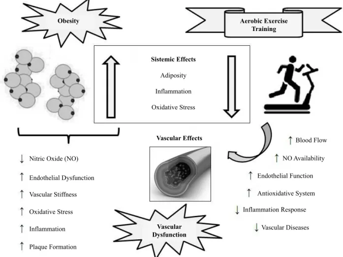
body weight, visceral obesity, and plasma glucose, as well as beneicial effects in cardiovascular parameters in obese rats and mice subjected to aerobic exercise training4. The aim of this mini-review is to summarize and integrate animal studies on the physiological mechanisms of vascular dysfunction in obesity and how they are inluenced by chronic aerobic exercise training (Figure 1). Special attention will be taken to describe the protocols of physical exercise on the treadmill, including itness training and strength analysis of the effectiveness of physical training in rodent models of obesity.
Vascular Dysfunction in Obesity
Normal blood vessels have a structure consisting of three layers: tunica intima (endothelial cells), tunica media (smooth muscle cells) and tunica adventitia (extracellular matrix). Endothelial cells are responsible for the synthesis, metabolism and release of a large variety of mediators that regulate platelet and leukocyte activity, vascular tone, vascular permeability and the metabolism of endogenous and exogenous substances. The integrity of the endothelial cells is of fundamental importance in the maintenance and control of the cardiovascular system.
The inluence of the endothelium in vascular tone is modu -lated by the synthesis and release of vasoconstriction (endothe-lin-1, prostaglandins, tromboxane A2 – TXA2, angiotensin II and the reactive oxygen species – ROS) and vasodilator substances (nitric oxide – NO, prostacyclin – PGI2 and endothelium-derived hyperpolarizing factors – EDHF). NO requires special attention, because of its important role in regulatory functions. In the regula-tion of vascular tone, NO has a signiicant vasodilator funcregula-tion, being able to spread easily between biological membranes5.
The nitric oxide enzymes (NOS), expressed in various tissues of the body, catalyze the oxidation of L-arginine in the presence of nicotinamide-adenine-dinucleotide phosphate (NADPH) Mini-Review
Vascular dysfunction in obesity: Beneicial effects
of aerobic exercise training in animal models
Amanda Christine da Silva Sponton Andressa Silva Sousa Maria Andréia Delbin
Universidade Estadual de Campinas, Campinas, SP, Brasil
Abstract — Cardiovascular disease (CVD) is the leading cause of mortality in the world and several risk factors for developing CVD have been pointed out, including obesity and physical inactivity. Endothelial dysfunction as a consequence of metabolic and inlammatory disorders plays an important role in the onset of vascular complications in obesity. In addition, it is well established that aerobic exercises promote beneicial effects on CVD by increasing nitric oxide (NO) production or its bioavailability in human and experimental models. The interest in exercise studies increased signiicantly, with promising results. Considering the importance of this ield, the purpose of this mini-review is to summarize the animal studies that investigated the physiological mechanisms of vascular dysfunction in obesity and how aerobic exercise training inluenced these alterations.
in NO and L-citruline. In mammals, NO can be generated by three different isoforms of the NO synthase enzyme: neuronal NOS (nNOS or NOS I), inducible NOS (iNOS or NOS II), and endothelial NOS (eNOS or NOS III). All isoforms of NOS use lavin adenine dinucleotide (FAD), lavin mononucleotide (FMN), and (6R-) – 5,6,7,8-tetrahydro-L-biopterin (BH4) as cofactors. Endothelial NOS (eNOS) is mostly expressed in
endothelial cells, which are very sensitive to the chemical stimu-lation of agonists such as bradykinin, acetylcholine, histamine, and adenosine triphosphate, among others. However, the best established stimulus is shear stress (mechanical stimulation of blood low); its activation is mediated by the phosphorylation of the enzyme and it does not produce sustained increases in intracellular Ca2+, but still induces a long-lasting release of NO5.
Obesity Aerobic Exercise
Training
Sistemic Effects
Adiposity
Inflammation
Oxidative Stress
Nitric Oxide (NO)
Endothelial Dysfunction
Vascular Stiffness
Oxidative Stress
Inflammation
Plaque Formation
Vascular Dysfunction
Vascular Effects Blood Flow
NO Availability
Endothelial Function
Antioxidative System
Inflammation Response
Vascular Diseases
Figure 1. Chronic effects of aerobic exercise training on vascular dysfunction in obese animals.
Endothelial dysfunction (ED) is characterized by an imbal-ance between endothelium-derived relaxing and contracting factors, and is mostly associated with lower production and/or bioavailability of NO by endothelial cells3. ED as a consequence of metabolic and inlammatory disorders plays an important role in the initiation of vascular complications in obesity6, and it is the hallmark of obesity associated with erectile dysfunction, coronary artery disease and peripheral artery disease7,8. Previous studies have demonstrated an impairment of relaxant responses in the aorta, mesenteric9, femoral, and coronary arteries10 in experi-mental models of obesity, that were associated with diminished NO release. The impaired vascular function and an increased vascular oxidative stress observed in obesity were associated with increased levels of inlammatory adipocytokines such as leptin, and tumor necrosis factor alpha – TNF-α, as well as decreased levels of anti-inlammatory adipocytokines such as adiponectin11.
and aortic endothelium function, has been demonstrated16. It has been shown that there is an imbalance between the release of in-lammatory and anti-inin-lammatory adipocytokines in obesity, and that it contributes for vascular oxidative stress17. This alteration is involved in the increased production of ROS such as superoxide anions (O2-), and its reaction with endothelial-derived NO reduces its bioavailability and leads to a highly unstable molecule, per-oxynitrite (ONOO-). The damage in cells and tissues by ROS has been associated with the development of endothelial dysfunction, and consequently with cardiovascular diseases18. There are many studies that associate obesity and its oxidative and inlammatory disorders with vascular dysfunction, however the mechanisms involved are complex and not fully understood19. Therefore, stud-ies involving obese animal models are important to elucidate and develop new approaches in this ield, specially the involvement of obesity with cardiovascular diseases.
Physical Exercise and Aerobic Protocols in Animal Models
According to the Centers for Disease Control and Prevention (CDC), physical activity is deined as any bodily movement produced by the contraction of skeletal muscles that increases energy expenditure above the basal level. It generally refers to the subset of physical activities that enhance health. In turn, physical exercises are deined as a subcategory of physical activity that is planned, structured, repetitive, and purposive in the sense that it has the improvement or maintenance of one or more components of physical itness as objective20. On the other hand, physical inactivity is deined as physical activity levels lower than those required for optimal health and prevention of premature death, and functional capacity is the ability of a cell, organ, system, or body to maintain homeostasis within their narrow limits of survival in response to a speciied stress21. It is well established that regular exercise not only enhances functional capacity but also brings about beneicial health outcomes22. Physical exercise leads to a variety of morphological and functional adaptations, which are known as the chronic effects of exercise23.
Speciically, cardiorespiratory endurance or aerobic en -durance is the ability of the whole body to sustain prolonged and rhythmic exercise24. This type of adaptation involves an increase in the capacity of the muscles for aerobic metabolism with an increase in endurance and is found in its most highly developed form in the muscles of competitive athletes, such as long-distance runners, long-distance cross-country skiers, bicyclists, and swimmers. It is well established that endurance exercises involving large muscle groups are recommended for the maintenance or improvement of cardiovascular itness25. Considering the cardiovascular beneits of exercise have been attributed to aerobic exercise training, in this review we will focus on this type of exercise training.
The intensity of exercise could determine the degree of beneit to the cardiovascular system. Two tests of cardiorespi -ratory endurance – submaximal and maximal – appear in the literature. The test of submaximal (cardiorespiratory) endurance capacity is more closely related to actual competitive endurance
performance and is likely determined by both the individual’s maximum oxygen consumption (VO2max) and his or her lac-tate threshold. Maximal cardiorespiratory endurance capacity (aerobic power – VO2peak) is deined as the highest rate of oxygen consumption attainable during maximal or exhaustive exercise24. Well-controlled training studies in animal models are important, as they form the basis for translational research.
In rats and mice, accurate training intensity may be obtained by directly measuring the maximum oxygen consumption (VO2max). For animal studies to mimic human standards for maximal cardiovascular endurance tests a determination of the VO2max or VO2peak may be obtained, as previously described26. This approach, however, is time consuming and expensive, es-pecially in comprehensive and long-lasting studies. Therefore, several other methods have been proposed to estimate exercise intensity without directly measuring the VO2max. Heart rate increases linearly with power output and VO2 both in humans6 and rats7. In humans, this method is easy to use through a num-ber of commercial systems. In rats and mice, control by heart rate is more challenging, as monitoring requires implantation of transducers that impose a signiicant extra weight, especially in mice. Controlling training intensity by critical velocity8, and lactate threshold has also been proposed27. The lactate threshold normally changes during a period of regular training, and frequent assessments are necessary for it to be used as a means of control-ling the training stimulus6. In addition, frequent blood sampling is likely to affect the performance because of increased stress levels and reduced hemoglobin. Moreover, a correlation between the VO2max and maximal running speed in rats and mice has been established. A previous study demonstrated that maximal running speed may be used both as a tool to adjust training intensity and to estimate the VO2max at low and moderate intensity exercise training26. Thus, this method may be a great way to control the intensity of physical exercise training in animal models.
in terms of duration, frequency and intensity is therefore lost. Therefore, the question is whether the intensity of voluntary wheel running would provide the same protective beneits as 1 hour/day of controlled/forced exercise training. Mice run most intensely during the irst several hours of the beginning of the active dark cycle, and it has been shown that voluntary wheel running for one hour a day ive days a week generates physiological responses in mice in the long term31. However, more studies are needed to assess the health beneits of voluntary wheel running in mice, particularly in chronic disease models.
Chronic Effects of Aerobic Exercise Training on Vascular Dysfunction in Obesity
The association between obesity and physical inactivity is the most important risk factor for the development of cardiovascular diseases (CVD). Actually, physical inactivity is now regarded as one of the most prevalent cardiovascular risk factors2. Numerous studies, conirmed by meta-analyses, indicate that exercise training reduces cardiovascular mortality and cardiovascular events32, particularly stroke, coronary heart disease, heart failure, and atherosclerosis33. Moreover, exercise training is an effective therapeutic strategy for patients with peripheral arterial diseases, coronary heart disease, heart failure, atherosclerosis, and hy-pertension32. Speciically for the endothelial function, the time course of endothelial adaptation following acute and regular exercise in rats was demonstrated. They found that a single bout of exercise improves endothelium-dependent dilation for about 2 days, with peak effect after 12-24 h, and regular exercise further improves adaptation and promotes an approximately fourfold increase in the sensitivity to acetylcholine, which slowly returns to sedentary levels within a week of detraining34.
The cardiovascular beneits of exercise have been frequently attributed to the reduction of many classical cardiovascular risk factors including blood lipids, high blood pressure, obesity, glucose, and type 2 diabetes as well as novel risk factors such as inlammation, and oxidative stress32,35. In the endothelial cells one of the most important molecular consequences of exercise training is the increase of vascular NO concentration. NO is responsible for vasodilation, which results in the lowering of peripheral resistance. The eNOS is up-regulated by an increase in low-mediated shear stress associated with physical exercise due to a complex pattern of intracellular regulation like acety-lation36 phosphorylation37 and translocation to the caveolae38. It is now clearly documented that exercise or increased shear
stress up-regulates eNOS activity in animal models39. Besides the cardiovascular beneits of exercise training associated with a variety of cellular and molecular alterations including up-regulation of eNOS, an increase in the expression and/or activity of antioxidant enzymes, as well as a decrease in pro-oxidant enzyme systems have been demonstrated in animal models40. The antioxidant defense systems consist of enzymes such as superoxide dismutase (SOD), catalase and glutathione peroxidase. These enzymes are scavengers of ROS resulting in an increase of NO bioavailability to the vascular smooth muscle and enhancement of endothelium-dependent vasodilatation9. However, it should be noted that exercise intensity seems to be a crucial variable in this response. A recent study evaluated the impact of exercise training on vascular gene expression proiles, using a transcriptome-wide RNA-Seq analysis in obese rats41. In particular, the analysis was performed on the soleus and gastrocnemius muscle feed arteries (SFA and GFA, respectively) and aortic endothelial cell-enriched samples from obese Otsuka Long-Evans Tokushima Fatty (OLETF) rats that underwent an endurance exercise training program (EndEx) or interval sprint training program (IST). They found that the number of genes which had their expression altered in response to both EndEx and IST was greater in the SFA compared with the GFA. Considering the fact that exercise produces greater relative increases in blood low in the gastrocnemius muscle compared with the soleus muscle, these data suggest an interest-ing disassociation between the magnitude of exercise-induced vascular transcriptional changes and the magnitude of exercise-induced blood low stimulus to which the arteries are exposed. They suggested that the signals produced by bouts of exercise that result in altered gene expression are not totally driven by hemodynamic forces associated with increased blood low, suggesting that other signals produced by exercise may also be involved41. Thus, it is still unclear which mechanisms explain the beneicial effects of exercise in vascular cells.
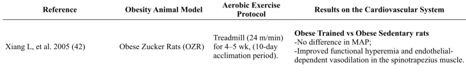
Therefore, well-controlled training studies in animal models are important, as they form the basis for translational research. Considering the increased number of studies which investigate the effects of exercise training in pathological conditions, such as cardiovascular diseases and obesity, in recent years, it is important to know which methodologies are employed to study exercise or exercise training, and also if the exercise protocols were properly described by the authors. In this review we focused on animal models of obesity studies that investigated the effects of aerobic exercise training on the cardiovascular system, which were summarized in Table 1.
Table 1: Chronic effects of aerobic exercise training on the cardiovascular system in obese animals: summary of studies.
Reference Obesity Animal Model Aerobic Exercise
Protocol Results on the Cardiovascular System
Xiang L, et al. 2005 (42) Obese Zucker Rats (OZR)
Treadmill (24 m/min) for 4–5 wk, (10-day acclimation period).
Obese Trained vs Obese Sedentary rats
-No difference in MAP;
Reference Obesity Animal Model Aerobic Exercise
Protocol Results on the Cardiovascular System
De Moraes C, et al. 2008 (9) Male Wistar rats, high-caloric diet
Treadmill (70-80% VO2max), 1h/day, 5 days/wk for 12 wk.
Obese Trained vs Obese Sedentary rats
-No difference in NOx – levels;
-Improved relaxation responses to acetylcholine in aortic and mesenteric rings;
-Increased protein expression of CuZn-SOD in aorta and mesenteric arteries.
Sebai M, et al. 2011 (43) Obese Zucker Rats (OZR)
Treadmill (24 m/min), 30 min/day, 5 days/wk for 6 wk.
Obese Trained vs Obese Sedentary rats
-Increased VO2max and workload;
-Improved functional vasodilation in spinotrapezius and cremaster muscles.
Touati S, et al. 2011 (44) Male Sprague-Dawley rats, high-fat diet for 12 wk.
Treadmill (27 m/min), 60 min/day, 5 days/wk for 12 wk.
Obese Trained vs Obese Sedentary rats
-Decreased SAP and atherogenic index;
-Improved endothelium-dependent relaxation to ace-tylcholine and insulin in aorta;
-Increased contents of aortic p-Akt at Serin473 and
peNOS at Serin1177.
Barbosa VA, et al. 2012 (45) Male Wistar rats, high fat-diet for 8 wk
Treadmill (1.0 km/h), 50 min/day, 5 days/wk for 8 wk.
Obese Trained vs Obese Sedentary rats
-Decreased TBARS and superoxide in myocardial tissue;
-Decreased TBARS, superoxide and carbonyl con-tent in aorta;
-Increased CuZn-SOD, CAT and GPx activities in myocardial and aorta.
Barretti DL, et al. 2012 (46) Obese Zucker Rats (OZR)
Swimming sessions (1h/day, 5 days/wk, for 10 wk), wearing caudal dumbbells weigh-ing 5% of their body weight.
Obese Trained vs Obese Sedentary rats
-No difference in the systemic ACE activity, resting heart rate and cardiac function;
-Prevented increase in cardiac hypertrophy, cardiac ACE activity, Ang II and AT2 receptor;
-Increased cardiac protein expression and activity of ACE2.
Mostarda C, et al. 2012 (47)
Male Wistar rats, overload of D-fructose (100 g/l) in drinking water for 10 wk
Treadmill (50-70% of the maximum running speed), 1h/day, 5 days/ wk for 10 wk.
Obese Trained vs Obese Sedentary rats
-Improvement in SAP, DAP, MAP, and diastolic function;
-No difference in HR, and systolic function.
Moraes-Silva IC, et al. 2013 (48)
Male Wistar rats overload of D-fructose (100 g/l) in drinking water for 10 wk
Treadmill (50 –70% maximal running speed, 0.6 –1.3 km/h, 0% inclination), 1h/ day, 5 days/wk for 10 wk.
Obese Trained vs Obese Sedentary rats
-Improvement in SAP, DAP, MAP, HRV, SAPV, RMSSD, TR and BR;
-No difference in HR.
Boa BC, et al. 2014 (49)
Male hamsters, high-fat diet starting on the 21st day of birth, for 12 wk.
Treadmill (50–70% of VO2max), 1h/day, 5days/ wk for 8 wk.
Obese Trained vs Obese Sedentary rats
-Decreased MAP and HR;
-Improved endothelial-dependent vasodilation in microcirculatory and macromolecular permeability after ischemia/reperfusion in three 2nd to 3rd order arterioles;
-Increased eNOS protein expression in thoracic aorta.
Oharomari LK, et al. 2014 (50) Male Wistar rats, highly palatable diet for 11 wk
Treadmill (60% maxi-mal running speed), 1h/day, 5 days/wk for 7 wk.
Obese Trained vs Obese Sedentary rats
-Improved relaxation response in aorta;
-Increased protein expression of Ec-SOD, CuZn-SOD and reduced gp91phox in aorta;
-Decreased superoxide formation in aorta.
Pieri BL, et al. 2014 (51) Diet-induced obesity (DIO) Wistar rats
Treadmill 50-min/day, 5 days/wk, velocity of 1.0 km/h for 8 wk.
Obese Trained vs Obese Sedentary rats
Conclusion
The association between obesity and physical inactivity is the most important risk factor for the development of cardiovascular diseases. Endothelial dysfunction as a consequence of metabolic and inlammatory disorders plays an important role in the initiation of vascular complications in obesity. On the other hand, many studies have shown the importance and effectiveness of exercise training in the prevention and treatment of cardiovascular disease in obesity. However, it is still unclear which mechanisms explain the beneicial effect of exercise in vascular cells. Therefore, well-controlled training studies in animal models are important, as they form the basis for translational research.
In summary, animal models of aerobic exercise training against obesity show promising beneits to the cardiovascular system. We found that most studies investigating the effects of aerobic exercise training on the cardiovascular system in animal models of obesity were concerned with the exercise training pro-tocol used and also have been reported properly by the authors.
References
1. Besnard P. Lipids and obesity: Also a matter of taste? Rev Endocr Metab Disord [Internet]. Reviews in Endocrine and Metabolic Disorders. 2016;May10 Available from: http://dx.doi.org/10.1007/ s11154-016-9355-2.
2. Word Health Organization. Global recommendationson physical activity for heathy. Word Health Organization. Geneva, out. 2010. 3. Tschöp M, Heiman HL. Rodent obesity models: an overview. Exp
Clin Endocrinol & Diabetes. 2001;109:307-319.
4. Pons S, Martin V, Portal L, Zini R, Morin D. Regular treadmill ex-ercise restores cardioprotective signaling pathways in obese mice independently from improvement in associated co-morbidities. J Mol Cell Cardiol. 2013;54:82–89.
5. Forstermann U, Sessa WC. Nitric oxide synthase: regulation and function. Eur Heart J. 2011;33(7):829-37.
6. Astrand P, Rodahl K. Textbook of work physiology; physiological bases of exercise. Ed. New York, McGraw-Hill, 1986.
7. Morhardt JE, Morhardt SS. Correlations between heart rate and oxygen consumption in rodents. Am J Physiol. 1971; 221:1580-86. 8. Trask AJ, Delbin MA, Katz PS, Zanesco A, Lucchesi PA.
Differential coronary resistance microvessel remodeling between type 1 and type 2 diabetic mice: impact of exercise training. Vasc Pharmacol. 2012;57:187-93.
9. de Moraes C, Davel AP, Rossoni LV, Antunes E, Zanesco A. Exercise training improves relaxation response and SOD-1 ex-pression in aortic and mesenteric rings from high caloric diet-fed rats. BMC Physiol. 2008;8:12.
10. Bender SB, Laughlin MH. Modulation of endothelial cell phenotype by physical activity: impact on obesity-related endothelial dysfunc-tion. Am J Physiol Heart Circ Physiol. 2015;309(1):H1-8. 11. Donato AJ, Henson GD, Morgan RG, Enz RA, Walker AE,
Lesniewski LA. TNF-α impairs endothelial function in adipose
tissue resistance arteries of mice with diet-induced obesity. Am J Physiol Heart Circ Physiol. 2012;303:H672-9.
12. Haynes WG. Interaction between leptin and sympathetic nervous system in hypertension. Curr Hypertens Rep. 2000;2(3):311-8. 13. Korda M, Kubant R, Patton S, Malinski T. Leptin-induced
endo-thelial dysfunction in obesity. Am J Physiol Heart Circ Physiol. 2008;295:H1514-21.
14. Kleinbongard P, Heusch G, Schulz R. TNFalpha in atherosclerosis, myocardial ischemia/reperfusion and heart failure. Pharmacol Ther. 2010;127:295-314.
15. Chen H, Montagnani M, Funahashi T, Shimomura I, Quon MJ. Adiponectin stimulates production of nitric oxide in vascular endothelial cells. J Biol Chem. 2003;278(45): 45021-6. 16. Zhang H, Park Y, Zhang C. Coronary and aortic endothelial
func-tion affected by feedback between adiponectin and tumor
necro-sis factor α in type 2 diabetic mice. Arterioscler Thromb Vasc
Biol. 2010;30(11):2156-63.
17. Berg AH, Scherer PE. Adipose tissue, inlammation, and cardio -vascular disease. Circ Res. 2005;96(9):939-49.
18. Galili O, Versari D, Sattler KJ, Olson ML, Mannheim D, McConnell JP, et al. Early experimental obesity is associated
Reference Obesity Animal Model Aerobic Exercise
Protocol Results on the Cardiovascular System
Touati S, et al. 2015 (52) Male Sprague-Dawley rats, high-fat diet for 12 wk.
Treadmill (27 m/min), 60 min/day, 5 days/wk for 12 wk.
Obese Trained vs Obese Sedentary rats
-Decreased NADPH oxidase activity in aorta; -No difference in Nox1 and Nox2 expression in aorta.
-Decreased protein expression of Nox4, p47phox
translocation, VCAM-1, pERK 1/2 (Thr202/Tyr204)
and pJNK/SAPK (Thr183/Tyr185) in aorta;
-Increased CuZn-SOD protein expression in aorta.
with coronary endothelial dysfunction and oxidative stress. Am J Physiol Heart Circ Physiol. 2007;292(2):H904-11.
19. Hajer GR, Van Haeften TW, Visseren FL. Adipose tissue dys-function in obesity, diabetes, and vascular diseases. Eur Heart J. 2008;29:2959-71.
20. Centers for Disease Control and Prevention. Available from: https://www.cdc.gov/physicalactivity/
21. Booth FW, Roberts CK, Laye MJ. Lack of exercise is a major cause of chronic diseases. Compr Physiol. 2012;2(2):1143-211. 22. Haskell WL, Lee IM, Pate RR, Powell KE, Blair SN, Franklin BA,
et al. Physical activity and public health: updated recommendation for adults from the American College of Sports Medicine and the J Am Heart Assoc. 2007;116(9):1081-93.
23. da Nobrega. The subacute effects of exercise: concept, char-acteristics, and clinical implications. Exerc Sport Sci Rev. 2005;33:84-87.
24. Wilmore JH, Costill DL. Physiology of Sport and Exercise. Ed. (3rd) Champion, Illinois: Human Kinetics, 2004.
25. Garber CE, Blissmer B, Deschenes MR, Franklin BA, Lamonte MJ, Lee IM, et al. American College of Sports Medicine posi-tion stand. Quantity and quality of exercise for developing and maintaining cardiorespiratory, musculoskeletal, and neuromotor
itness in apparently healthy adults: guidance for prescribing
exercise. Med Sci Sports Exerc. 2011;43:1334-59.
26. Hoydal MA, Wisloff U, Kemi OJ, Ellingsen O. Running speed and maximal oxygen uptake in rats and mice: practical implications for exercise training. Eur J Cardiovasc Prev Rehabil. 2007;14(6):753-60. 27. Delbin MA, Davel AP, Couto GK, de Araújo GG, Rossoni LV,
Antunes E, et al. Interaction between advanced glycation end prod-ucts formation and vascular responses in femoral and coronary arteries from exercise rats. PLoS One. 2012;7:e53318.
28. Goh J, Ladiges W. Voluntary wheel running in mice. Curr Protoc Mouse Biol. 2015;5(4):283-90.
29. Moraska A, Deak T, Spencer RL, Roth D, Fleshner M. Treadmill running produces both positive and negative physiological adapta-tions in Sprague-Dawley rats. Am J Physiol Regul Integr Comp Physiol. 2000;279:R1321-29.
30. de Bono JP, Adlam D, Paterson DJ, Channon KM. Novel quan-titative phenotypes of exercise training in mouse models. Am J Physiol Integr Comp Physiol. 2006;290:R926-R934.
31. Goh J, and Ladiges WC. A novel long term short interval physical activity regime improves body composition in mice. BMC Res Notes. 2013;6:66.
32. Taylor RS, Brown A, Ebrahim S, Jolliffe J, Noorani H, Rees K, et al. Exercise-based rehabilitation for patients with coronary heart disease: systematic review and meta-analysis of randomized controlled trials. Am J Med. 2004;116(10):682-92.
33. Thompson PD, Buchner D, Pina IL, Balady GJ, Williams MA, Marcus BH, et al. Exercise and physical activity in the pre-vention and treatment of atherosclerotic cardiovascular disease. Circulation. 2003;107(24):3109-3116.
34. Haram PM, Adams V, Kemi OJ, Brubakk AO, Hambrecht R, Ellingsen O, Wisløff U. Time-course of endothelial adapta-tion following acute and regular exercise. Eur J Cardiovasc Prev Rehabil. 2006;13(4):585-91.
35. Bacchi E, Negri C, Zanolin ME, Milanese C, Faccioli N, Trombetta M, et al. Metabolic effects of aerobic training and resistance
training in type 2 diabetic subjects: a randomized controlled trial (the RAED2 study). Diabetes Care. 2012;35(4):676-682. 36. Busconi L, Michel T. Endothelial nitric oxide synthase; N-terminal
myristoylation determines subcellular localization. J Biol Chem. 1993;268:8410-8413.
37. Kolluru GK, Siamwala JH, Chatterjee S. eNOS phosphorylation in health and disease. Biochimie. 2010;92:1186-1198.
38. Ortiz PA, Garvin JL. Traficking and activation of eNOS in epi -thelial cells. Acta Physiol Scand. 2003;179:107-114.
39. Woodman CR, Muller JM, Laughlin MH, Price EM. Induction of nitric oxide synthase mRNA in coronary resistance ar-teries isolated from exercise-trained pigs. Am J Physiol. 1997;273:H2575-H2579.
40. Zanesco A, Antunes E. Effects of exercise training on the cardio-vascular system: pharmacological approaches. Pharmacol Ther. 2007;114(3):307-317.
41. Padilla J, Jenkins NT, Thorne PK, Martin JS, Rector RS, Davis JW, Laughlin MH. Transcriptome-wide RNA sequencing analysis of rat skeletal muscle feed arteries. II. Impact of exercisetrain-ing in obesity. J Appl Physiol (1985). 2014;116(8):1033-47. 42. Xiang L, Naik J, Hester RL. Exercise-induced increase in skeletal
muscle vasodilatory responses in obese Zucker rats. Am J Physiol Regul Integr Comp Physiol. 2005;288(4):R987-91.
43. Sebai M, Lu S, Xiang L, Hester RL. Improved functional vaso-dilation in obese Zucker rats following exercise training. Am J Physiol Heart Circ Physiol. 2011;301(3):H1090-6.
44. Touati S, Meziri F, Devaux S, Berthelot A, Touyz RM, Laurant P. Exercise reverses metabolic syndrome in high-fat diet-induced obese rats. Med Sci Sports Exerc. 2011;43(3):398-407. 45. Barbosa VA, Luciano TF, Vitto MF, Cesconetto PA, Marques
SO, Souza DR, et al. Exercise training plays cardioprotection through the oxidative stress reduction in obese rats submitted to myocardial infarction. Int J Cardiol. 2012;157(3):422-4. 46. Barretti DL, Magalhães FC, Fernandes T, do Carmo EC, Rosa
KT, Irigoyen MC, et al. Effects of aerobic exercise training on cardiac renin-angiotensin system in an obese Zucker rat strain. PLoS One. 2012;7(10):e46114.
47. Mostarda C, Moraes-Silva IC, Salemi VMC, Machi JF, Rodrigues B, De Angelis K, et al. Exercise training prevents diastolic dys-function induced by metabolic syndrome in rats. Clinics (Sao Paulo). 2012;67(7):815-820.
48. Moraes-Silva IC, Mostarda C, Moreira ED, Silva KA, dos Santos F, de Angelis K, et al. Preventive role of exercise training in au-tonomic, hemodynamic, and metabolic parameters in rats under high risk of metabolic syndrome development. J Appl Physiol (1985). 2013;114(6):786-91.
49. Boa BC, Souza Md, Leite RD, da Silva SV, Barja-Fidalgo TC, Kraemer-Aguiar LG, Bouskela E. Chronic aerobic exer-cise associated to dietary modification improve endothelial function and eNOS expression in high fat fed hamsters. PLoS One. 2014;9(7):e102554.
50. Oharomari LK, Garcia NF, Freitas EC, Jordão Júnior AA, Ovídio PP, Maia AR, et al. Exercise training and taurine supplementation reduce oxidative stress and prevent endothelium dysfunction in rats fed a highly palatable diet. Life Sci. 2015;139:91-6. 51. Pieri BL, Souza DR, Luciano TF, Marques SO, Pauli JR, Silva
14-3-3 pathways in the myocardium of diet-induced obesity rats. Horm Metab Res. 2014;46(9):621-7.
52. Touati S, Montezano AC, Meziri F, Riva C, Touyz RM, Laurant P. Exercise training protects against atherosclerotic risk factors through vascular NADPH oxidase, extracellular signal – regulated kinase 1/2 and stress-activated protein kinase/c-Jun N-terminal kinase downregulation in obese rats. Clin Exp Pharmacol Physiol. 2015;42(2):179-85.
Acknowledgments
This work was supported in part by the Conselho Nacional de Desenvolvimento
Cientíico e Tecnológico – CNPq and the Coordenação de Aperfeiçoamento de Pes -soal de Nível Superior – CAPES.
Corresponding author
Maria A Delbin
Assistant Professor, Institute of Biology, Department of Structural and Functional Biology. Rua Monteiro Lobato, 255 – Cidade Universitária Zeferino Vaz. Campi-nas, São Paulo, Brazil.
Email: [email protected]
Manuscript received on September 14, 2016 Manuscript accepted on November 03, 2016