Mini Review
Hippocampal insulin signaling and
neuroprotection mediated by physical exercise in
Alzheimer´s Disease
Gabriel Keine Kuga
Universidade Estadual Paulista “Júlio de Mesquita Filho”, Rio Claro, SP, Brasil
José Diego Botezelli Rafael Calais Gaspar
Universidade Estadual de Campinas, Limeira, SP, Brasil
Ricardo José Gomes
Universidade Federal de São Paulo, Santos, SP, Brasil
José Rodrigo Pauli
Universidade Estadual de Campinas, Limeira, SP, Brasil
José Alexandre Curiacos de Almeida Leme Centro Universitário Auxilium Unisaleasiano, Lins, SP, Brasil
Abstract — Epidemiological studies indicate continuous increases in the prevalence of Alzheimer’s Disease (AD) in the next few decades. The key feature of this disease is hippocampal neurodegeneration. This structure has an important role in learning and memory. Intense research efforts have sought to elucidate neuroprotective mechanisms responsible for hippocampal integrity. Insulin signaling seems to be a very promising pathway for the prevention and treatment of AD. This hormone has been described as a powerful activator of neuronal survival. Recent research showed that
reduced insulin sensitivity leads to low-grade inlammation, and both phenomena are closely related to AD genesis. Concomitantly, exercise has been shown to exert anti-inlammatory effects and to promote improvement in insulin signaling in the hippocampus, which supports neuronal survival and constitutes an interesting non-pharmacological
alternative for the prevention and treatment of AD. This review examines recent advances in understanding the molecular mechanisms involved in hippocampal neuroprotection mediated by exercise.
Keywords: insulin, hippocampus, inlammation, physical exercise.
Introduction
Alzheimer's disease (AD) is the most common form of de-mentia manifestation1 and its prevalence increases with aging
population2. Alzheimer's Disease International3 estimates that
dementias reach around 46.8 million people in the world in 2015. Projections indicate that this number will double every 20
years, reaching a 74 million in 2030 and 131 million in 2050 3.
In 2050, it is expected that at least 100 million of these patients
will suffer from AD4,5. In the last year, 818 billion dollars were invested to treat the main forms of dementia. This cost includes
hospital expenses, caregivers services and personal expenses, and in 2030, it is estimated a budget of 2 trillion dollars3. Among
the reasons to justify this exorbitant cost it is the absence of an effective cure or therapeutic treatment that really slows the progress of AD and other dementias.
Despite signiicant progress in elucidating the molecular
mechanisms involved in neuron survival and the main agents involved in neuronal protection or neuron degeneration in the
hippocampus, the scientiic community still has big gaps to
translate these indings across species: therapies that work in
rodents often do not succeed in humans6.
In hippocampal neurons, insulin and growth factors such as BDNF (brain derived neurotrophic factor), IGF-1 and 2
(Insulin-like growth factors 1 and 2) and VEGF (vascular endothelial growth factor) transmit the intracellular signal for neuronal
integrity. Under regular physiological conditions, insulin and growth factors act properly to promote neuronal, survival keep -ing the hippocampus functionality. When insulin and growth factors are inhibited to properly exercise its effects we have a favorable condition for the establishment of AD. Changes in insulin and growth factors signaling disrupts neuronal survival contributing to AD pathogenesis. The neuropathology hallmark
of AD is characterized by accumulation of β-amyloid, synaptic dysfunction, neuronal inlammation, phosphorylation of tau protein and neuroibrillary tangle formation1,6.
Exercise has been shown to attenuate inlammation in the
hippocampus7. The insulin sensitizing effects of physical ex-ercise in peripheral tissues are well described in the literature8.
the central nervous system (SNC), especially in hippocampal
functions and a possible neuroprotective role in AD4. Therefore,
the objective of this review is to present the molecular aspects involved in the development of AD and the role of physical exercise in the prevention and treatment of this disorder. This
study is a narrative mini-review, including relevant work in the author’s ield of expertise and current literature. Articles
that describe the molecular mechanisms of cellular signaling
in AD were selected, with special attention to insulin signaling pathway. In addition, were included papers that highlight the
relevant role of physical exercise to prevent and attenuate the
inlammatory and neurodegenerative process of this disease.
Hippocampal Insulin Signaling
Insulin exerts intracellular signaling by binding to the insulin
receptor (IR). The IR is composed of two α-subunits and two β-subunits connected through disulide bonds. Insulin binding to α-subunit induces receptor conformational change and acti
-vates the tyrosine kinase activity of the β-subunit. The β subunit has an intrinsic tyrosine kinase activity, having the ability to
phosphorylate itself and other tyrosine substrates. Tyrosine phosphorylation of major insulin receptor substrates (IRS-1 and IRS-2) by IR creates binding sites for the enzyme
phos-phatidylinositol-3 kinase (PI3K), activating it. Once activated,
PI3K converts the membrane phospholipid phosphatidylinositol
4,5bisphosphate to phosphatidylinositol 3,4,5-trisphosphate. This
conversion mediates the activation of the protein kinase B (PKB/ Akt) through the 3-phosphoinositide dependent kinase (PDK). The protein kinase b PKB/Akt signaling coordinates diverse cellular responses8,9.One of the main actions of this protein in the hippocampus is to promote neuronal survival. Akt enhances
phosphorylation of FOXO transcription factor (forkhead box
transcriptional factor family) and promotes its extrusion from the cell nucleus for subsequent proteasomal degradation. The
FOXO presence in the nucleus acts as transcription factor of
pro-apoptotic proteins and contributes to degenerative process10.
In addition, Akt is responsible for inactivating protein of GSK3-β (glycogen synthase kinase 3 β). When activated in hippocampal neurons, GSK3-β promotes the phosphorylation of
tau protein a major contributor to AD. By reducing the ability of
insulin to propagate its intracellular signal, the continuous activa
-tion of GSK3-β promotes hyperphosphoryla-tion of tau protein9
and the subsequent formation of neuroibrillary tangles. All this
mechanism results deleterious effects on synaptic dysfunction and cognitive impairment1.
The accumulation of β-amyloid oligomers is another patho -genic aspect of AD. These oligomers are main component of
senile plaques seen in brains with AD. The β-amyloid protein is
a peptide originated from cleavage of amyloid precursor protein (APP) by gamma-secretase enzyme11 and leads to
neurotoxic-ity. Several studies have provided evidence between insulin
metabolism and β-amyloid accumulation. The hyperinsulinemia may increase the extracellular concentration of β-amyloid
protein by increasing its release from neurons and modulation of gamma-secretase enzyme activity12. Other indings have
shown that increased insulin concentration in the extracellular
medium competes with β-amyloid protein as substrate of the
insulin degradation enzyme (IDE). This protein may degrade
insulin or β-amyloid protein. By promoting the degradation of insulin instead of β-amyloid protein, this mechanism hits the neuron in two different ways. First, by reducing the degrada
-tion of β-amyloid protein and second, by reducing the binding of insulin IR and, thus, its ability to spread its neuroprotective
signal intracellularly12.
Inside the cell, the phosphorylation of IRS 1 and 2 also acti -vates the mitogen activated protein kinase (MAPK) cascade of Ras/MEK/ERK13, triggering multiple cell responses. The ERK
phosphorylation leads growth, differentiation and proliferation of hippocampal neurons, being essential for neuronal survival
and neurogenesis14 through the activation of cytosolic proteins
and transcription factors in the nucleus15. The main target protein
phosphorylation of ERK (extracellular signal-regulated kinase) is CREB (cAMP response binding protein). This protein func-tion in hippocampus results the formafunc-tion of long-term memory and increases synaptic plasticity16. These modiications generate
activity-dependent responses of synaptic transmission eficiency
(functional plasticity) and changes in the structure and number of synaptic connections (structural plasticity)17.
Understanding insulin signaling is important to understand
the connection between the mechanisms underlying inlam
-mation and the reduced action of this hormone. Moreover, this
accumulated knowledge involving the insulin signal transduction in the hippocampus has allowed the understanding of the effect of exercise to cooperate in mitigating the disease.
Inlammation and Insulin Resistance
The low-grade inlammation is a molecular process that reduces the capacity of insulin to propagate their intracellular signal,
culminating in a state of insulin resistance. Compensatory body's response to reduced insulin action consists of the chronic conditions of hyperinsulinemia18, providing the basis for the
establishment of Diabetes Mellitus Type 2 (DM2). It was found
that the AD has features in common with DM2, leading some
researchers to announce the AD as Diabetes Type 3. The insulin resistance and decrease of growth factors are closely associated with typical neurodegeneration in AD9. During inlammation,
hippocampal levels of pro-inlammatory cytokines such as TNF-α (tumor necrosis factor alpha) and β-IL1 (interleukin 1 β) are
elevated7. One factor that is highly related to increased cytokines is aging7,19 and this process is denominated "Inlammaging"20,21. This condition has high prevalence in the elderly population and AD carriers3. The cytokines bind to their receptors
trig-ger the inlammatory pathway through the JNK protein (c-Jun
N-terminal kinase) and IKK (I kappa B kinase) activation. Both
proteins disrupt insulin signaling and exacerbate inlammation by increasing the transcription of inlammatory genes22.
JNK is a serine kinase activated by TNF-α binding to the
receptor or through the activation of TLR-4 receptors (Toll-like receptor 4)21. When activated by these pathways, the JNK pro
of its signaling cascade and constituting a major mechanism of insulin resistance22. JNK also acts as activator of the transcription
factor AP1 leading to the transcription inlammatory genes and
cytokines production22. The post-mortem brains from AD patients
showed increased phosphorylation of IRS-1 in the serine307
residue and JNK expression, just as like in T2DM patients23.
Other studies have shown that both oligomers of β-amyloid
protein23 as a high fat diet activate JNK phosphorylation in
hippocampal neurons24. Both indings led to increased IRS-1
serine307 phosphorylation and tau protein levels. This mechanism
brings a strong association between AD and obesity due to the reduction of insulin signaling in neuronal survival. Although
the aging process is associated with increased inlammation,
this situation is even worse in the obese elderly.
The other mediator of insulin resistance is IKK. When
activated by the signaling cascade of TNF-α and TLR-4, the IKK cleaves the NF-kB/Iκβ complex19. This complex disruption
releases the transcription factor NF-kB (nuclear factor kappa
B), which migrates to the nucleus and initiates transcription proteins such as TNF-α, IL1-β and iNOS7,19,22. In addition, IKK
is also capable of promoting IRS-1 serine307 phosphorylation in
hippocampal neurons upon activation by oligomers of β-amyloid
protein23. The aging contributes to increased activation of
NF-kB transcription factor in hippocampus and may be one of the factors by which AD is mainly related to elderly population7.
The numerous events that lead to activation of inlammatory
signaling involve formation and clearance of several different proteins. The crucial organelle for this machinery is the endo-plasmic reticulum (ER). This structure is a major regulator of
inlammation feedback. This important site is responsible for
the proper synthesis and protein folding22. This organelle is also
highly sensitive to disturbances in cellular homeostasis and ER
malfunction increases the concentration of misfolded proteins,
altering the normal functionality of these proteins. This state of unbalance of protein folding is denominated endoplasmic reticulum stress25. The circulating lipids has a powerful effect of
endoplasmic reticulum stress, linking the excessive consumption
of this macronutrient and obesity25. As cellular response to restore
the level of unfolded proteins properly there is a mechanism denominated UPR (unfolded protein response). This mechanism consists in the activation of proteins IRE1 (inositol-requiring
enzyme 1), PERK (protein kinase (PKR) -like endoplasmic
reticulum kinase) and ATF6 (activating transcription factor 6).
This regulation reduces the overall transcription of inlamma
-tory agents and increase the synthesis of chaperones, restoring
the proper function of endoplasmic reticulum22. However, with
persistent ER stress, the UPR ultimately triggers cellular de
-fense mechanisms by inlammation and programmed cell death
(apoptosis) in last case. This role process involves the previous described proteins JNK and NF-kB.
In summary, these mechanisms (JNK, IKK and ER stress) are the major agents of low-grade inlammation and insulin
resistance. Activation of these pathways in the hippocampus results in reduced ability of insulin to exerts its neuroprotective
effect and the consequences are accumulation of β-amyloid oligomers, hyperphosphorylation of tau protein and neuronal apoptosis, creating favorable conditions for the genesis of AD.
The next step is to discover how exercise works in this context
to reduce the expression and activity of inlammatory and
apoptotic proteins.
Neuroprotection Mediated by Physical Exercise
The effects of exercise on insulin signaling pathway have been
intensively investigated in the last few years. On peripheral tissues, exercise stimulates the glucose uptake independent
of insulin via activation of protein kinase activated by AMP
(AMPK) and mTOR (mammalian target of rapamycin)26. In
addition, the exercise reduces the activation of serine kinase
JNK and IKK27, attenuating the subclinical inlammation and
the deleterious effects pro-inlammatory cytokines on the insulin signal transduction. However, physical exercise exerts different effects on the CNS. Several beneicial results demonstrated that
physical exercise improves synaptic plasticity and cognition13,28, regulates the production and degradation of neurotransmitters29,30 and increases angiogenesis31 and neurogenesis32. Although the
molecular mechanisms responsible for these events are not
completely understood, there are strong evidences showing
the exercise mediating the production and sensitivity to growth
factors such as BDNF, IGF-1, IGF-2 and VEGF1,28,32,33.
Diegues and colleagues13 conducted an elegant study in which
investigated the molecular mechanisms of physical training in diabetic Wistar rats. After induction of diabetes by administration
of Alloxan, these researchers have found impaired performance
in Morris water maze (spatial memory test) in sedentary diabetic rats compared to diabetic animals subjected to physical training
(6 weeks of swimming, 1 hour daily for 5 days per week and the animal supporting load corresponding to 90% of the anaerobic
threshold). Further analysis in hippocampus showed increased IGF-1 receptor expression and phosphorylation of Akt-1/2
iso-forms in trained groups. The sedentary diabetic rats, however, presented increased β-amyloid phosphorylation. Furthermore,
the exercise reduced both the expression of APP and the
phos-phorylation of tau protein, which is consistent evidence about
the neuroprotective effects of exercise on hippocampal neurons and prevention of AD.
The hippocampal neuroprotection mediated by exercise has been under investigation of Um and colleagues34. In this
remarkable study, Tg-NSE / hPS2m mice (transgenic model of
AD) performed physical training for 3 months on a treadmill (5 days per week with a daily 60 minutes and speed of 12 m/min).
Analyzing hippocampal samples, they found reduced expression of β-amyloid protein, Cox-2 (cyclooxygenase 2) and caspase-3
(pro-apoptotic protein) in trained animals. These alterations were concomitant to reduced JNK and tau phosphorylation
and increased ERK phosphorylation, CREB, PI3K, Akt and GSK3-β, all involved in insulin signaling pathway. They also
found reduced apoptotic state of hippocampal neurons in mice trained through a TUNEL assay to detect DNA fragmentation
typical of apoptotic process. In addition, they have found a
substantial increase of BDNF expression. These results demon-strate in mice the powerful effect of exercise training to reduce
or even reverse the inlammation and apoptosis of hippocampal
The high fat diet consumption and obesity are one of the main cause of ER stress. Feeding Sprague-Dawley rats with high fat
diet for 8 weeks was suficient to induce obesity and ER stress
in hippocampus35. A study conducted by Cai and colleagues35
supported this fact, revealing increased IRE- 1 and eIF2a phos -phorylation/total extract protein ration in obese rats. Physical
training treadmill (8 weeks, 5 days per week with 40 minutes
daily and speed of 18m/min) have shown a powerful tool in this scenario. Physical exercise recovered the ERK and CREB phosphorylation in high fat animals in comparison to sedentary
chow-fed animals. In fact, the trained chow-fed animals showed
the highest levels of ERK and CREB phosphorylation among
all groups studied. Obesity increased expression of apoptotic proteins (caspase-12, CHOP and Bax) and reduced Bcl-2(anti-apoptotic) content. However, it was signiicantly restored in the obese trained group in comparison to obese sedentary group,
but not to levels of lean animals. While physical exercise
con-tributed to reduction of ER stress, these results demonstrate that
the losses caused by obesity are not fully restored. This gap can
be an important target for development of more eficient proto
-cols of physical training. Further, reinforcing the importance of
weight control and reduced intake of saturated fats to prevent the degeneration of hippocampal neurons.
The activation of the transcription factor NF-kB appears to have close relation to aging and the AD development. Aged Wistar animals (20 months) showed increased activation of NF-kB in hippocampus compared to young animals (3 months)
7. In the same study, it was investigated the effect of two weeks
of exercise on a treadmill (20 minutes per day to 60% of the
estimated intensity of maximum consumption of animal oxygen)
on the activation of NF-kB in the hippocampus, and there was no signiicant difference between young animals that were or not subjected to exercise. However, in older animals (20 months),
physical exercise was able to reduce the NF-kB activation in the hippocampus 1 hour after the last exercise bout. In this
par-ticular study, Wistar rats were not induced obesity, diabetes or DA, leading us to believe that exercise may attenuates the effect
of aging7. Future studies shall be conducted to investigate the effects of exercise on the NF-kB activation in the hippocampus of animals at different ages.
Recent results indicate that physical exercise promotes increased phosphorylation of AMPK in the hippocampus36,37. AMPK is an essential regulator of autophagy leading degrada-tion and inhibidegrada-tion of poorly folded proteins. This mechanism prevents and stops the ER stress contributing for the cell func-tion. The pharmacological activation of AMPK in cell culture
was able to promote the degradation of β-amyloid protein38.
However, further researches need to investigate the AMPK
activation mediated by physical exercise in vivo.
The effects of exercise on insulin signaling and neuroinlam -mation are truly relevant in the hippocampus. Physical training
reduces the expression of β-amyloid (34) and tau phosphoryla -tion13, classic hallmarks in development of AD. The increased
signal transduction of the insulin pathway of key proteins in
the hippocampus, as Akt and ERK, is an indication that there is direct positive effect on neuronal survival. Furthermore, the insulin signaling acts on inlammation, reducing the JNK
phosphorylation, NF-kB activation and transcription of both pro-inlammatory and pro-apoptotic proteins. For all these ac
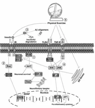
-tions, it is possible that the exercise is still an underestimated method, but promising method for prevention and treatment of AD. In the same context, Figure 1 shows an original and schematic illustration of the effects of exercise, which promote
neuroprotection in the hippocampus.
Figure 1. In hippocampal neurons, binding of insulin to
the IR active routes of PI3-K/Akt and MAPKs. 1) PI3K/Akt
activation leads to inhibition of GSK3 beta activity, reducing phosphorylation of tau protein, a protein strictly involved in AD.
This pathway also promotes neuronal survival by an
antiapop-totic effect through FOXO phosphorylation and deactivation. FOXO is an important transcriptional factor of genes involved
in apoptosis and it is active when dephosphorylated. 2) The MAPKs pathway (Ras/ERK) induces CREB phosphorylation
and activation, increasing the transcription of BDNF, an impor -tant growth factor. 3) The IDE is a major degradative enzymes
β-amyloid. Higher levels of circulating insulin competes as substrate for degradation by IDE, favoring the concentration of β-amyloid. The hyperphosphorylation of tau protein and β-amyloid accumulation are typical characteristics of brain with Alzheimer's disease. 4) Pro-inlammatory mechanisms, such as activation of JNK and IKK enzymes by TNF-α, TLR-4 and Endoplasmic Reticulum Stress, reduce the ability of
insulin to propagate their intracellular signal and contribute to
the pathogenesis of Alzheimer's disease. 5) On the other hand,
physical exercise has been shown to be effective in attenuating
treatment of AD. AD, Alzheimer disease; IR, insulin receptor; IDE, insulin degrading enzyme; IRS-1/2, 1/2 subtract insulin receptor; PI3-K, phosphatidylinositol 3 kinase; Akt protein kinase B; GSK3-β, glycogen synthase kinase 3 beta; ERK, extracellular signal-regulated kinase; CREB: cAMP response element binding protein; BDNF, brain-derived neurotrophic factor TLR4, toll like receptor 4; TNF-R, tumor necrosis factor receptor; IKK, I kappa B kinase; JNK, c-Jun N-terminal kinase; NF-kB, nuclear factor kappa B; AP1, activator protein 1 tran
-scriptional factor; ER, endoplasmic reticulum. This igure is
original and made by the authors with the purpose of illustrate the molecular mechanisms involved in AD.
In a recent meta-analysis39, it was estimated that physical
exercise could reduce the risk of AD development by up to 45%.
Despite the limitations of performing this measurement due to
different exercise protocols used, the studies showed that vari
-ables related to physical exercise as type, frequency, intensity,
duration and progression could produce different physiological and metabolic responses. Although that is very well established
that physical exercise promotes beneicial effects in AD, there is no consensus on which dose between volume, intensity or type
of exercise is better to treat40. Nevertheless, it is accept that both
moderated or high-intensity as resistance and strength training promotes neuroprotective effects40.
The greater challenge of the scientiic community in the future is overcome the dificulties of translation between species
in studies of AD. Elucidate the mechanisms involved in neuro-protection mediated by exercise will help our understanding in the AD and the understanding of each result may provide the missing information that we need to treat or prevent this disease.
Conclusion
Exercise has been shown to promote neuronal survival and
protection, especially in the hippocampus. This important
structure responsible for learning and memory functions is severely affected by AD. The positive effects of exercise are
known to reduce low-grade inlammation, increase insulin
sensitivity and augment the expression of growth factors in hippocampal neurons.
References
1. Gomes RJ, de Oliveira CAM, Ribeiro C, Mota CS de A, Moura LP, Tognoli LMMC, et al. Effects of exercise training on hip -pocampus concentrations of insulin and IGF-1 in diabetic rats. HPC. 2009 Oct;19(10):981–7.
2. De Felice FG, Ferreira ST. Inflammation, defective insulin signaling, and mitochondrial dysfunction as common molecular denominators connecting type 2 diabetes to Alzheimer disease. Diabetes. 2014 Jul;63(7):2262–72.
3. Alzheimer’s Disease International. World Alzheimer Report 2015: The Global Impact of Dementia [Internet]. 2015 [cited 2016 Jul 9]. Available from: https://www.alz.co.uk/research/ WorldAlzheimerReport2015.pdf
4. Intlekofer KA, Cotman CW. Exercise counteracts declining hip -pocampal function in aging and Alzheimer’s disease. Neurobiol Dis. 2013;57:47–55.
5. Brookmeyer R, Johnson E, Ziegler-Graham K, Arrighi HM. Forecasting the global burden of Alzheimer’s disease. Alzheimers Dement. Elsevier; 2007 Jul;3(3):186–91.
6. De Felice FG, Munoz DP. Opportunities and challenges in de -veloping relevant animal models for Alzheimer’s disease. Ageing Res Rev. 2016;26:112–4.
7. Lovatel GA, Elsner VR, Bertoldi K, Vanzella C, Moysés F dos S, Vizuete A, et al. Treadmill exercise induces age-related changes in aversive memory, neuroinlammatory and epigenetic processes in the rat hippocampus. Neurobiol Learn Mem. 2013;101:94–102. 8. Pauli JR, Cintra DE, de Souza CT, Ropelle ER. Novos me -canismos pelos quais o exercício físico melhora a resistência à insulina no músculo esquelético. Arq Bras Endocrinol e Metab. 2009;53(4):399–408.
9. de la Monte SM, Wands JR. Alzheimer’s disease is type 3 diabetes-evidence reviewed. J Diabetes Sci Technol. 2008 Nov;2(6):1101–13.
10. 10. Wang H, Quirion R, Little PJ, Cheng Y, Feng Z-P, Sun H-S, et al. Forkhead box O transcription factors as possible mediators in the development of major depression. Neuropharmacology. 2015;99:527–37.
11. Osenkowski P, Ye W, Wang R, Wolfe MS, Selkoe DJ. Direct and Potent Regulation of -Secretase by Its Lipid Microenvironment. J Biol Chem. American Society for Biochemistry and Molecular Biology; 2008 Aug 15;283(33):22529–40.
12. Luo D, Hou X, Hou L, Wang M, Xu S, Dong C, et al. Effect of pioglitazone on altered expression of Aβ metabolism-associated molecules in the brain of fructose-drinking rats, a rodent model of insulin resistance. Eur J Pharmacol. 2011 Aug 16;664(1-3):14–9. 13. 13. Diegues JC, Pauli JR, Luciano E, de Almeida Leme JAC, de Moura LP, Dalia RA, et al. Spatial memory in sedentary and trained diabetic rats: molecular mechanisms. HPC. 2014 Jun;24(6):703–11.
14. Bruel-Jungerman E, Veyrac A, Dufour F, Horwood J, Laroche S, Davis S. Inhibition of PI3K-Akt signaling blocks exercise-medi-ated enhancement of adult neurogenesis and synaptic plasticity in the dentate gyrus. PLoS One. 2009;4(11):e7901.
15. Blázquez E, Velázquez E, Hurtado-Carneiro V, Ruiz-Albusac JM. Insulin in the brain: its pathophysiological implications for States related with central insulin resistance, type 2 diabetes and Alzheimer’s disease. Front Endocrinol (Lausanne). 2014;5:161. 16. Kandel ER, Dudai Y, Mayford MR. The Molecular and Systems
Biology of Memory. Cell. 2014;157(1):163–86.
17. Korte M, Schmitz D. Cellular and System Biology of Memory: Timing, Molecules, and Beyond. Physiol Rev. 2016 Apr;96(2):647–93.
18. Mehran AE, Templeman NM, Brigidi GS, Lim GE, Chu K-Y, Hu X, et al. Hyperinsulinemia drives diet-induced obesity in -dependently of brain insulin production. Cell Metab. 2012 Dec 5;16(6):723–37.
20. Giunta B, Fernandez F, Nikolic W V, Obregon D, Rrapo E, Town T, et al. Inlammaging as a prodrome to Alzheimer’s disease. J Neuroinlammation. 2008;5:51.
21. Franceschi C, Bonafè M, Valensin S, Olivieri F, De Luca M, Ottaviani E, et al. Inlamm-aging. An evolutionary perspective on immunosenescence. Ann N Y Acad Sci. 2000 Jun;908:244–54. 22. Velloso LA, Folli F, Saad MJ. TLR4 at the Crossroads of Nutrients, Gut Microbiota, and Metabolic Inlammation. Endocr Rev. 2015 Jun;36(3):245–71.
23. Bomim TR, Forny-Germano L, Sathler LB, Brito-Moreira J, Houzel J-C, Decker H, et al. An anti-diabetes agent protects the mouse brain from defective insulin signaling caused by Alzheimer’s disease- associated Aβ oligomers. J Clin Invest. 2012 Apr;122(4):1339–53.
24. Ma Q-L, Yang F, Rosario ER, Ubeda OJ, Beech W, Gant DJ, et al. Beta-amyloid oligomers induce phosphorylation of tau and inac-tivation of insulin receptor substrate via c-Jun N-terminal kinase signaling: suppression by omega-3 fatty acids and curcumin. J Neurosci. 2009 Jul 15;29(28):9078–89.
25. Boden G. Endoplasmic reticulum stress: another link between obesity and insulin resistance/inlammation? Diabetes. American Diabetes Association; 2009 Mar;58(3):518–9.
26. Pauli JR, Ropelle ER, Cintra DE, de Souza CT. Efeitos do Exercício Físico na Expressão e Atividade da AMPKα em Ratos Obesos Induzidos por Dieta Rica em Gordura. Rev Bras Med do Esporte. 2009;15(2):98–103.
27. Pauli JR, Ropelle ER, Cintra DE, De Souza CT, da Silva ASR, Moraes JC, et al. Acute exercise reverses aged-induced impair -ments in insulin signaling in rodent skeletal muscle. Mech Ageing Dev. 2010 May;131(5):323–9.
28. Vaynman S, Ying Z, Gomez-Pinilla F. Hippocampal BDNF medi -ates the eficacy of exercise on synaptic plasticity and cognition. Eur J Neurosci. 2004 Nov;20(10):2580–90.
29. Gligoroska JP, Manchevska S. The effect of physical activ -ity on cognition - physiological mechanisms. Mater Sociomed. 2012;24(3):198–202.
30. Paillard T. Preventive effects of regular physical exercise against cognitive decline and the risk of dementia with age advancement. Sport Med - open. 1(1):4.
31. Van der Borght K, Kóbor-Nyakas DE, Klauke K, Eggen BJL, Nyakas C, Van der Zee EA, et al. Physical exercise leads to rapid adaptations in hippocampal vasculature: temporal dynamics and relationship to cell proliferation and neurogenesis. HPC. 2009 Oct;19(10):928–36.
32. Nokia MS, Lensu S, Ahtiainen JP, Johansson PP, Koch LG, Britton SL, et al. Physical exercise increases adult hippocampal neurogenesis in male rats provided it is aerobic and sustained. J Physiol. 2016 Apr 1;594(7):1855–73.
33. Lou S, Liu J, Chang H, Chen P. Hippocampal neurogenesis and gene expression depend on exercise intensity in juvenile rats. Brain Res. 2008;1210:48–55.
34. Um H-S, Kang E-B, Koo J-H, Kim H-T, Jin-Lee, Kim E-J, et al. Treadmill exercise represses neuronal cell death in an aged transgenic mouse model of Alzheimer’s disease. Neurosci Res. 2011;69(2):161–73.
35. Cai M, Wang H, Li J, Zhang Y-L, Xin L, Li F, et al. The signal-ing mechanisms of hippocampal endoplasmic reticulum stress affecting neuronal plasticity-related protein levels in high fat diet-induced obese rats and the regulation of aerobic exercise. Brain Behav Immun. 2016;in press.
36. Yau SY, Li A, Hoo RLC, Ching YP, Christie BR, Lee TMC, et al. Physical exercise-induced hippocampal neurogenesis and antidepressant effects are mediated by the adipocyte hormone adiponectin. Proc Natl Acad Sci. National Academy of Sciences; 2014 Nov 4;111(44):15810–5.
37. Marosi K, Bori Z, Hart N, Rga LS, Koltai E, Rad Z, et al. Long-term exercise treatment reduces oxidative stress in the hippocam-pus of aging rats. Neuroscience. 2012;226:21–8.
38. Vingtdeux V, Giliberto L, Zhao H, Chandakkar P, Wu Q, Simon JE, et al. AMP-activated protein kinase signaling activation by resveratrol modulates amyloid-beta peptide metabolism. J Biol Chem. 2010 Mar 19;285(12):9100–13.
39. Hamer M, Chida Y. Physical activity and risk of neurodegenera-tive disease: a systematic review of prospecneurodegenera-tive evidence. Psychol Med. 2009 Jan;39(1):3–11.
40. Chen W-W, Zhang X, Huang W-J. Role of physical exercise in Alzheimer's disease. Biomedical Rep. 2016 Apr; 4(4):403-7.
Corresponding author
José Diego Botezelli
Universidade Estadual de Campinas, Limeira, SP, Brasil
Email: [email protected]
Manuscript received on August 26, 2016 Manuscript accepted on October 27, 2016