429
Prostatic rhabdomiosarcoma in an adult patient: a case report
Radiol Bras 2007;40(6):429–432
Case Report
PROSTATIC RHABDOMIOSARCOMA IN AN ADULT PATIENT: A CASE
REPORT*
Fábio Abílio Gomes de Almeida1
, Carlos Leite de Macêdo Filho1
, Ernesto Lima Araújo Melo2 , Luciana Mendes de Oliveira Cerri2
, Giovanni Guido Cerri3
Prostatic rhabdomyosarcoma is an aggressive tumor predominantly found in children. The present paper reports a case of a 27-year-old-patient who had an extensive lesion invading periprostatic planes, with distant metastases found at the moment of the diagnosis. Imaging findings on ultrasound, computed tomography, magnetic resonance imaging, and positron emission tomography/computed tomography were evaluated and correlated with the ones already described in the literature.
Keywords: Rhabdomyosarcoma; Prostate; Adult.
Rabdomiossarcoma prostático em adulto: relato de caso.
O rabdomiossarcoma prostático é um tumor agressivo, encontrado predominantemente na infância, sendo bastante raro na vida adulta. Apresentamos o caso de um paciente de 27 anos de idade que tinha lesão extensa, com invasão dos planos periprostáticos e metástases a distância no momento do diagnóstico. Es-tudamos os achados aos exames de ultra-som, tomografia computadorizada, ressonância magnética e to-mografia por emissão de pósitrons/toto-mografia computadorizada, correlacionando-os com os casos já descri-tos na literatura.
Unitermos: Rabdomiossarcoma; Próstata; Adultos. Abstract
Resumo
* Study developed at the Center of Imaging Diagnosis, Hos-pital Sírio-Libanês, São Paulo, SP, Brazil.
1. MDs, Residents in Radiology at Center of Imaging Diagno-sis, Hospital Sírio-Libanês, São Paulo, SP, Brazil.
2. Physician Assistants at Center of Imaging Diagnosis, Hos-pital Sírio-Libanês, São Paulo, SP, Brazil.
3. Head of Center of Imaging Diagnosis, Hospital Sírio-Libanês, Titular Professor of Radiology, Faculdade de Medicina da Universidade de São Paulo (FMUSP), São Paulo, SP, Brazil.
Mailing address: Dr. Fábio Abílio Gomes de Almeida. Avenida Jandira, 226, ap. 221-B, Moema. São Paulo, SP, Brazil, 04080-000. E-mail: [email protected]
Received April 11, 2006. Accepted after revision, May 17, 2006.
INTRODUCTION
Prostatic rhabdomyosarcoma is a malig-nant tumor originating from mesenchymal cells with sarcomatous differentiation(1,2).
This disease progresses very fast and ag-gressively, and is predominantly found in children in whom it corresponds to the main tumor of the prostate(3,4).
In adult individuals, this tumor is ex-tremely rare, with only 30 cases described in the English-language literature(4–6). The
major series described includes only nine cases(1), demonstrating the lack of
accumu-lated experience on the disease in the worldwide literature.
The present study is aimed at describ-ing imagdescrib-ing finddescrib-ings of a case diagnosed in our institution, analyzing them in asso-ciation with other studies already published in the literature.
CASE REPORT
A 27-year-old male patient was admit-ted with recidivating episodes of pain in the left flank. The clinical hypothesis of urinary lithiasis was considered, and the patient was submitted to computed tomography (CT).
The tomographic images (Figure 1) demonstrated a large, solid and heteroge-neous prostatic mass, with a predominantly peripheral contrast-enhancement. The le-sion extended posterosuperolaterally to the left, involving the left ureter and causing dilatation of the upper caliceal system. After CT examination, the patient was re-ferred for being submitted to ultra-sonog-raphy (US)-guided transrectal biopsy, mag-netic resonance imaging, and hybrid positron emission tomography/computed tomography (PET/CT) imaging for local and systemic staging.
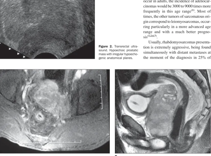
At transrectal ultrasound (Figure 2) for puncture guidance, a solid, hypoechogenic mass was observed affecting the whole prostate, causing architectural distortion, interruption of the anatomic capsule and infiltration of adjacent planes. The seminal vesicle presented increased in volume be-cause of the tumor infiltration. The anato-mopathological and immunohistochemical
study showed the presence of an embryo-nal rhabdomyosarcoma.
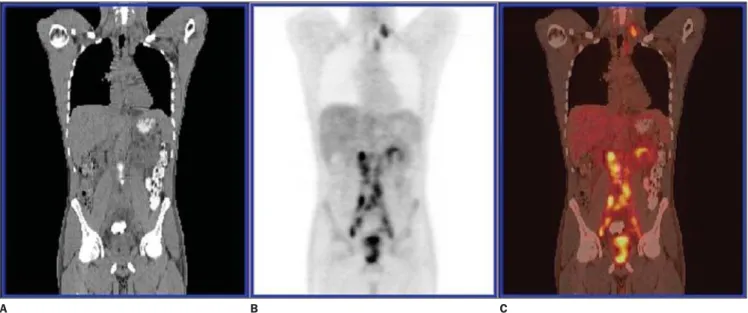
Endorectal coil magnetic resonance imaging (Figure 3) was performed for lo-cal staging, demonstrating an ill-defined mass with infiltrative aspect, intermediary signal on T1-, hypersignal on T2-weighted sequences, with intense and heterogeneous gadolinium-enhancement involving the left vasculonervous bundle, left seminal vesicle, and the medial portion of the right seminal vesicle. The lesion presented a contact surface with the vesical wall at the level of the trigone, with a probable in-volvement of the bladder. PET/CT study (Figure 4) also demonstrated hypermeta-bolic lesions in left, posterior cervical, left lower paratracheal, left supraclavicular fossa and retroperitoneal lymph nodes ex-tending from the renal hila to the pelvic cavity. Lymph node metastases were con-firmed by biopsy of supraclavicular lymph node.
DISCUSSION
430
Gomes de Almeida FA et al.
Radiol Bras 2007;40(6):429–432
Figure 1. Computed tomography. Expansive prostatic growth with invasion of the left seminal vesicle.
A B
Figure 3. Endorectal coil magnetic resonance imaging, axial, contrast-enhanced T1-weighted fat-saturated sequence (A) and sagittal T2-weighted(B). Mass with an infiltrative aspect, with heterogeneous gadolinium enhancement involving the vasculonervous bundle and left seminal vesicle.
A B
cases occurs in children, and is difficult to estimate the incidence in adult individuals. Assuming that about 10% of these cases occur in adults, the incidence of adenocar-cinomas would be 3000 to 9000 times more frequently in this age range(1). Most of
times, the other tumors of sarcomatous ori-gin correspond to leiomyosarcomas, occur-ring particularly in a more advanced age range and with a much better progno-sis(1,2,6,7).
Usually, rhabdomyosarcomas presenta-tion is extremely aggressive, being found simultaneously with distant metastases at the moment of the diagnosis in 25% of Figure 2. Transrectal
431
Prostatic rhabdomiosarcoma in an adult patient: a case report
Radiol Bras 2007;40(6):429–432 patients(4). Lungs are the most frequent site,
followed by bones, lymph nodes, liver and serosas(1,3,8). Bone metastases are
predomi-nantly disseminated osteolytic, in contrast to adenocarcinomas that typically present osteoblastic metastases concentrating espe-cially in the axial skeleton(1).
Locally, rhabdomyosarcomas present periuretral, perivesical and perirectal infil-tration, displacing the bladder and the rec-tum(1–3). Histologically, these tumors can be
divided into embryonal, alveolar, a quite aggressive subtype predominantly occur-ring in soft tissues of children; and pleo-morphous, a rare and aggressive subtype found in adult individuals(2,9).
The embryonal pattern is found in about two thirds of all cases of rhabdomyosarco-mas(2), and can be divided into botryoid
type, with vegetative growth, most fre-quently affecting bladder and vaginal cu-pula; spinous cells and conventional type, with a better prognosis in the first two cases(2,3).
Differential histologic diagnosis in-cludes lymphoma and small-cell carcinoma of the prostate and may be resolved by means of an appropriate panel of immuno-histochemical stains(4).
The most frequent clinical presentation is associated with obstructive urinary symptoms resulting from a significant in-crease in the prostatic volume. Addition-ally, hematuria and incontinence may
oc-cur. Pelvic pain is a frequent finding, and usually requires rigorous analgesia. Peri-rectal planes compression or invasion may result in a significant intestinal obstipa-tion(1,3,4,8).
Imaging studies are quite scarce, and there is no pattern defining the diagnosis that is confirmed by means of US-guided transrectal biopsy(4,7). However, some
find-ings may suggest the diagnosis, such as the presence of a large mass on the prostatic bed, infiltrating adjacent planes in a young patient who has not presented any increase in the levels of tumor markers or prostate-specific antigen (PSA)(1,7).
At ultrasound, the lesion may present predominantly hyper or hypoechogenic, with sonolucent areas corresponding to hemorrhage or necrosis sites(3). The authors
could not find any previous report on dopplervelocimetric profile characteristic of the lesion. Reports describe hyperemia and high diastolic flow velocity in parates-ticular rhabdomyosarcomas of children(3).
In the case presently described, US has also allowed the diagnosis of the tumor exten-sion to the left seminal vesicle, proved by anatomopathological study.
CT demonstrates an infiltrative, many times ill-defined mass with heterogeneous attenuation, whose prostatic of vesical site of origin cannot be identified(3,10).
At MRI, the primary tumor shows non-specific low signal intensity on T1-weighted
sequences, and high sinal intensity on T2-weighted sequences. Heterogeneous signal intensity representing hemorrhagic foci may be identified(3). MRI allows a more
accurate evaluation of the local tumor ex-tent and involvement of adjacent planes as compared with US and CT(3,5,11); this could
be observed in the present study.
The utilization of PET/CT for rhab-domyosarcoma staging in adult individuals is still to be consolidated in the literature, however, reports on cases involving chil-dren have demonstrated the relevance of this method in the detection of the primary focus in metastatic disease, obscure me-tastases and in unusual sites(12). In the
present case, PET/CT was extremely use-ful in the detection of distant lymph node metastases.
The treatment of this disease is per-formed with chemotherapy in association, or not, with surgical resection and/or radia-tion therapy. The prognosis is poor, with a five-year survival rate of 30%-35% for all rhabdomyosarcomas in adult individu-als(6,13); this rate is probably lower in cases
of primary tumor on the prostatic bed(1).
CONCLUSION
Prostatic rhabdomyosarcoma is much more aggressive, rare, and presents a worst prognosis in adults as compared with its presentation in children. New technologies Figure 4. PET/CT image. Multiple, retroperitoneal and left supraclavicular metastatic lymph nodes.
432
Gomes de Almeida FA et al.
Radiol Bras 2007;40(6):429–432 in imaging diagnosis may aid in the correct
diagnosis and also provide an accurate staging of the disease, allowing a better therapeutic planning.
REFERENCES
1. Waring PM, Newland RC. Prostatic embryonal rhabdomyosarcoma in adults. A clinicopathologic review. Cancer 1992;69:755–762.
2. Nigro KG, MacLennan GT. Rhabdomyosarcoma of the bladder and prostate. J Urol 2005;173:1365. 3. Agrons GA, Wagner BJ, Lonergan GJ, Dickey GE, Kaufman MS. From the archives of the AFIP. Genitourinary rhabdomyosarcoma in children: ra-diologic-pathologic correlation. RadioGraphics 1997;17:919–937.
4. Logani S, Cabrera RA, Amin MB. Embryonal
rhabdomyosarcoma of the adult prostate: differ-ential diagnostic considerations. Path Cas Rev 2003;8:82–87.
5. Hawkins WG, Hoos A, Antonescu CR, et al. Clini-copathologic analysis of patients with adult rhab-domyosarcoma. Cancer 2001;91:794–803. 6. Ferrari A, Dileo P, Casanova M, et al.
Rhabdo-myosarcoma in adults. A retrospective analysis of 171 patients treated at a single institution. Can-cer 2003;98:571–580.
7. Sexton WJ, Lance RE, Reyes AO, Pisters PW, Tu SM, Pisters LL. Adult prostate sarcoma: the M.D. Anderson Cancer Center Experience. J Urol 2001;166:521–525.
8. Bos SD, Slaa ET. An adult man with a rhabdo-myosarcoma of the prostate. A case report. Scand J Urol Nephrol 1991;25:329–330.
9. Miettinen M. Rhabdomyosarcoma in patients
older than 40 years of age. Cancer 1988;62:2060– 2065.
10. Baker ME, Silverman PM, Korobkin M. Com-puted tomography of prostatic and bladder rhab-domyosarcomas. J Comput Assist Tomogr 1985; 9:780–783.
11. Bartolozzi C, Selli C, Olmastroni M, Menchi I, Di Candio G. Rhabdomyosarcoma of the prostate: MR findings. AJR Am J Roentgenol 1988;150: 1333–1334.
12. McCarville MB, Christie R, Daw NC, Spunt SL, Kaste SC. PET/CT in the evaluation of childhood sarcomas. AJR Am J Roentgenol 2005;184: 1293–1304.