Rev. bras. ortop. vol.50 número1
Texto
Imagem
Documentos relacionados
E para obviar ao esquecimento foi logo dizendo á moça que aquelle era o Mar- condes — o tal companheiro de collegio de que lhe fallava tantas vezes. Então os dous
O presente estudo, através de uma escolha aleatória, foi realizado numa parte do setor nordeste do estado de São Paulo (Fig. 1), numa área constituída por três
tive analgesia and the need for tramadol in patients undergoing reconstruction of the anterior cruciate ligament with spinal an- esthesia, fentanyl and femoral nerve block.. METHODS
In this scenario several measurements will be performed to test different aspects of the implementation, namely aspects relative to the overall multihoming performance (through-
Objective: To establish the radiographic distances from posterior cruciate ligament (PCL) tib- ial insertions centers to the lateral and medial tibial cortex in the
Tibial plateau fracture following gracilis-semitendinosus anterior cruciate ligament reconstruction: the tibial tunnel stress-riser. Delcogliano A, Chiossi S, Caporaso A, Franzese
LTE, lateral tibial eminence; MTE, medial tibial eminence; SWF, shiny white fibers located in the posterior horn of the medial meniscus; ACL, anterior cruciate ligament insertion;
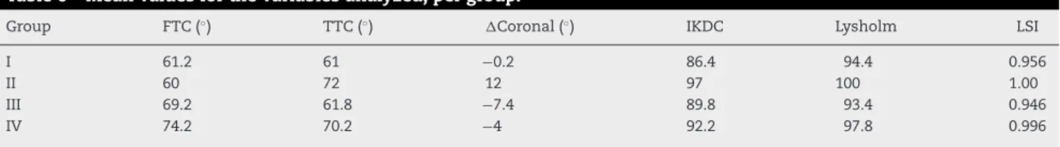
Conclusions: There were no significant differences in the subjective and objective clinical assessments among patients submitted to anterior cruciate ligament reconstruction using
