Radiol Bras vol.47 número4
Texto
Imagem

Documentos relacionados
For the diagnosis, it is necessary to use imaging methods during the investigation, especially computed tomography and magnetic resonance imaging, both with emphasis on
OBJECTIVE: To determine the frequency and localization of parenchymal abnormalities in cerebral venous thrombosis on magnetic resonance imaging and magnetic resonance angiography
In the present study, the authors describe several non-dysfunctional conditions affecting the temporomandibular joints through computed tomography and magnetic resonance
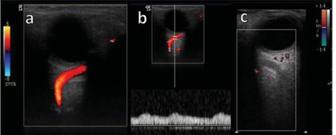
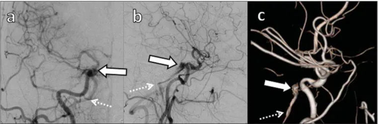
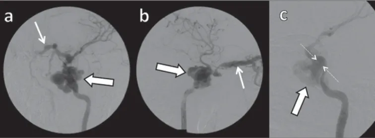
This finding was confirmed by magnetic resonance angiography of the cervical vessels, and axial computed tomography angiography showed agenesis of the right carotid canal..
Technological developments in imaging, such as elastography, magnetic resonance imaging-guided biopsy, magnetic resonance imaging-ultrasonography fusion, can revolutionize the
Cardiac magnetic resonance imaging (CMRI) and cardiac computed tomography (CCT) are noninvasive imaging methods that serve as useful tools in the diagnosis of coronary artery
With the development of imaging studies, the enterogra- phy, either by computed tomography or magnetic resonance, is replacing intestinal transit and enteroclysis procedures in
Computed tomography scan and magnetic resonance imaging revealed a pituitary tumor invading the left sphenoidal and cavernous sinuses.. Laboratory data excluded
