Effect of supplementary zinc on orthodontic tooth
movement in a rat model
Mohammad Sadegh Ahmad Akhoundi1, Rezvaneh Ghazanfari2, Shahroo Etemad-Moghadam3, Mojgan Alaeddini3, Azam Khorshidian4, Shahram Rabbani5, Ahmad Reza Shamshiri6, Nafiseh Momeni4
Introduction: Osteoclasts and osteoblasts are responsible for regulating bone homeostasis during which the trace ele-ment zinc has been shown to exert a cumulative effect on bone mass by stimulating osteoblastic bone formation and inhibiting osteoclastic bone resorption.
Objective: The aim of the present study was to investigate the effects of zinc (Zn) on orthodontic tooth movement (OTM) in a rat model.
Material and Methods: A total of 44 male Wistar rats were divided into four groups of 11 animals each and received 0, 1.5, 20 and 50 ppm Zn in distilled water for 60 days. In the last 21 days of the study, nickel-titanium closed coil springs were ligated between maxillary right incisors and first molars of all rats, and tooth movement was measured at the end of this period. Histological analysis of hematoxylin/eosin slides was performed to assess root resorption lacunae, osteoclast number and periodontal ligament (PDL) width.
Results: Mean OTM was calculated as 51.8, 49.1, 35.5 and 45 µm in the 0, 1.5, 20 and 50 ppm zinc-receiving groups, respectively. There were no significant differences in neither OTM nor histological parameters among the study groups (p > 0.05).
Conclusion: According to the results obtained in the current investigation, increase in supplementary zinc up to 50 ppm does not affect the rate of OTM neither bone and root resorption in rats.
Keywords: Dietary supplement. Orthodontics. Tooth movement. Zinc.
1 Professor, Tehran University of Medical Sciences, Dental Research Center, Orthodontic Department, Tehran, Iran.
2 Postgraduate student, Tehran University of Medical Sciences, Prosthodontic Department, Tehran, Iran.
3 Associate Professor, Tehran University of Medical Sciences, Dental Research Center, Dentistry Research Institute, Tehran, Iran.
4 Dentist, Tehran University of Medical Sciences, Dental Research center, Dentistry Research Institute, Tehran, Iran.
5 Head of Experimental Research Laboratory, Tehran University of Medical Sciences, Tehran Heart Center, Tehran, Iran.
6 Tehran University of Medical Sciences, Research Center for Caries Prevention, Dentistry Research Institute, Department of Community Oral Health, Tehran, Iran. DOI: http://dx.doi.org/10.1590/2177-6709.21.2.045-050.oar
How to cite this article: Akhoundi MSA, Ghazanfari R, Etemad-Moghad-am S, Alaeddini M, Khorshidian A, Rabbani S, Shamshiri AR, Momeni N. Ef-fect of supplementary zinc on orthodontic tooth movement in a rat model. Dental Press J Orthod. 2016 Mar-Apr;21(2):45-50.
DOI: http://dx.doi.org/10.1590/2177-6709.21.2.045-050.oar
Submitted: January 13, 2015 - Revised and accepted: August 17, 2015
» The authors report no commercial, proprietary or financial interest in the products or companies described in this article.
INTRODUCTION
Zinc (Zn) is an essential trace element that serves as a cofactor for more than 200 enzymes, and being a constit-uent of nearly all human cell types, plays a major role in a number of basic biological processes including
prolifera-tion, wound healing, immunity and osteogenesis.1
De-ficiency of this fundamental mineral is a universal health issue, especially during adolescence due to the occur-rence of growth spurts. This has led to the recognition of a need for improved public health programs to support individuals with Zn deficiency known to comprise half
the world’s population.2 Delayed bone maturation and
impaired growth are two of the major consequences of insufficient Zn intake in pubescent individuals who are being treated worldwide by prescription of Zn
supple-ments as part of their treatment regimen.3 In addition to
the bone-related applications of this substance, different compounds, such as zinc gluconate glycine and zinc
ac-etate, are routinely used as anti-cold agents.4
Zn impacts bone metabolism via augmentation of osteoblastic activity and down regulation of osteoclas-tic bone resorption, a fact reported by numerous
in-vestigations.5-8 Bone remodeling is the foundation of
orthodontic treatment and tooth movement relies on this
phenomenon.9 Following the application of orthodontic
forces, the periodontium responds by an inflammatory reaction leading to reorganization of its cellular compo-nents and a modification in its equilibrium in favor of bone remodeling, the end result of which would be tooth
movement.10,11,12
Several authors have studied the effects of local and systemic medicaments, including dietary supplements
on orthodontic tooth movement (OTM).13-16 A
con-siderable number of patients seeking orthodontic treat-ment may be using medications due to general health problems; moreover, regarding the prevailing trend to-wards the increased use of dietary supplements among these individuals, having some notion of the effect of various drugs on OTM would be helpful for treatment planning and predicting the length of these treatment
modalities.17 Considering the osteogenic potential of
Zn, along with its inhibiting impact on bone
resorp-tion,7 it was hypothesized that this substance might
negatively regulate the rate of OTM in rats. Since Zn supplementation is becoming prevalent among patients,
MATERIAL AND METHODS Animals
The experimental protocol of the current study was approved by the Ethics Committee of Tehran Univer-sity of Medical Sciences (code: 91-01-70-17586-55909). A total of 44 male Wistar rats (200-250 g) were housed in plastic cages, maintained on a 12/12 hour light-dark cycle and randomly divided into four groups (n = 11) with free access to standard laboratory chow. Their drinking water consisted of double distilled water with Zn sulfate added at concentrations of 0, 1.5, 20 and 50 ppm for use in the
control group and groups 1, 2 and 3, respectively.18,19,20
All animals were weighed at the beginning of the study (day 1), on the first day of appliance placement (day 40) and immediately before sacrifice (day 60).
Orthodontic treatment and measurement of tooth movement
On day 40th of the study period, each rat was
anaes-thetized with an intraperitoneal injection of xylazine HCL (6 mg/kg body weight) and ketamine (50 mg/kg body weight) in order to receive orthodontic appliances. Based
on the method suggested by Nilforoushan et al,22
nickel-titanium (NiTi) closed coil springs (NiTi, 3M Unitek, Monrovia, CA, Hitek, 0.006 × 0.022-in) were ligated be-tween left maxillary first molars and incisors of all rats with 0.010-in stainless steel ligature wires to deliver a force of 60 g without further activation throughout the duration of the investigation. Labial and distal grooves cut, in ap-proximation to the gingival margins of incisors, were used to retain the wires. The mesiolingual undercut of the first molar provided necessary retention in the posterior seg-ment of the appliance. Two incisors were attached together by means of composite resin (Transbond XT, 3M Unitek, Monrovia, Calif) to achieve anterior anchorage and ensure
mesial movement of molars.21 Moreover, composite resin
covered the anterior ligatures to preserve the wires during the study period. This was followed by 1.5 mm reduction of mandibular incisors with a high-speed handpiece in
or-der to prevent severance of the ligature wires.22,23 At the
beginning of the study, none of the animals demonstrated any kind of space between first and second molars and all contacts were intact.
mini-All animals were sacrificed on day 60th of the study period by ether overdose followed by decapitation. A feeler gauge was employed to assess mesial move-ment of first molar by measuring the space between first and second molars before removal of the appli-ances, so as to prevent any possible distal relapse of the first molar. All measurements were repeated twice by the same operator blinded to the study groups, and the means were used for statistical analysis.
Histological evaluation
The maxillae were separated, fixed in 10% formalin for five days and immersed in 5% formic acid until ad-equately decalcified (an average of five days). Sequential 5-µm serial sections were prepared from each paraffin block and the five sections containing the largest root area were chosen and analyzed histomorphometrically, as
described previously.24,25 The final value was expressed as
the mean of the selected sections.26 The mesial root was
histomorphometrically evaluated on the section contain-ing the full length of the root from the cemento-enamel junction (CEJ) to the apex, by means of a double-headed Olympus BX-41 light microscope equipped with a digi-tal camera (DP25 Olympus) and analysis software (DP2-BSW, Olympus). The number of osteoclasts, periodontal ligament (PDL) width, number of resorption lacunae and their depths and widths were analyzed by two observers,
and disagreements were solved by consensus.27 The width
of the PDL was determined coronally and apically on
both mesial and distal aspects of the mesial root.27,28
All sections were measured twice by both observers on the double-headed microscope, and the mean of the two values was used in all consecutive calculations.
Statistical analysis
Differences among groups were analyzed by one-way ANOVA followed by Tukey post-hoc tests for multiple
comparisons. Probability values p < 0.05 were considered
statistically significant.
RESULTS
There was a gradual increase in rats’ weight during our investigation, and none of them died or demonstrated weight loss throughout the study period. No significant differences in mean overall weights were found among
the four study groups (p = 0.25). All treated first molars
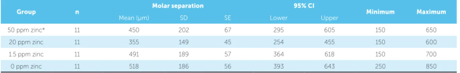
shifted mesially into the space between molars and inci-sors (Table 1). The highest and lowest amounts of tooth movement were observed in the control and 20 ppm Zn groups, respectively; but there were no significant
differ-ences among groups (p = 0.18).
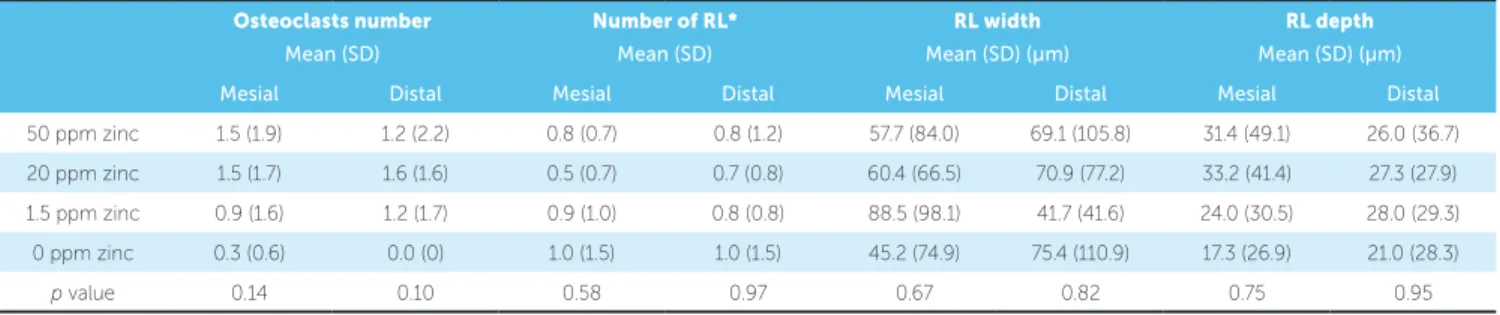
Descriptive histological data are shown in Table 2. Osteoclast numbers and resorptive lacunae widths and depths showed no significant differences among study groups (Table 2). Of the four measured locations of PDL width, significant difference was found only in the disto-apical area (Table 2) among groups. Based on multiple comparisons, PDL width was significantly higher in group 1 (1.5 ppm) compared to the control
group (p = 0.02).
Group n Molar separation 95% CI Minimum Maximum
Mean (µm) SD SE Lower Upper
50 ppm zinc* 11 450 202 67 295 605 150 650
20 ppm zinc 11 355 149 45 254 455 150 600
1.5 ppm zinc 11 491 189 57 364 618 150 700
0 ppm zinc 11 518 186 56 393 643 250 850
Table 1 - Molar separation over 21 days.
DISCUSSION
Zn supplements are prescribed for adults for numer-ous reasons. Among the varinumer-ous nutritional attributes of Zn, inhibition of bone resorption and stimulation of
bone growth and mineralization7 might directly
influ-ence OTM induced by orthodontic treatment. Several studies have investigated the effects of Zn on various
as-pects of bone quality,5,29 and its deficiency has been
sug-gested to play a role in the development of osteoporosis.30
In the present study, we measured the amount of tooth movement in rats receiving 0 to 50 ppm zinc sulfate, followed by application of a simple orthodon-tic appliance, and did not observe significant differ-ence among groups, which was also confirmed by our histomorphometric analysis. The present result was in
agreement with a study conducted by Abrisham et al8
who also did not find Zn to be effective in bone healing and reported no significant relationship between this substance and bone formation in rabbits. Similarly, a periodontal study in rats failed to demonstrate differ-ences in pocket depths between animals receiving Zn-containing diets and those deficient in this
micronutri-ent.31 In contrast to our findings, Zn has been shown
to prevent osteoporosis32 and induce osteogenesis.5
Previous investigations have indicated that the du-ration of Zn application may have an impact on its expected bone effects. Accordingly, any possible posi-tive function of this micronutrient diminishes with
time.8,33 This may explain the lack of difference in
OTM between control and Zn-receiving rats;
conse-may have suppressed the activity of this element. For the same reason, it has been suggested that Zn should be prescribed at the initial stages of
inflamma-tory reactions,34,35 which is known to play a major role
in tooth movement.36 Additionally, a number of
stud-ies have pointed out that Zn can only perform where
its deficiency exists;37 thus, pretreatment
supplemen-tation administered in the present study might have reduced any chance of Zn inadequacy and, therefore, eliminated its possible impact on OTM.
Despite the insignificant difference among our study groups, a decrease in OTM occurred from the first dose of Zn up to 20 ppm, but increased in Group 3 in which rats received 50 ppm. The decrease could be justified based on the proposed anti-resorptive ef-fects of Zn, but the reason for the increase may not
be as simple to explain. Cerovic et al38 also reported
similar findings regarding alkaline phosphatase activity and in vitro bone nodule formation with increasing Zn concentrations, suggesting “biphasic effects” for this element in addition to other possible processes includ-ing cytotoxicity at higher doses.
Our histomorphometric findings showed no significant difference in osteoclast number among groups. This supports our clinical OTM data, but may seem contradictory to a number of former in-vestigations demonstrating a down-regulating influ-ence of Zn on osteoclastic resorptive potential and
differentiation.7,39,40 Nevertheless, osteoclastic number
may not necessarily have a positive association with
Table 2 - Descriptive histological data.
* RL = Resorption lacuna.
Osteoclasts number
Mean (SD)
Number of RL*
Mean (SD)
RL width
Mean (SD) (µm)
RL depth
Mean (SD) (µm) Mesial Distal Mesial Distal Mesial Distal Mesial Distal
50 ppm zinc 1.5 (1.9) 1.2 (2.2) 0.8 (0.7) 0.8 (1.2) 57.7 (84.0) 69.1 (105.8) 31.4 (49.1) 26.0 (36.7)
20 ppm zinc 1.5 (1.7) 1.6 (1.6) 0.5 (0.7) 0.7 (0.8) 60.4 (66.5) 70.9 (77.2) 33.2 (41.4) 27.3 (27.9)
1.5 ppm zinc 0.9 (1.6) 1.2 (1.7) 0.9 (1.0) 0.8 (0.8) 88.5 (98.1) 41.7 (41.6) 24.0 (30.5) 28.0 (29.3)
0 ppm zinc 0.3 (0.6) 0.0 (0) 1.0 (1.5) 1.0 (1.5) 45.2 (74.9) 75.4 (110.9) 17.3 (26.9) 21.0 (28.3)
osteoblastic/osteoclastic co-culture, but found in-creased osteoclastic numbers. However, the study
sit-uation (in vitro versus in vivo), detection methods and
Zn administration in their research were different from those used in the present investigation.
The methods used in this study were selected based
on previous research in this field;7,43 and, according
to our findings, neither clinical nor histopathological changes were observed following Zn application in rats. Future studies using serum and/or urine analysis could help clarify the role of Zn in OTM and possi-bly confirm the conclusions of the current investiga-tion. Additionally, further researches with immuno-histochemical and molecular techniques are needed to understand the effect of Zn supplementation on bone remodeling during orthodontic treatment. If the pres-ent results are supported by future studies in animals and humans, the specific modifications in orthodontic treatment planning might not be necessary for patients receiving Zn as a dietary supplementation.
Regarding the four locations of PDL width measured in our study, significant difference was found only in the distoapical aspect among groups. The reason for this difference is not clear; further
studies on various histopathologic features of OTM following Zn treatment are suggested to help clarify the role of this important micronutrient in orth-odontic treatment.
CONCLUSIONS
According to the result obtained in the present study, systemic Zn supplementation up to 50 ppm does not affect OTM, neither bone nor root resorp-tion in rats. Extrapolaresorp-tion of these findings to human subjects would require extensive research using more sophisticated techniques and drug concentrations.
Acknowledgments
This study has been funded and supported by Tehran University of Medical Sciences (TUMS); Grant # 132/872.
Author contributions
1. Czerwinski AW, Clark ML, Serafetinides EA, Perrier C, Huber W. Safety and eicacy of zinc sulfate in geriatric patients. Clin Pharmacol Ther. 1974 Apr;15(4):436-41.
2. Kawade R. Zinc status and its association with the health of adolescents: a review of studies in India. Glob Health Action. 2012;5:7353.
3. Dekker LH, Villamor E. Zinc supplementation in children is not associated with decreases in hemoglobin concentrations. J Nutr. 2010 May;140(5):1035-40.
4. Taylor DM, Liyanage JA, Williams DR, Harding KG. A new approach to
monitoring trace element concentrations and speciation in wounds and wound luids. Appl Radiat Isot. 1998 May-Jun;49(5-6):677-9.
5. Yamaguchi M. Nutritional factors and bone homeostasis: synergistic efect with zinc and genistein in osteogenesis. Mol Cell Biochem. 2012 July;366(1-2):201-21.
6. Igarashi A, Yamaguchi M. Increase in bone protein components with
healing rat fractures: enhancement by zinc treatment. Int J Mol Med. 1999 Dec;4(6):615-20.
7. Hadley KB, Newman SM, Hunt JR. Dietary zinc reduces osteoclast
resorption activities and increases markers of osteoblast diferentiation, matrix maturation, and mineralization in the long bones of growing rats. J Nutr Biochem. 2010 Apr;21(4):297-303.
8. Abrisham SM, Yaghmaei M, Abbas FM, Sharii D, Abrisham SM. Efect of
oral zinc therapy on osteogenesis in rabbits. J Oral Maxillofac Surg. 2010 July;68(7):1676-80.
9. Meikle MC. The tissue, cellular, and molecular regulation of orthodontic tooth movement: 100 years after Carl Sandstedt. Eur J Orthod. 2006 Jun;28(3):221-40.
10. Pizzo G, Licata ME, Guiglia R, Giuliana G. Root resorption and orthodontic treatment. Review of the literature. Minerva Stomatol. 2007 Jan-Feb;56(1-2):31-44.
11. Brezniak N, Wasserstein A. Orthodontically induced inlammatory root resorption. Part I: The basic science aspects. Angle Orthod. 2002 Apr;72(2):175-9.
12. Storey E. The nature of tooth movement. Am J Orthod. 1973 Mar;63(3):292-314.
13. Gameiro GH, Pereira-Neto JS, Magnani MB, Nouer DF. The inluence of drugs and systemic factors on orthodontic tooth movement. J Clin Orthod. 2007 Feb;41(2):73-8; quiz 71.
14. Tyrovola JB, Spyropoulos MN. Efects of drugs and systemic factors on orthodontic treatment. Quintessence Int. 2001 May;32(5):365-71. 15. Krishnan V, Davidovitch Z. The efect of drugs on orthodontic tooth
movement. Orthod Craniofac Res. 2006;9(4):163-71.
16. Bartzela T, Türp JC, Motschall E, Maltha JC. Medication efects on the rate of orthodontic tooth movement: a systematic literature review. Am J Orthod Dentofacial Orthop. 2009 Jan;135(1):16-26.
17. Isaacson JR. Your patients are on drugs [editorial]. Angle Orthod. 2000;70(2):96.
18. Brzóska MM, Galazyn-Sidorczuk M, Rogalska J, Roszczenko A, Jurczuk M, Majewska K, et al. Beneicial efect of zinc supplementation on biomechanical properties of femoral distal end and femoral diaphysis of male rats chronically exposed to cadmium. Chem Biol Interact. 2008 Feb 15;171(3):312-24.
19. Brzóska MM, Rogalska J, Galazyn-Sidorczuk M, Jurczuk M, Roszczenko A, Kulikowska-Karpińska E, et al. Efect of zinc supplementation on bone metabolism in male rats chronically exposed to cadmium. Toxicology. 2007 July 31;237(1-3):89-103.
20. Fong LY, Jiang Y, Rawahneh ML, Smalley KJ, Croce CM, Farber JL, et al. Zinc supplementation suppresses 4-nitroquinoline 1-oxide-induced rat oral carcinogenesis. Carcinogenesis. 2011 Apr;32(4):554-60.
21. Shirazi M, Khosrowshahi M, Dehpour AR. The efect of chronic renal insuiciency on orthodontic tooth movement in rats. Angle Orthod. 2001 Dec;71(6):494-8.
REFERENCES
22. Nilforoushan D, Shirazi M, Dehpour AR. The role of opioid systems on orthodontic tooth movement in cholestatic rats. Angle Orthod. 2002 Oct;72(5):476-80.
23. Sirisoontorn I, Hotokezaka H, Hashimoto M, Gonzales C, Luppanapornlarp S, Darendeliler MA, et al. Orthodontic tooth movement and root resorption in ovariectomized rats treated by systemic administration of zoledronic acid. Am J Orthod Dentofacial Orthop. 2012 May;141(5):563-73.
24. Hakami Z, Kitaura H, Kimura K, Ishida M, Sugisawa H, Ida H, et al. Efect of interleukin-4 on orthodontic tooth movement and associated root resorption. Eur J Orthod. 2015 Feb;37(1):87-94.
25. Taddei SR, Andrade I Jr, Queiroz-Junior CM, Garlet TP, Garlet GP, Cunha FQ, et al. Role of CCR2 in orthodontic tooth movement. Am J Orthod Dentofacial Orthop. 2012 Feb;141(2):153-60.
26. Leiker BJ, Nanda RS, Currier GF, Howes RI, Sinha PK. The efects of exogenous prostaglandins on orthodontic tooth movement in rats. Am J Orthod Dentofacial Orthop. 1995 Oct;108(4):380-8.
27. Sekhavat AR, Mousavizadeh K, Pakshir HR, Aslani FS. Efect of misoprostol, a prostaglandin E1 analog, on orthodontic tooth movement in rats. Am J Orthod Dentofacial Orthop. 2002 Nov;122(5):542-7.
28. Tengku BS, Joseph BK, Harbrow D, Taverne AA, Symons AL. Efect of a static magnetic ield on orthodontic tooth movement in the rat. Eur J Orthod. 2000 Oct;22(5):475-87.
29. Kara C, Orbak R, Dagsuyu IM, Orbak Z, Bilici N, Gumustekin K. In vivo assessment of zinc deiciency on craniofacial growth in a rat model. Eur J Dent. 2009 Jan;3(1):10-5.
30. Eberle J, Schmidmayer S, Erben RG, Stangassinger M, Roth HP. Skeletal efects of zinc deiciency in growing rats. J Trace Elem Med Biol. 1999;13(1-2):21-6. 31. Orbak R, Kara C, Ozbek E, Tezel A, Demir T. Efects of zinc deiciency on oral
and periodontal diseases in rats. J Periodontal Res. 2007;42(2):138-43. 32. Yamaguchi M. Role of nutritional zinc in the prevention of osteoporosis. Mol Cell
Biochem. 2010;338(1-2):241-54.
33. Yamaguchi M, Mochizuki A, Okada S. Stimulatory efect of zinc on bone growth in weanling rats. J Pharmacobiodyn. 1982;5(8):619-26.
34. Kaplan B, Gönül B, Dinçer S, Dinçer Kaya FN, Babül A. Relationships between tensile strength, ascorbic acid, hydroxyproline, and zinc levels of rabbit full-thickness incision wound healing. Surg Today. 2004;34(9):747-51. 35. Savlov ED, Strain WH, Huegin F. Radiozinc studies in experimental wound
healing. J Surg Res. 1962 May;2:209-12.
36. Alhashimi N, Frithiof L, Brudvik P, Bakhiet M. Orthodontic tooth movement and de novo synthesis of proinlammatory cytokines. Am J Orthod Dentofacial Orthop. 2001 Mar;119(3):307-12.
37. Hallböök T, Lanner E. Serum-zinc and healing of venous leg ulcers. Lancet. 1972 Oct 14;2(7781):780-2.
38. Cerovic A, Miletic I, Sobajic S, Blagojevic D, Radusinovic M, El-Sohemy A. Efects of zinc on the mineralization of bone nodules from human osteoblast-like cells. Biol Trace Elem Res. 2007 Apr;116(1):61-71.
39. Yamaguchi M, Weitzmann MN. Zinc stimulates osteoblastogenesis and suppresses osteoclastogenesis by antagonizing NF-κB activation. Mol Cell Biochem. 2011 Sept;355(1-2):179-86.
40. Park KH, Park B, Yoon DS, Kwon SH, Shin DM, Lee JW, et al. Zinc inhibits osteoclast diferentiation by suppression of Ca2+-Calcineurin-NFATc1 signaling pathway. Cell Commun Signal. 2013 Oct 2;11:74.
41. Karsdal MA, Martin TJ, Bollerslev J, Christiansen C, Henriksen K. Are nonresorbing osteoclasts sources of bone anabolic activity? J Bone Miner Res. 2007 Apr;22(4):487-94.
42. Holloway WR, Collier FM, Herbst RE, Hodge JM, Nicholson GC. Osteoblast-mediated efects of zinc on isolated rat osteoclasts: inhibition of bone resorption and enhancement of osteoclast number. Bone. 1996 Aug;19(2):137-42. 43. Watanabe T, Nakada H, Takahashi T, Fujita K, Tanimoto Y, Sakae T, et al. Potential
ERRATUM
In the article Effect of supplementary zinc on orthodontic tooth movement in a
rat model, published at Dental Press J Orthod. 2016 Mar-Apr;21(2):45-50,
where it is written:
Ahmad Akhoundi Mohammad Sadegh, Ghazanfari Rezvaneh,
Etemad-Moghadam Shahroo, Alaeddini Mojgan, Khorshidian Azam, Rabbani Shahram,
Shamshiri Ahmad Reza, Momeni Nafiseh.
Should be written:
Mohammad Sadegh Ahmad Akhoundi, Rezvaneh Ghazanfari, Shahroo
Etemad-Moghadam, Mojgan Alaeddini, Azam Khorshidian, Shahram Rabbani,