Brachycephalic, dolichocephalic and mesocephalic: Is it
appropriate to describe the face using skull patterns?
Fernanda Catharino Menezes Franco1, Telma Martins de Araujo2, Carlos Jorge Vogel3, Cátia Cardoso Abdo Quintão4
The use of a standardized terminology in the medical sciences is essential for both clinical practice and scientiic research. In addition to facilitating communication between professionals, it enhances the reliability of comparisons made between studies from diferent areas, thereby contributing to a higher level of scientiic evidence. Examples of attempts made to standardize the terminology in other areas dedicated to the study of craniofacial morphology can be found in the litera-ture. On the other hand, one can ind in the orthodontic literature a variety of terms that render the consensus and com-munication between orthodontists and other researchers even more problematic. As an example, one could cite the use of the terms brachyfacial, mesofacial and dolichofacial, which form part of a cranial index terminology used to describe facial types. Thus, a relection on the origin and diferences of the terms used to describe the human facial phenotype may pave the way toward a consensus regarding the meaning that best represents the craniofacial patterns.
Keywords:Face. Terminology. Classiication.
How to cite this article: Franco FCM, Araujo TM, Vogel CJ, Quintão CCA. Brachycephalic, dolichocephalic and mesocephalic: Is it appropriate to describe the face using skull patterns? Dental Press J Orthod. 2013 May-June;18(3):159-63.
Submitted: July 29, 2011 - Revised and accepted: December 20, 2011 » The specimens shown in the present article belong to the collection of skulls with normal occlusion of the Prof. José Édimo Soares Martins Orthodontics Cen-ter (UFBA), having been donated by Prof. Carlos Jorge Vogel.
» The authors report no commercial, proprietary or financial interest in the products or companies described in this article.
Contact address: Fernanda Catharino Menezes Franco
Av. Araújo Pinho, 62/ 7º andar - Faculdade de Odontologia da UFBA – Brazil CEP: 40.110-150 - Canela, Salvador/BA - E-mail: [email protected]
1 Professor of Orthodontics, Bahiana School of Medicine and Public Health
(EBMSP). Professor, Specialization Program in Orthodontics, Federal University of Bahia (UFBA). Diplomate, Brazilian Board of Orthodontics and Dentofacial Orthopedics (BBO). MSc in Orthodontics, Federal University of Rio de Janeiro (UFRJ). Doctoral student in Orthodontics, Rio de Janeiro State University (UERJ).
2 Professor of Orthodontics at UFBA. Coordinator of the Prof. José Édimo
Soares Martins Orthodontics Center, UFBA. Former chairwoman of the Brazilian Board of Orthodontics and Dentofacial Orthopedics.
3 Former chairman of the Brazilian Board of Orthodontics and Dentofacial
Orthopedics.
4 PhD and MSc in Orthodontics, UFRJ. Associate Professor of Orthodontics,
UERJ.
A padronização da nomenclatura utilizada nas ciências médicas é fundamental tanto para a prática clínica quanto para a pesquisa cientíica. Além de facilitar a comunicação entre os proissionais, aumenta a coniabilidade da comparação entre trabalhos de diferentes áreas, favorecendo um melhor nível de evidência cientíica. Exemplos de esforços, em áreas também voltadas ao estudo da morfologia craniofacial, no sentido de uniformização da terminologia podem ser encontrados na literatura médica. Por outro lado, observa-se na literatura ortodôntica uma diversidade de termos que torna mais difícil o consenso e a comunicação entre ortodontistas e demais pesquisadores. Como exemplo, pode-se citar o uso dos termos braquifacial, mesofacial e dolicofacial, terminologia relativa ao índice craniano utilizada para descrever o tipo facial. Sendo assim, a relexão sobre a origem e diferenças dos termos utilizados para descrever o fenó-tipo facial humano pode ser útil ao consenso do signiicado que melhor representa o padrão craniofacial.
Figure 1 - Cranial index: Calculated using the ratio between the maximum width and length of the head. In A, dolichocephalic skull and in B, brachy-cephalic skull.
INTRODUCTION
Facial type assessment is in many aspects crucial for the planning and prognosis of orthodontic treatment. Facial morphology is related to factors such as volume and shape of pharyngeal airspace,1 anatomy of
mastica-tory muscles,2,3 dentoalveolar anatomy4 and occlusion
type.5,6 Moreover, the facial pattern indicates the
direc-tion of growth of the craniofacial complex7,8 and must
be taken into consideration when selecting the orth-odontic biomechanics.9
The terminology used to describe the craniofacial complex stemmed from classical anthropometry, which employs measurements taken in living individuals and human skulls as well as indices that represent facial pro-portions.10,12 The most common among these is the
cra-nial index (which classiies skull types as brachycephalic, mesocephalic and dolichocephalic), and the facial index (which classiies the face as euryprosopic, mesoprosopic and leptoprosopic).11,13 Despite the need to standardize
the terminology, in orthodontics one still notes a wide range of terms to describe the various facial types.14 The
standardization of these terms in diferent ields of knowl-edge is essential to facilitate communication between re-searchers and allow reliable comparisons between difer-ent studies.15 In view of these issues, this article aimed to
clarify the concept, origin and diferences between the terms that describe the human facial phenotype.
CEPHALIC AND FACIAL INDICES
The irst classiication based on cranial morphology is attributed to the professor of anatomy Anders Retzius (1840). Retzius described as gentes dolichocephalae those individuals who had an elongated skull shape, and gen-tes brachycephalae those whose skulls were short. Howev-er, he assigned no numerical values to set the boundar-ies between individual types in both groups and neither did he use the intermediate term mesocephalae, which was introduced at a later time.14 The measures used by
Retzius — when applied to living individuals — are known as cephalic index, and when referring to dry skulls, cranial index.12,13 These indices are calculated by
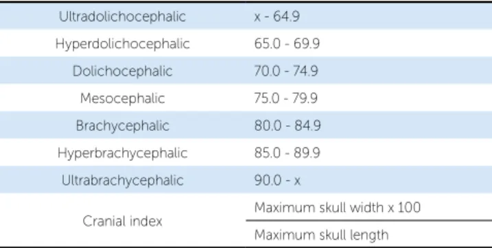
determining the ratio between maximum width and maximum length of the head (Fig 1).12,13 The concept
was subsequently enhanced with the deinition of inter-mediate values,14 which provide a classiication system
and relect more accurately the diversity found in hu-man facial morphology (Table 1).16 Both the cephalic
and cranial indices are therefore measures related to the shape of the skull. The index used in anthropometry to describe the face proportions is the facial index, a prod-uct of morphological facial height, measured from the Nasion (N) to Gnathion (Gn) anatomical landmarks, divided by the bizygomatic width, measured from the right to the let Zygion (Zyr-Zyl) (Fig 2).11,13
Semanti-cally, the terms used in the facial index are derived from Greek, where the word for face is prosopon.14
Accord-ing to this classiication system, numerical values are as-signed which establish the euryprosopic, mesoprosopic and leptoprosopic categories (Table 2).11,13
A B
Table 1 - Head classiication according to the cephalic index.
Table 2 - Face classiication according to the facial index.
Ultradolichocephalic x - 64.9
Hyperdolichocephalic 65.0 - 69.9
Dolichocephalic 70.0 - 74.9
Mesocephalic 75.0 - 79.9
Brachycephalic 80.0 - 84.9
Hyperbrachycephalic 85.0 - 89.9
Ultrabrachycephalic 90.0 - x
Cranial index Maximum skull width x 100
Maximum skull length
Hypereuryprosopic x - 79.9
Euryprosopic 80.0 - 84.9
Mesoprosopic 85.0 - 89.9
Leptoprosopic 90.0 - 94.9
Hyperleptoprosopic 95.0 - x
Facial index
Morphological facial height (N-Gn) x 100
FACIAL MORPHOLOGY IN
PHYSICAL ANTHROPOLOGY
The description of the human body has been a major concern since ancient times. In ancient Greece, canons based on ratio rules were used to describe the ideal hu-man igure. These canons were once again employed by Renaissance artists such as Leonardo da Vinci and Al-brecht Dürer.17,18 Many of these neoclassical principles
are used today in the arts and medicine.17,18,19 The
physi-cal anthropology, or anthropometry, provide a scientiic foundation to these concepts with a view to evaluating the dimensions and proportions of the human body.17
It was only when anthropometric methods were adopted in clinical practice to quantify changes in the craniofacial structure that a wide diversity of human phenotypes and speciic features that diferentiate indi-viduals and ethnic groups emerged.16,20 In diferent areas
of clinical care, standardized anthropometric data have become indispensable for an accurate assessment of the degree of deviation from normality.15,16 Examples can
be found in plastic surgery, during the treatment of con-genital or post-traumatic deformities16,17,21, or in legal or
forensic medicine when identifying individuals,22 or in
medical genetics for the diagnosis of dysmorphisms or craniofacial abnormalities.15
Attempts to build a comprehensive database cover-ing diferent populations have been made by the national scientiic community. One could cite an inter-national group of scientists led by Leslie Farkas,16 who
compiled measurements of the face of 1,470 healthy subjects aged between 18 and 30 years, covering the European, Asian and African continents as well as the Middle East.Farkas himself, a plastic surgeon living in Canada,23 has devoted much of his career to gleaning
Figure 2 - Facial index: Calculated as the ratio between the morphological face height (N-Gn) and bizygomatic width (Zyr-Zyl). In A, euryprosopic face; in B, mesoprosopic face, and in C, leptoprosopic face.
A B C
Figure 3 - Skull classiied as hyperleptoprosopic (fa-cial index = 96.2%) in relation to its predominantly vertical facial morphology (A), and hyperbrachyce-phalic (cranial index = 87.3%) in terms of shape (B).
that also address facial morphology, such as anthropol-ogy, plastic surgery and genetics.14 An important issue to
consider is that in studies evaluating craniofacial dysmor-phisms the terms brachycephaly and dolichocephaly are used to describe deformations of the cranial vault.15
THE EFFECTS OF GROWTH
Any relection on the meaning and validity of the dif-ferent systems of facial morphology classiication must take into account changes that occur in facial growth.14
One particularly signiicant factor regards evaluating the inluence exerted by the head shape on the shape of the face, since the base of the skull is considered a primarily stable structure, from which the face develops in an inferior and anterior direction.8
Some studies conducted by Enlow et al,8,27,28 are
based on the premise that face morphology can be de-termined by the cranial base, which acts as a mold or “template.” According to these studies, individuals with a dolichocephalic head shape have a brain that is long in the anteroposterior direction and narrow in the trans-verse direction, which results in a longer, latter skull base, i.e., the angle formed by the loor of the skull is wider. As a result, the entire nasomaxillary complex as-sumes a lower, more protrusive position, inducing an inferior and posterior rotation of the mandible. Thus, a dolichocephalic head would favor the development of a predominantly long morphology of the face, with a tendency toward a retrognathic mandible and a Class II molar relationship compatible with a leptoprosopic fa-cial type. The same reasoning may be applied to patients with a brachycephalic head shape. Their brains would be wider and more rounded, with a shorter, more angu-lar cranial base, causing a relative retrusion of the naso-maxillary complex and anterior rotation of the mandi-ble. Therefore, these individuals would exhibit features that are closer to a euryprosopic facial pattern.8
The inluence of cranial morphology on facial type is still not fully understood and few studies directly assess the impact of these variables. Bhat and Enlow27 investigated
the relationship between facial types and head shape in in-dividuals with Class I and Class II malocclusions that had not been treated orthodontically. They noted that the lep-toprosopic facial type and a tendency toward developing a Class II are characteristic of mesocephalic and dolichoce-phalic skulls; whereas the tendency to develop a protruded mandible is related to brachycephalic skulls. Results from facial anthropometric data to set the standards for U.S.
Caucasian individuals.16,23 It should be emphasized that
today the advent of globalization and the emergence of multicultural societies have strengthened the im-portance of diferentiating ethnic characteristics in the selection of samples in scientiic studies.17 Yet another
noteworthy concern, especially in medical genetics, re-gards the standardization of the terminology used to de-scribe craniofacial dysmorphisms or anomalies. In this sense, the objectives are to standardize the terminology and establish consensus regarding deinitions and devia-tions from a standard of normality.15
In the medical ield, most studies make use of a no-menclature to describe facial patterns in accordance with anthropometry.15,17 The term brachycephaly, for
example, describes individuals with a cephalic index greater than 81% and the skull shortened in its an-teroposterior dimension. Dolichocephaly, on the other hand, consists of anomalies with a cephalic index below 71% and an elongated cranial vault.15
FACIAL MORPHOLOGY IN ORTHODONTICS In orthodontics, the assessment of facial morphology difers from other medical areas, especially by taking as reference the facial proile or side view, rather than the front view of the face. Therefore, the face width is not considered in most classiication systems.14 This trend
can be understood in light of the importance of radio-graphic cephalometry in modern orthodontics, with the prevalence of analyses based on lateral cephalomet-ric radiographies.7 Some of the terminology used to
de-scribe the facial pattern are: Dolichofacial, mesofacial or brachyfacial;24 hyperdivergent, neutral or hypodivergent;6
long, medium or short;7 and skeletal open bite or skeletal
deep bite.20 It should be noted that the terms
brachyfa-cial, dolichofacial and mesofabrachyfa-cial, which are commonly used by orthodontists, were introduced in the orthodon-tic literature in an arorthodon-ticle by Ricketts in 1960.14,24 Some
orthodontics textbooks describe the face by resorting to terms such as brachycephalic, dolichocephalic and meso-cephalic, and associate speciic types of facial morphology with speciic dental arch forms. This association should be avoided, since a direct relationship between the shape of the face, skull shape and arch form does not occur in all individuals (Fig 3).25,26 The terms euryprosopic,
dontics to describe facial patterns, a terminology that often differs from that used in other medical fields. This is due in large measure to a strong influence exerted by cephalometrics as a method to study cra-niofacial growth, notably based on studies that em-phasize the role of skull morphology in determining the shape of the face. Investigating the influence of skull shape on face shape can provide a benchmark to validate the nomenclature used in orthodontics. If the assertion that skull type determines face type is true then it would not be wrong to use terms derived from the cephalic index, such as “brachyfacial”, “mesofa-cial” and “dolichofacial,” to describe the face. On the other hand, in the event that it is not possible to de-termine this correlation, the use of this nomencla-ture, as well as hindering communication with other medical specialties, would not be justified — and the terms “euryprosopic”, “mesoprosopic” and “lepto-prosopic” should be incorporated into orthodontic terminology. These issues point to the need for fur-ther research on this topic.
other investigations28,29 also give grounds to infer a positive
relationship between skull morphology and face morphol-ogy. However, there is no consensus concerning this as-sociation, given that studies25,26 using diferent
methodolo-gies have failed to reach the same conclusions. In a study to investigate the craniofacial morphology of bruxist and non-bruxist individuals, Menapace et al26 found no
rela-tionship between head shape and craniofacial morphology. In this sample, a frequent association was found between dolichocephalic head shape and euryprosopic facial type.
FINAL CONSIDERATIONS
The growing presence of orthodontics in the con-text of scientiic research makes it necessary to adopt a language consistent with other biological ields.
Terminology standardization is essential to facilitate communication among professionals, enabling compar-isons to be made between diferent studies and afording increasingly evidence-based outcomes.
Nevertheless, it is a fact that currently a non-ho-mogeneous nomenclature is still employed in
ortho-1. Grauer D, Cevidanes LSH, Styner MA, Ackerman JL, Proit WR. Pharyngeal airway volume and shape from cone-beam computed tomography: relationship to facial morphology. Am J Orthod Dentofacial Orthop. 2009;136(6):805-14.
2. Pepicelli A, Woods M, Briggs C. The mandibular muscles and their importance in orthodontics: a contemporary review. Am J Orthod Dentofacial Orthop. 2005;128(6):774-80.
3. Chan HJ, Woods M, Stella D. Mandibular muscle morphology in children with diferent vertical facial patterns: a 3-dimensional computed tomography study. Am J Orthod Dentofacial Orthop. 2008;133(1):10e1-13.
4. Tsunori M, Mashita M, Kasai K. Relationship between facial types and tooth and bone characteristics of the mandible obtained by CT scanning. Angle Orthod. 1998;68(6):557-62.
5. Dibbets JM. Morphological associations between the Angle classes. Eur J Orthod. 1996;18(2):111-8.
6. Siriwat PP, Jarabak JR. Malocclusion and facial morphology Is there a relationship? Angle Orthod. 1985;55(2):127-38.
7. Bishara SE, Jakobsen JR. Longitudinal changes in three normal facial types. Am J Orthod. 1985;88(6):466-502.
8. Enlow DH. Facial growth. 3rd ed. Philadelphia: WB Saunders; 1990. 9. Horn AJ, Thiers-Jegou I. Class II deep bite faces: one-phase or two-phase
treatment? World J Orthod. 2005;6(2):171-9.
10. Edler R, Agarwal P, Wertheim D, Greenhill D. The use of anthropometric proportion indices in the measurement of facial attractiveness. Eur J Orthod. 2006;28(3):274-81.
11. Farkas, LG, Munro IR. Anthropometric facial proportions in Medicine. Springield: Charles C. Thomas Publisher; 1986.
12. Sicher H. Oral anatomy. 6th ed. St Louis: Mosby; 1975.
13. Rakosi T, Jonas I, Graber T. Orthodontic diagnosis (Color Atlas of Dental Medicine). 1st ed. Thieme; 1993.
14. Collett AR, West VC. Terminology of facial morphology in the vertical dimension. Aust Dent J. 1993;38(6):480-1.
15. Allanson JE, Cunnif C, Hoyme HE, McGaughran J, Muenke M, Neri G. Elements of morphology: standard terminology for the head and face. Am J Med Genet. 2009;149A(1):6-28.
REFERENCES
16. Farkas LG, Katic MJ, Forrest CR, Alt KW, Bagic I, Baltadjiev G, et al. International anthropometric study of facial morphology in various ethnic groups/races. J Craniofac Surg. 2005;16(4):615-46.
17. Arslan SG, Genç C, Odabaş B, Kama JD. Comparison of facial proportions and anthropometric norms among Turkish young adults with diferent face types. Aesthetic Plast Surg. 2008 Mar;32(2):234-42.
18. Kolar JC, Salter EM. Craniofacial anthropometry: practical measurement of the head and face for clinical, surgical, and research use. Springield: C.C. Thomas; 1997.
19. Edler R. The use of anthropometric proportion indices in the measurement of facial attractiveness. Eur J Orthod. 2005;28(3):274-81.
20. Sassouni V. A classiication of skeletal facial types. Am J Orthod. 1969;55(2):109-23.
21. Ward RE, Jamison PL, Farkas LG. Craniofacial variability index: a simple measure of normal and abnormal variation in the head and face. Am J Med Genet. 1998;80(3):232-40.
22. Mane DR, Kale AD, Bhai MB, Hallikerimath S. Anthropometric and anthroposcopic analysis of diferent shapes of faces in group of Indian population: a pilot study. J Forensic Leg Med. 2010;17(8):421-5.
23. Naini FB. Leslie G. Farkas, 1915-2008. Am J Orthod Dentofacial Orthop. 2009;136(4):614.
24. Ricketts R. A foundation for cephalometric communication. Am J Orthod. 1960;46(1):230-57.
25. Kerr WJ, Hirst D. Craniofacial characteristics of subjects with normal and postnormal occlusions — a longitudinal study. Am J Orthod Dentofacial Orthop. 1987;92(3):207-12.
26. Menapace SE, Rinchuse DJ, Zullo T, Pierce CJ, Shnorhokian H. The dentofacial morphology of bruxers versus non-bruxers. Angle Orthod. 1994;64(1):43-52. 27. Bhat M, Enlow DH. Facial variations related to headform type. Angle Orthod.
1985;55(4):269-80.
28. Enlow DH, McNamara JA. The neurocranial basis for facial form and pattern. Angle Orthod. 1973;43(3):256-70.