Long-term stability of maxillary anterior
alignment in non-extraction cases
Luiz Filiphe Gonçalves Canuto1, Marcos Roberto de Freitas2, Karina Maria Salvatore de Freitas3, Rodrigo Hermont Cançado4, Leniana Santos Neves5
Objective: The purpose of this retrospective study was to evaluate long-term stability of maxillary incisors alignment in cases submitted to non-extraction orthodontic treatment. Methods: The sample comprised 23 patients (13 female; 10 male) at a mean initial age of 13.36 years (SD = 1.81 years), treated with ixed appliances. Dental cast measurements were obtained at three diferent time points (T1 – pretreatment, T2 – posttreatment and T3 – long-term posttreatment). Variables assessed in maxillary arch were Little Irregularity Index, intercanine, interpremolar and intermolar widths, arch length and perimeter. The statistical analysis was performed by one-way ANOVA and Tukey tests when neces-sary. Pearson’ correlation coeicients were used to investigate possible associations between the evaluated variables.
Results: There was no signiicant change in most arch dimension measurements during and ater treatment, however, during the long-term posttreatment period, it was observed a signiicant maxillary incisors crowding relapse. Conclu-sion: The maxillary incisors irregularity increased signiicantly (1.52 mm) during long-term posttreatment. None of the studied clinical factors demonstrated to be predictive of the maxillary crowding relapse.
Keywords: Relapse. Corrective orthodontics. Malocclusion.
How to cite this article: Canuto LFG, Freitas MR, Freitas KMS, Cançado RH, Neves LS. Long-term stability of maxillary anterior alignment in non-extraction cases. Dental Press J Orthod. 2013 May-June;18(3):46-53.
Submitted: August 12, 2009 - Revised and accepted: December 17, 2010
» The authors report no commercial, proprietary or financial interest in the products or companies described in this article.
Contact address: Luiz Filiphe Gonçalves Canuto Alameda Octávio Pinheiro Brisolla, 9-75 CEP: 17.012-901 – Bauru/SP – Brazil E-mail: [email protected]
1 Post-Doctor in Orthodontics, FOB-USP. Professor, Specialization Course in
Orthodontics, ABO-PE and FACSETE.
2 Professor, Orthodontics, FOB-USP.
3 PhD in Orthodontics, FOB-USP. Post-Doctor in Orthodontics, Toronto
Dentistry University.
4 Adjunct Professor, Uningá. Professor, Specialization Course in Orthodontics,
UFVJM.
5 Professor, Specialization Course in Orthodontics, UFVJM.
Objetivo:o presente estudo avaliou, por meio de uma análise retrospectiva, a estabilidade pós-tratamento do ali-nhamento dos incisivos anterossuperiores de pacientes submetidos ao tratamento ortodôntico sem extrações. Méto-dos: a amostra foi constituída de 23 pacientes (13 do sexo feminino e 10 do sexo masculino), com idade inicial de 13,36 ± 1,81 anos. Mediu-se nos modelos de estudo das fases inicial (T1), inal (T2) e pós-tratamento (T3) de aproxima-damente de 5 anos, a irregularidade dos incisivos superiores, as distâncias intercaninos e entre os primeiros e segundos pré-molares, a distância intermolares, o comprimento e o perímetro da arcada superior. Após a obtenção dos dados, realizou-se a análise estatística. Para a análise das alterações ao longo dos três tempos estudados, utilizou-se a análise de variância (ANOVA) a um critério de seleção e, em caso de resultado signiicativo, o teste de Tukey. Para veriicar a presença de correlação entre a recidiva do apinhamento anterossuperior e a recidiva das variáveis distâncias intercani-nos, interpré-molares, intermolares, comprimento e perímetro da arcada, utilizou-se o teste de correlação de Pearson.
Resultados: os resultados não evidenciaram alterações dimensionais signiicativas ao inal do tratamento; entretanto, durante o período de pós-tratamento, foram observadas alterações signiicativas em relação à quantidade de irregulari-dade dos incisivos superiores. Conclusão: concluiu-se que houve recidiva estatisticamente signiicativa (+1,52mm) na irregularidade anterossuperior durante o período de pós-tratamento. Entretanto, nenhuma das variáveis aferidas nos modelos pôde ser clinicamente associada à recidiva anterossuperior.
INTRODUCTION
The primary purpose of orthodontic treatments is malocclusion correction; however, treatment stability shows considerable variability during post-retention phase. Despite the literature consensus that some oc-clusal changes will inevitably occur ater orthodontic treatment,15,19,28 it is noted that long-term stability of
the aligned teeth is highly variable and unpredictable.17
Greater research emphasis has been placed on re-lapse of mandibular anterior crowding and little em-phasis has been given to investigating the maxillary crowding relapse and parameters that may be helpful in predicting its long-term stability.2,3,9,12,18,23,25
Alignment stability of mandibular incisors is less
than that of the maxillary anterior teeth.8,10,22,26,29,30
Factors such as pretreatment crowding severity25 and
gingival fibers traction5,6,7 are considered risk factors
for maxillary incisors crowding relapse. However, there is an association between a prolonged period of retention and greater stability of maxillary teeth
alignment.23 Maxillary incisors tend to rotate in the
direction of their initial positions,25,26 despite
bucco-lingual relapse being unpredictable.25 Furthermore,
palatal contacts between maxillary and mandibular incisors preclude lingual movement of the maxillary teeth and any vestibular movement is probably
deter-mined by the lips position and function.12
Accordingly to Little,14 evidence of progressive
in-stability is often first noted by progressive crowding of mandibular incisors following removal of retain-ing devices. Whatever the multiplicity of causes for relapse, mandibular incisor irregularity is often the precursor of maxillary crowding, deepening of the overbite, and generalized deterioration of orthodon-tic treated cases.
Kahl-Nieke, Fischbach and Schwarze,13
evalu-ated pretreatment, posttreatment, and post-retention models of 226 cases with all types of anomaly. Find-ings indicated that relapse of incisors crowding oc-curred in approximately half of the sample and that post-retention crowding increased more frequently in mandible than in the maxilla. Pretreatment vari-ables such as severe crowding and incisors irregular-ity, arch length deficiency, arch constriction and in-creased overbite were found to be associated factors in the process of post-retention increase of crowding and incisors irregularity. Premolars extraction
treat-ment exhibited greater maxillary and mandibular crowding relapse than non-extraction protocol.
In a longitudinal study, Moussa, O’Reilly and
Close,18 evaluated 55 non-extraction orthodontic
pa-tients that were previously submitted to rapid palatal
expansion (RPE). The authors18 observed that
maxil-lary incisors irregularity increased 0.60 mm during post-retention. They suggested that RPE procedure may be helpful in long-term stability; however, due to the absence of a control group there was no clear evidence about a possible influence of RPE proce-dure on the crowding relapse. However, Canuto
et al,3 compared the long-term stability of maxillary
incisor alignment in patients treated with and with-out rapid maxillary expansion. They concluded that RME did not influence long-term maxillary anterior alignment stability.
Vaden, Harris and Gardner,30 concluded that most
(96%) of the maxillary incisor irregularity correction was maintained after 15 years of treatment. At the post-retention recall, the maxillary irregularity index
increased only 0.30 mm. Surbeck et al25 evaluated
whether pretreatment misalignment of the maxillary anterior teeth are of significance for post-retention relapse of alignment. The results suggested that an-atomic contact point displacement of the maxillary anterior teeth and maxillary incisor rotation relative to the dental arch are significant risk factors for post-retention relapse of alignment and that the pattern of rotational displacement relative to the dental arch has a strong tendency to repeat itself.
Taner et al27 evaluated the effects of fiberotomy in
alleviating dental relapse of incisors after orthodon-tic treatment. The authors described that there was significant increase of irregularity index in the con-trol group, for both maxillary and mandibular ante-rior segments. Meanwhile, in the group where cir-cumferential supracrestal fiberotomy was performed, no significant increase of the irregularity index was
noted. One year later, Huang and Artun,12 evaluated
whether post-retention relapse of maxillary and man-dibular incisor alignment were associated. The
au-thors suggested12 that the occlusal contacts with the
that the post-retention movement of the mandibu-lar incisors may be influenced by the position of the maxillary incisors and vice versa and indicated that an association between the post-retention misalignment of the incisors in the 2 arches might exist.
Ferris et al9 investigated the long-term
post-reten-tion stability of RPE-lip bumper therapy followed by full fixed appliances. The sample comprised 20 pa-tients at the late mixed dentition that were recalled to obtain post-retention records. The subjects were out of retention for an average of 7.9 years. The majority of treatment increases in maxillary and mandibular arch dimensions were maintained during post-re-tention phase. Post-repost-re-tention incisor irregularity in-creased 0.5 mm in the maxillary arch and 1.1 mm in
the mandibular arch. The authors9 concluded that use
of RPE–lip bumper therapy in the late mixed denti-tion followed by full fixed appliances is an effective form of treatment for patients with up to moderate tooth size-arch length discrepancies.
Erdinc, Nanda and Isiksal,8 evaluated long-term
sta-bility of incisor crowding in orthodontic patients treated with and without premolar extractions. Minimal inci-sor crowding relapse occurred (0.19 mm and 0.12 mm for extraction and non-extraction groups, respectively). Maxillary incisor irregularity relapse was smaller than mandibular incisor relapse for both groups. Intercanine width expanded during treatment. Incisor positions in both groups tended to return to pretreatment values.
Clinically acceptable stability was obtained.8
Because of insufficient studies on maxillary an-terior tooth alignment and parameters that may be helpful in predicting its long-term stability, this study aimed to evaluate the long-term maxillary incisors crowding relapse and possible factors that may influ-ence tooth alignment stability.
MATERIAL AND METHODS
Material
The sample was obtained from the files of Bauru Dental School, University of São Paulo, Bauru, São Paulo, Brazil, and consisted of Class I and II mal-occlusion patients treated orthodontically without extractions.
The criteria for sample selection included the presence of all permanent teeth at treatment begin-ning (at least first permanent molars) and the absence
of shape and/or number dental anomalies. All patients had complete orthodontic records, including study
models of the initial phase (T1), end of treatment (T2)
and post-retention (T3). None of the subjects
under-went rapid maxillary expansion.
Sample comprised 69 dental casts of 23 subjects (13 girls and 10 boys; initial mean age: 13.36 years; SD = 1.81 years) who received full maxillary and mandibular fixed edgewise appliances. These patients underwent orthodontic treatment for a mean period of 2.18 years (SD = 0.93) and were satisfactorily fin-ished at a mean age of 15.54 years (SD = 1.86). The post-retention study models were taken after a mean period of 4.92 years (SD = 1.11).
Regarding initial malocclusion, ten patients had Class I, 8 had quarter-cusp Class II, and 5 had half Class II anteroposterior molar relationships. None of
the patients exhibited posterior crossbite at T1.
After active treatment, all patients wore a full time Hawley retainer in the maxillary arch for 12 months. A lingual canine-to-canine mandibular bonded re-tainer was placed and left for a mean period of 3 years.
Methods
Dental cast measurements
The T1, T2 and T3 maxillary dental casts were
used. All dental cast measurements were made with a centesimal precision digital caliper (Mitutoyo Amer-ica, Aurora, Ill, São Paulo, Brazil).
All were linear measurements, in millimeters, de-scribed as follows:
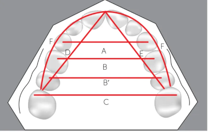
A) Maxillary incisor irregularity14 (LITTLE) (Fig 1).
B) Intercanine width (A; INTERC): The linear dis-tance between the cusp tips of the maxillary ca-nines. When there was a facet, the cusp tip was estimated (Fig 2).
C) Inter-premolar widths (INTERPB and
IN-TERPB’): The linear distance between left and right central fossae of the maxillary first (B) and second (B’) premolars (Fig 2).
D) Intermolar width (C; INTERMOL): The lin-ear distance between the mesiobuccal cusps tips of the maxillary first molars. When there was a facet, the cusp tip was estimated (Fig 2).
RESULTS
Dahlberg’s formula and Paired t tests showed no significant casual and systematic errors.
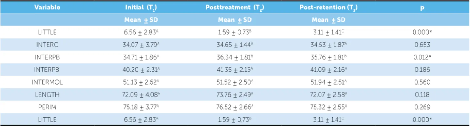
The Table 1 exhibits results of one-way analysis of variance (ANOVA) with the post-hoc Tukey test (different letters means a statistically significant dif-ference between variables) that were used to deter-mine whether there was a significant difference
be-tween the measured variables during T1, T2 and T3.
The results showed that the incisors irregularity had significant changes not only during treatment but also at posttreatment. Maxillary crowding relapse oc-curred in most patients with a mean percentage of 30.64% of the treatment correction. However, no significant differences were detected to the dimen-sional variables evaluated during the 3 phases, except for the inter-first-premolar width (INTERPB), that exhibited a statistically significant increase from
pre-treatment (T1) to posttreatment (T2).
Results of the Pearson correlation tests are in the Tables 2 and 3. There was a significant and nega-tive correlation between maxillary incisors crowding relapse and the relapse of the intercanine and inter-first-premolar widths.
DISCUSSION
Although incisors alignment relapse in maxillary arch is less prevalent than in mandibular arch, the evaluation of possible factors that may influence max-illary tooth alignment stability has validity. Relapse of crowding in this region may also results in esthetic and functional occlusal deficiencies. Mainly due to its F) Arch perimeter (F; PERIM): The distance in
millimeters from the mesial dental contact of the left first molars to the mesial dental contact of the right first molars to (Fig 2).
Statistical analysis Method error
Within a month interval from the first
measure-ment, ten dental casts from T1, T2 and T3 phases were
randomly selected and remeasured. The casual er-ror was calculated according to Dahlberg’s formula
(Se2= Σd2/2n).4 The systematic error was calculated
with dependent t tests, according to Houston.11
Statistical method
One-way dependent ANOVA and Tukey tests were used to evaluate the behavior of the measured
variables during the three phases (Initial – T1;
Post-treatment – T2; Post-retention – T3).
The Pearson correlation coefficient was calculated by using the whole sample to investigate a significant correlation between maxillary incisors crowding re-lapse and the pretreatment irregularity or the amount of crowding correction.
Pearson correlation coeicient was also calculated to investigate a association between maxillary incisors crowding relapse and the relapse of intercanine, interpre-molar or interinterpre-molar widths, arch length and perimeter.
The results were considered statistically significant at p < 0.05. All statistical analyses were performed with the software Statistica for Windows, version 6.0, Statsoft, Tulsa, Okla, USA.
Figure 1 - Little Irregularity Index (modiied for the upper arch) = A+B+C+D+E. Figure 2 - Variables studied on dental casts: A, intercanine width; B, inter-irst-premolar width; B’, inter-second-inter-irst-premolar width; C, intermolar width; D + E, arch length; F, arch perimeter.
A B
C
D
E F F
D
B
E
B′
Variable Initial (T1) Posttreatment (T2) Post-retention (T3) p
Mean ± SD Mean ± SD Mean ± SD
LITTLE 6.56 ± 2.83A 1.59 ± 0.73B 3.11 ± 1.41C 0.000*
INTERC 34.07 ± 3.79A 34.65 ± 1.44A 34.53 ± 1.87A 0.653
INTERPB 34.71 ± 1.86A 36.34 ± 1.81B 35.76 ± 1.81B 0.012*
INTERPB’ 40.20 ± 2.31A 41.35 ± 2.15A 41.09 ± 2.16A 0.186
INTERMOL 51.13 ± 2.62A 51.52 ± 2.50A 51.94 ± 2.51A 0.560
LENGTH 72.09 ± 4.08A 73.76 ± 2.49A 72.07 ± 2.58A 0.118
PERIM 75.18 ± 3.77A 76.52 ± 2.66A 75.32 ± 2.55A 0.269
LITTLE 6.56 ± 2.83A 1.59 ± 0.73B 3.11 ± 1.41C 0.000*
Table 1 - Results of one-way analysis of variance (ANOVA) with the post-hoc Tukey test (diferent letters means a statistically signiicant diference between vari-ables) for the variables measured on dental casts (N = 23), at the three stages studied (T1, T2 and T3).
Table 2 - Results of the Pearson correlation test. Table 3 - Results of the Pearson correlation test.
*Statistically signiicant at p < 0.05.
Variable r p
LITTLE1 x LITTLE3 0.252 0.071
LITTLE1 x LITTLE3-2 0.241 0.084
LITTLE2-1 x LITTLE3-2 -0.264 0.055
Variable r p
LITTLE3-2 x INTERC3-2 -0.459 0.000*
LITTLE3-2 x INTERPB3-2 -0.419 0.001*
LITTLE3-2 x INTERPB’3-2 -0.269 0.053
LITTLE3-2 x INTERMOL3-2 -0.064 -0.649
LITTLE3-2 x LENGTH3-2 0.028 0.842
LITTLE3-2 x PERIM3-2 -0.012 0.930
location, maxillary incisors crowding relapse tends to become more visible and therefore promote greater esthetic impacts than mandibular irregularity.
Results for one-way analysis of variance (ANOVA) with the post-hoc Tukey test (Table 1) showed that occurred statistically signiicant changes in the Little irregularity index during the three phases studied. It was observed a signiicant maxillary crowding reduc-tion during treatment. However, there was a signii-cant relapse of the incisors irregularity ater treatment. Regarding changes in maxillary arch dimensions dur-ing treatment, there was only a signiicant change in the variable INTERPB (Inter-irst-premolar width), suggesting that most maxillary arch dimensions were maintained during treatment, and remained stable
during post-retention. Sadowsky et al23 evaluating
stability in maxillary and mandibular dental arches of patients treated without extractions and Edgewise mechanics, observed no signiicant changes in the intercanine and inter-premolars widths, ive years
post-retention. Erdinc, Nanda and Isiksal,8 evaluated
stability of the orthodontic treatment, with and with-out extractions. Similarly to the present study, it was
observed signiicant decreases in maxillary incisors irregularity during treatment. Patients treated with-out extractions exhibited signiicant increases of the intercanine and inter-premolars widths during treat-ment. The maxillary arch dimensional measurements showed no signiicant changes ater treatment, how-ever, relapse of maxillary crowding was signiicant.
These studies8,23 and the present suggests a favorable
prognosis regarding maxillary arch long-term dimen-sional stability of orthodontic cases treated without premolars extractions.
In the present study, mean post-retention
crowd-ing relapse was 1.52 mm. Sadowsky et al,23 assessing
stability of subjects treated non-extraction, report-ed a relatively similar amount of relapse (1.1 mm), 5 years post-retention. However, Moussa, O’Reilly
and Close18 observed more favorable results
regard-ing crowdregard-ing relapse, 8 to 10 years post-retention, in a sample comprising 18 subjects treated with rapid maxillary expansion and ixed appliances. It was ob-served a mean maxillary crowding relapse of 0.6 mm
(SD = 1.30). Vaden, Harris and Gardner,30 noted that
stable 15 years ater treatment. The amount of crowd-ing increased from 1.5 mm at posttreatment to 1.8
mm at post-retention. Ferris et al9 also evaluated the
maxillary crowding relapse in non-extraction cases. It was observed only 0.47 (SD = 1.19) of maxillary irregularity increase during the post-retention (7.9 years). The increased stability observed in these stud-ies may be explained by the prolonged retention
pro-tocol ater orthodontic treatment.23 In the Sadowsky
et al23 study, retainers were placed and let for a mean
period of 8.4 years. Moussa, O’Reilly and Close,18
described a mean period of 6.6 years of retention for the mandibular arch and full time Hawley retainer in the maxillary arch for 2 years. The study conducted
by Vaden, Harris and Gardner,30 reports that patients
used these retainers in the maxillary and mandibular arches or these retainers in the maxillary arch and a lingual canine-to-canine mandibular bonded retainer. The irst posttreatment control was carried out only
ater six years. The study by Ferris et al9 described a
retention protocol that included the use of Hawley in the maxillary for 3 years (full time during one year) and lingual bonded retainer or Hawley plates in the mandibular arch for a mean period of 3 years. In the present study, all subjects wore a full time Hawley re-tainer in the maxillary arch for 12 months. A lingual canine-to-canine mandibular bonded retainer was placed and let for a mean period of 3 years.
Erdinc, Nanda and Isiksal8 described an increase in
maxillary incisors irregularity of 0.19 mm and 0.12 mm for patients treated with or without extractions, respec-tively, 4 years and 11 months ater treatment. The ex-traction group showed 4.4 mm of pretreatment crowd-ing. However, the non-extraction group exhibited only 1.94 mm of initial irregularity. The initial crowding was signiicantly less severe than that observed in our sample (6.56 mm). Maxillary and mandibular retainers were re-moved at least two years before the post-retention mea-surements. The exceptional stability of this study may be related to the amount of initial crowding and due to a short interval between retainer removal and the post-retention evaluation.
Although the results indicate a posttreatment maxillary crowding relapse greater than that reported in previous studies,8,9,18,23,30 the mean irregularity
in-dex at posttreatment (3.12 mm) is considered
clini-cally acceptable according to Little.14
Results of the Pearson correlation tests showed no significant correlations to most variables evaluated (Tables 2 and 3). It was observed that the amount of initial crowding had no effect on relapse, as described in previous studies.1,17 Surbeck et al,25 in contrast,
re-ported a positive correlation between the amount of maxillary incisors irregularity and the amount of
in-cisors crowding relapse. The authors reported25 that
the tendency to maxillary crowding relapse increases 2.3 times for each 0.2 mm of incisors contact point displacement in relation to the dental arch. Further-more, each 4° of tooth rotation at pretreatment has increased by 2.7 times the probability of
irregular-ity relapse. The authors25 also pointed out that
par-tially aligned tooth exhibits significant risk of relapse. They suggested the use of individualized retention protocols and that patients should be aware about the possibility of relapse accordingly to the initial
irregu-larity.25 However, a positive correlation between the
amount maxillary incisors crowding at pretreatment and the crowding relapse after treatment seems un-likely when analyzing our results and previous studies. For example, ours results indicated that the experi-mental group exhibited 6.56 mm of initial irregulari-ty and had a mean post-retention relapse of 1.52 mm. The mean crowding relapse observed in the present
study was higher than that reported by Ferris et al,9
Sadowsky et al23 and Vaden, Harris and Gardner,30
with samples that exhibited more maxillary incisors irregularity at pretreatment (10.45 mm, 8.0 mm and 7.9 mm, respectively). Despite more initial crowding, these studies reported less posttreatment irregularity relapse (0.47 mm, 1.1 mm, 0.3 mm, respectively).
The amount of maxillary crowding relapse (LIT-TLE3-2) showed a statistically signiicant and negative correlation (p < 0.05) with the post-retention changes in intercanine (INTERC3-2) and inter-irst-premolars (INTERPB3 -2) widths (Table 3). These results suggest that the higher the post-retention decreases of interca-nine and inter-irst-premolars widths the higher the maxillary crowding relapse. However, although these correlations have statistical signiicance, the coeicients
values observed implicate in a weak correlation (r values
Moreover, it seems obvious that the reduction of these measurements tends to be consequence of maxillary arch constriction in the anterior region. Therefore, it is expected a space availability decrease and an increase in the amount of tooth crowding.
Despite numerous studies that evaluated a possible relationship between changes in intercanine width and mandibular incisors crowding relapse, the correlation between maxillary crowding relapse and maxillary intercanine width changes were only investigated by
Surbeck et al,25 and Erdinc, Nanda and Isiksal.8
Sur-beck et al25 found a signiicant association (p < 0.001)
between intercanine width decreases and maxillary incisors crowding relapse, however, the correlation test result also revealed a weak association (r < 0.70).
Erdinc, Nanda and Isiksal,8 found no correlation
be-tween post-retention changes in incisors irregularity and changes in intercanine width.
Clinical implications
Maxillary anterior alignment shows better prog-nosis regarding stability when compared to the same region in the mandibular arch, this fact may explain the scarce studies in literature about this issue. De-spite the greater stability, maxillary crowding relapse can compromise orthodontic results after retention
appliances removal. The post-retention relapse ob-served in this study (1.52 mm), although statistically
significant, may be considered clinically acceptable.14
Otherwise, this minimal amount of crowding relapse can lead to patient dissatisfaction.
Maxillary incisors crowding relapse shows some etiological factors as retention time, initial crowding severity; relapse of teeth in the opposite side, changes in arch dimensions, rotated teeth at pretreatment and lack of complete correction of rotated teeth resulting in absence of adequate interdental contacts. Thus, it becomes clear that more stable results can be obtained with a prolonged retention protocol and an adequate alignment of maxillary incisors during treatment.
CONCLUSION
The maxillary incisors irregularity increased sig-nificantly (1.52 mm) five years posttreatment.
None of the clinical factors studied in the dental casts demonstrated to be predictive of the maxillary crowding relapse.
1. Artun J, Garol JD, Little RM. Long-term stability of mandibular incisors following successful treatment of Class II, Division 1, malocclusions. Angle Orthod. 1996;66(3):229-38.
2. Busdrang PH, Horton-Reuland SJ, Legler L, Nevant C. Non-extraction approach to tooth size arch length discrepancies with the Alexander discipline. Semin Orthod. 2001;7(2):117-31.
3. Canuto LF, Freitas MR, Janson G, Freitas KM, Martins PP. Inluence of rapid palatal expansion on maxillary incisors alignment stability. Am J Orthod Dentofacial Orthop. 2010;137(2):164.e1-6.
4. Dahlberg G. Statistical methods for medical and biological students. New York: Interscience; 1940.
5. Edwards JG. A study of the periodontium during orthodontic rotation of teeth. Am J Orthod. 1968;54(6):441-61.
6. Edwards JG. A surgical procedure to eliminate rotational relapse. Am J Orthod. 1970;57(1):35-46.
7. Edwards JG. A long-term prospective evaluation of the circumferential supracrestal iberotomy in alleviating orthodontic relapse. Am J Orthod Dentofacial Orthop. 1988;93(5):380-7.
8. Erdinc AE, Nanda RS, Işiksal E. Relapse of anterior crowding in patients treated with extraction and non-extraction of premolars. Am J Orthod Dentofacial Orthop. 2006;129(6):775-84.
9. Ferris T, Alexander RG, Boley J, Buschang PH. Long-term stability of combined rapid palatal expansion-lip bumper therapy followed by full ixed appliances. Am J Orthod Dentofacial Orthop. 2005;128(3):310-25. 10. Heiser W, Niederwanger A, Bancher B, Bittermann G, Neunteufel N,
Kulmer S. Three-dimensional dental arch and palatal form changes after extraction and non-extraction treatment. Part 1. Arch length and area. Am J Orthod Dentofacial Orthop. 2004;126(1):82-90.
11. Houston WJ. The analysis of errors in orthodontic measurements. Am J Orthod. 1983;83(5):382-90.
12. Huang L, Artun J. Is the post-retention relapse of maxillary and mandibular incisor alignment related? Am J Orthod Dentofacial Orthop. 2001;120(1):9-19.
13. Kahl-Nieke B, Fischbach H, Schwarze CW. Post-retention crowding and incisor irregularity: a long-term follow-up evaluation of stability and relapse. Br J Orthod. 1995;22(3):249-57.
14. Little RM. The irregularity index: a quantitative score of mandibular anterior alignment. Am J Orthod. 1975;68(5):554-63.
15. Little RM. Stability and relapse of dental arch alignment. Br J Orthod. 1990;17(3):235-41.
REFERENCES
16. Little RM, Riedel RA, Artun J. An evaluation of changes in mandibular anterior alignment from 10 to 20 years post-retention. Am J Orthod Dentofacial Orthop. 1988;93(5):423-8.
17. Little RM, Wallen TR, Riedel RA. Stability and relapse of mandibular anterior alignment – irst premolar extraction cases treated by traditional edgewise orthodontics. Am J Orthod. 1981;80(4):349-65.
18. Moussa R, O’Reilly MT, Close JM. Long-term stability of rapid palatal expander treatment and edgewise mechanotherapy. Am J Orthod Dentofacial Orthop. 1995;108(5):478-88.
19. Parker WS. Retention: retainers may be forever. Am J Orthod Dentofacial Orthop. 1989;95(6):505-13.
20. Richardson ME. A review of changes in lower arch alignment from seven to ifty years. Semin Orthod. 1999;5(3):151-9.
21. Rossouw PE, Preston CB, Lombard CJ, Truter JW. A longitudinal evaluation of the anterior border of the dentition. Am J Orthod Dentofacial Orthop. 1993;104(2):146-52.
22. Sadowsky C, Sakols EI. Long-term assessment of orthodontic relapse. Am J Orthod. 1982;82(6):456-63.
23. Sadowsky C, Schneider BJ, BeGole EA, Tahir E. Long-term stability after orthodontic treatment: non-extraction with prolonged retention. Am J Orthod Dentofacial Orthop. 1994;106(3):243-9.
24. Sinclair PM, Little RM. Maturation of untreated normal occlusions. Am J Orthod. 1983;83(2):114-23.
25. Surbeck BT, Artun J, Hawkins NR, Leroux B. Associations between initial, posttreatment, and post-retention alignment of maxillary anterior teeth. Am J Orthod Dentofacial Orthop. 1998;113(2):186-95.
26. Swanson WD, Riedel RA, Danna JA. Post-retention study: incidence and stability of rotated teeth in humans. Angle Orthod. 1975;45(3):198-203. 27. Taner TU, Haydar B, Kavuklu I, Korkmaz A. Short-term efects of
iberotomy on relapse of anterior crowding. Am J Orthod Dentofacial Orthop. 2000;118(6):617-23.
28. Thilander B. Orthodontic relapse versus natural development. Am J Orthod Dentofacial Orthop. 2000;117(5):562-3.
29. Uhde MD, Sadowsky C, BeGole EA. Long-term stability of dental relationships after orthodontic treatment. Angle Orthod. 1983;53(3):240-52.