Sagittal changes in lower incisors
by the use of lingual arch
Helen Carolina Becker Letti1, Susana Maria Deon Rizzatto2, Luciane Macedo de Menezes2, Chalana Sangalli Reale3, Eduardo Martinelli de Lima2, Fernando Lima Martinelli2
Objective:The objective of this study was to evaluate a sagittal variation on the lower incisors with the use of the lingual arch on the transition from mixed to permanent dentition. Methods: The sample was constituted of 44 Caucasian patients (26 girls and 18 boys), divided in two groups: CG, control group, monitoring the lower arch space with no orthodontic/ orthopedic treatment during the rated period (n = 14); EG, experimental group, presenting anterior inferior mild crowd-ing and installation of the lcrowd-ingual arch for space maintenance (n = 30). The position of the lower incisors was analyzed on computerized cephalometric tracings performed at the beginning of the monitoring (T1) and at the end, on the permanent dentition (T2). In order to evaluate the position of the incisors it was used Tweed and Steiner measurements: IMPA, 1.NB and 1-NB. The alterations were analyzed through the “t” test for paired samples, while the diferences between the groups were analyzed through the “t” test for independent samples, as for sexual dimorphism. Results: The values in T2 were greater in relation to T1 for all measurements on EG (IMPA, p = 0.038; 1.NB, p = 0.007 and 1-NB, p < 0.0001). On com-paring the diferences (T2-T1) between CG and EG, it can be gauged diferences signiicantly superior for measure 1.NB (p = 0.002) and 1-NB (p < 0.0001) on EG. There was no statisticaly signiicant diference between genres. Conclusion: It was concluded that the lower incisors were projected ater using the lingual arch to control the space on the transition from mixed to permanent dentition, however, within acceptable standards, not having diference between genres.
Keywords:Incisor. Orthodontics. Cranial circumference.
How to cite this article: Letti HCB, Rizzatto SMD, Menezes LM, Reale CS, Lima EM, Martinelli FL. Sagittal changes in lower incisors by the use of lingual arch. Dental Press J Orthod. 2013 May-June;18(3):29-34.
Submitted: January 28, 2009 - Revised and accepted: April 27, 2011
» The patients displayed in this article previously approved the use of their facial and intraoral photographs.
Contact address: Helen Carolina Becker Letti
Av. Ipiranga, 6681 Prédio 6, Faculdade de Odontologia – Brazil
Bairro Partenon – Porto Alegre/RS - CEP: 90619-900 – Email: [email protected]
» The authors report no commercial, proprietary or financial interest in the products or companies described in this article.
1 Specialist in Orthodontics, PUCRS.
2 Professor at the Post-Graduation Course in Orthodontics and Facial
Orthopedics, PUCRS.
3 MSc in Orthodontics and Facial Orthopedics, PUCRS.
Objetivo:avaliar a alteração sagital ocorrida nos incisivos inferiores com o uso do arco lingual no período de transição da dentição mista para a permanente. Métodos: a amostra foi composta por 44 pacientes leucodermas (26 meninas e 18 meninos), divididos em dois grupos: (GC) grupo controle, no qual foi efetuado monitoramento do espaço da arcada in-ferior, sem tratamento ortodôntico/ortopédico no período avaliado (n = 14); (GE) grupo experimental, presença de suave apinhamento anteroinferior e instalação do arco lingual para manutenção do espaço (n = 30). A posição dos incisivos infe-riores foi analisada em traçados cefalométricos computadorizados realizados ao início (T1) e ao inal do acompanhamento, já na dentição permanente (T2). Para avaliar a posição dos incisivos foram utilizadas as medidas das análises cefalométricas de Tweed e de Steiner: IMPA, 1.NB e 1-NB. As alterações ocorridas foram analisadas pelo teste t para amostras parea-das, enquanto as diferenças entre os grupos foram avaliadas pelo teste t para amostras independentes, bem como para o dimorismo sexual. Resultados: os valores em T2 foram maiores em relação a T1 para todas as medidas no GE (IMPA, p = 0,038; 1.NB, p = 0,007; e 1-NB, p < 0,0001). Na comparação das diferenças (T2-T1) entre o GC e GE pôde-se aferir diferenças signiicativamente superiores para as medidas 1.NB (p = 0,002) e 1-NB (p < 0,0001) no GE. Não houve dife-rença signiicativamente estatística entre os sexos. Conclusão: concluiu-se que os incisivos inferiores foram projetados após a utilização do arco lingual para o controle do espaço no período de transição da dentição mista para a permanente, porém a projeção esteve dentro dos padrões aceitáveis, não havendo diferença entre sexos.
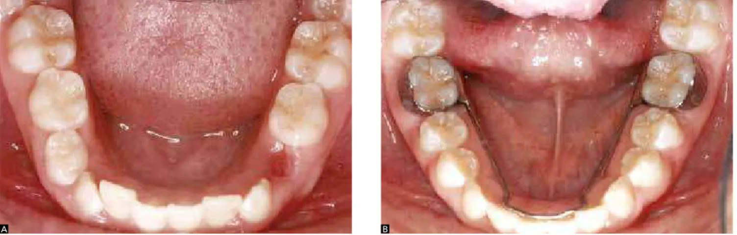
Figure 1 - A) T1 : mixed dentition, previously to installation of the lingual arch. B) T2: young permanent dentition with lingual arch installed. INTRODUCTION
The origin of the lingual arch was assigned by Dewey3
to Dr. Lourie in 1904, however, Mershon14,15 was the
re-sponsible for the popularization of the referred appliance. Originally it was used to expand the lower arch and pos-teriorly Nance16,17 described the indication of the lingual
arch for the treatment on mixed dentition, suggesting its use only on the maintenance of the distance between per-manent incisors and molars in speciic cases.
The lingual arch, as an appliance of passive appli-cation, similar to the recommended by Nance,16 is
widely used in orthodontic clinics until the present day. Its main utility is on controlling the perimeter of the lower arch, maintaining the distance between the first permanent molars and the lower incisors af-ter the loss of deciduous molars. It can also be used as assistant for intraoral anchorage on the permanent dentition, from the mixed dentition. This type of ap-pliance shows efficiency on the maintenance of the lower arch perimeter, preventing the molars move-ment to mesial and the incisors lingual inclination.22
The maintenance of the perimeter on the denti-tion development is fundamentally important be-cause the use of the Leeway Space for eruption of premolars guarantees space for the correct alignment of the lower teeth in up to 80% of the patients with mild crowding.1,6,10,18
If not provided with adequate space for alignment of the permanent teeth, it may be necessary a less conservative conduct when the corrective orthodon-tic treatment is performed.
This way, the lingual arch installed on appropriate moment can reduce the number of future premolars
extraction, lower arch stripping and other procedures to restore the space for adequate alignment of teeth.6
The cognition about the effects of the lingual arch on these teeth represents a necessity on the orth-odontic practice, being an appliance of wide clinical utilization. The purpose of this study was to evaluate the sagittal variation on lower incisors due to utili-zation of lingual arch on the transition from mixed dentition to permanent.
MATERIAL AND METHODS
The present study comprehended 44 Caucasian pa-tients (26 girls and 18 boys) in need of supervision of the space on the mixed dentition. The transition period from mixed dentition to permanent was monitored in two ways: Control group (CG, n = 14), space monitor-ing with no orthodontic/orthopedic treatment durmonitor-ing the rated period; Experimental group (EG, n = 30), pre-senting anterior inferior mild crowding and installation of the lingual arch, previously to exfoliation of decidu-ous second molar, for space maintenance (Fig 1A).
The lingual arch appliance was made with stainless steel wire 0.9 mm (Morelli, Sorocaba/SP) outlining the incisal third of the lower incisors crown. The ap-pliance was attached with glass ionomer cement (3M Unitek, CA/USA) in all patients from the experi-mental group (Fig 1B).
In the individuals from the control group it were performed lateral radiographs in two-step with a mean age of 9.7 years in T1 (±1.6) and 11.6 years in T2 (±1.3). In the patients from the EG, it were also performed two radiographs, in T1 obtained with mean age of 9.3 years (±1.1) and T2 with mean age of 11.9 years (±1.1).
Computerized cephalometric tracings were obtained from the Ortoview 2.5 sotware and analyzed at the beginning of the monitoring (T1) and at the end, ater eruption of permanent canines and premolars (T2), with lingual arch still installed (Figs 2A and 2B). In order to evaluate the sagittal variations of the incisor it was used Tweed22,23,24 and Steiner19 measurements:
» IMPA: angle between the long axis of the lower incisor and the base of the mandible (line join-ing the point “Me” and the “lower portion on the back of the mandible base”) – Tweed’s analysis; » 1.NB: angle between the long axis of the lower incisor and the line NB – Steiner’s analysis; » 1-NB: linear distance (mm) between the most prominent portion of the lower incisor crown and line NB – Steiner’s analysis.
Initially, it was performed a study of error. The measurements were redone by the same operator and compared through Student’s t test for paired samples. There was no significant difference between the val-ues of the 1st and the 2nd measurement for the evalu-ated measures (p > 0.05).
The data were, then, collected and statistically ana-lyzed comparing T1 to T2, evaluating the diference between genres and the diference between the two groups. The data were organized in tables and graph-ics, with mean values and standard deviation for pe-riod T1 and T2. To compare the two periods it was used the Student’s t test for paired data. To evaluate
the diference between genres and on comparing the two groups (experimental and control) it was used the Student’s t test for independent samples. The results were considered signiicant in a maximum level of sig-niicance of 5%. To verify the normality of the data it was used the Kolmogorov-Smirnov non-parametric test, concluding that they have normal distribution.
RESULTS
The obtained results revealed signiicant diferences between periods T1 and T2 in relation to the position of the lower incisors in the experimental group. In this group, it was noticed increasing values in T2 to the lin-ear measure 1-NB (p = 0.000), as to the angular mea-sures 1.NB (p = 0.007), IMPA (p = 0.038) (Table 1).
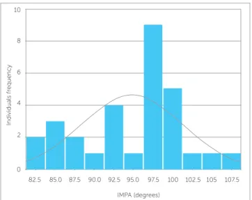
The data show the projection on the lower incisors with the utilization of the passive lingual arch. On EG, in T2, over 40% of the patients presented nor-mal variation to the angular measure IMPA and 85% did not exceeded 100° (Fig 3). In the control group (CG), there was no significant difference between T1 and T2 to any of the evaluated measures, according to Table 1. On comparing the differences (T1-T2) be-tween the control group and the experimental group, it can be gauged differences significantly superior on experimental group for measure 1.NB(p = 0.002) and 1-NB(p < 0.000) (Table 1). There was no significant difference between genres to the evaluated measures (Tables 2 and 3).
Figure 2 - A) Cephalometric tracing at T1: previously to installation of the lingual arch. B) Cephalometric tracing at T2: young permanent dentition with lingual
arch still installed.
Morphological variations in the symphysis men-talis region, due to growth, result in modifications on the position of lower incisors. The cognition of these variations is of great clinical importance, being fundamental data on orthodontic planning.8,11
Ac-cording to Enlow,5 as regards to lower incisors, there
is a physiological remodeling on the alveolar region, causing a lingual inclination. The lower dental arch is referenced as one of the main elements for the di-agnosis and for the orthodontic therapy. The dental positions will be established basically by the bone configuration, that is, the teeth should be aligned on the alveolar ridge.21
Tweed,22 through his studies, suggested the
incli-nation of the lower incisors in relation to the man-dibular ridge of 90° with a variation of 5° for further or for fewer, creating the IMPA, first angle of what would be the diagnostic facial triangle. Posteriorly, the same author, found the norm of 87° to the angu-lar measure IMPA with variation from 76° to 99°on the studied sample.23,24 In the experimental group
(in T2), in relation to this angle, over 40% of the pa-tients are within the normal variation and 85% did not exceeded 100° (Fig 3), being these values con-sidered according to acceptable standard of normal-ity and the minimum value found of 82.5°and the maximum of 106°.
Steiner20 suggested angular and linear mean values
to the position of lower incisors in relation to line NB considered as standard, being 25° to 1.NB and 4 mm to 1-NB. In this study, in T2, to the angular measure 1.NB the minimum value found was 18.8° and the maximum was 36.6°. To the linear measure, 1-NB, the values were 3.6 mm and 11.3 mm, being respectively minimum and maximum. However, val-ues conventionally adopted as representative of nor-mality not always can be used indistinctly, given that variation due to growth and craniofacial development are characteristic of different organisms.11
The normal mandibular growth tend to increase the SNB, with greater progress attributed to point B, since the point N does not progresses with the same intensity, altering the geometric relation of the line NB with the long axis of the lower incisor, with ar-tificial reduction of 1.NB and 1-NB. Therefore, the IMPA presents more reliable value for the analysis of the lower incisor position, since it experiences less
DISCUSSION
The length of the arch, as well as its perimeter, is reduced on the transition from mixed dentition to permanent, especially on the mandibular arch. This reduction on the arch perimeter is basically due to a mesial migration of the first permanent molar after the loss of the second deciduous molar.7 The
lin-gual arch shows efficiency on the maintenance of the lower arch perimeter, that is, preventing the molars movement to mesial and the linguoversion of the in-cisors, fact that can be associated to the reduction of mandibular crowding.2,25 It allows the lower
inci-sors to take an appropriate position by the perioral musculature and intraoral functional forces. Singer19
noticed distal inclination of lower molar, tenden-cy (not significant) to projection of incisors and a greater tendency of the incisors and molars to pres-ent smaller vertical developmpres-ent in patipres-ents treated with lingual arch.
Table 1 - Means, mean diferences and t test comparing the groups.
*p value: Paired sample; **p value: Independent samples.
T1 T2 *p T2-T1 **p
IMPA (CG) 94.8 94.2 0.468 -0.6
0.083
IMPA (EG) 92.9 94.8 0.038 1.9
1.NB (CG) 26.7 25.9 0.163 -0.8
0.002
1.NB (EG) 27.1 29.8 0.007 2.7
1-NB (CG) 4.9 5.1 0.669 0.2
0.000
1-NB (EG) 5.1 6.7 0.000 1.6
Table 2 - EG: Mean diferences ± standard deviation and t test comparing genders.
*p value: Independent samples.
Mean diferences T2-T1
n 1-NB
(mm)
1.NB (degrees)
IMPA (degrees)
Feminine 19 1.5 ± 1.1 1.6 ± 4.6 1.1 ± 4.8
Masculine 11 2.0 ± 1.2 4.7 ± 5.6 3.6 ± 5.2
*p 0.258 0.118 0.186
Table 3 - CG: Mean diferences ± standard deviation and t test comparing genders.
*p value: Independent samples.
Mean diferences T2-T1
n 1-NB
(mm)
1.NB
(degrees)
IMPA
(degrees)
Feminine 7 -0.1 ± 0.3 -0.4 ± 2.2 -0.1 ± 2.6
Masculine 7 0.2 ± 1.2 -1.2 ± 1.9 -1.1 ± 3.0
promote smaller chances of development of future occlusal problems.9 Besides the maintenance of the
Leeway space, using the lingual arch is possible to restore space in up to 2 mm of negative discrepan-cy, eliminating the crowding on the transition from mixed dentition to permanent when it is presented.10
Dugoni et al4 verified that using the lingual arch
on alignment of lower incisors was clinically accept-able in 76% of the cases on the post-restraint period. According to the same authors, the use of the Lee-way space to dissolution of crowding may result in better stability in long-term.4 The methods usually
adopted to calculate the discrepancy in the analysis of the mixed dentition can overestimate the values obtained. It is suggested, therefore, the utilization of size prediction methods of permanent teeth that have a coefficient of explanation (R²) closer to 1. For this, the utilization of oblique teleradiograph of 45° is an option with good degree of accuracy.12,13
Rabellato et al18 reported a slight projection of
the lower incisors, finding a mean value of increase on IMPA of 0.73°, having evaluated 14 patients with mean age of 11.5 years, that used only passive lingual arch in this period of the treatment, using the overlay method of cephalometric tracings for comparison pre and post-treatment.
Villalobos24 reported that the lower incisor
pre-sented a discrete retroclination of 0.52°, on aver-age, using the lingual arch through the same overlay method of cephalometric tracings. Despite the meth-odological difference about the comparison between the initial and the final position of the lower incisors, in the present study it was found a mean projection of 1.9° for the IMPA. The option for using computer-ized cephalometric tracings with angular and linear measures was based on standardization of the tech-nique, allowing longitudinal evaluations of the same individual with good reliability degree.
CONCLUSION
It was concluded that the lower incisors were project-ed ater using the lingual arch to control the space on the transition from mixed dentition to permanent, however within acceptable standards, not having statistic difer-ence between genres for the evaluated measures.
influence of the mandibular growth. Aiming to de-termine accurately the inclination of the lower inci-sors, the measures 1.NB, 1-NB and IMPA were used with reference in this work.
The variations on point B occur smoothly from 6 to 10 years and well-marked from 10 to 15 years, concomitantly with the lingual inclination of inci-sors.8 In agreement to the previous study, Watanabe
et al26 also verified a lingual inclination of mandibular
incisors between 8 and 15 years old. During puberty, therefore, the lower incisors tend to move lingual, if nothing prevents this movement. If the lingual in-clination does not occur, this can be attributed to crowding in the anterior region.
According to the results found in this study, the use of the lingual arch prevented the tendency of lin-gual inclination reported on previous studies, being observed some projection of lower incisors. The pro-jection of the lower incisors verified in this study can be considered clinically advantageous, noting that minimizes or even eliminates the necessity of future extractions or stripping during the treatment in cases of small negative discrepancies on the mixed denti-tion and depending of the cephalometric and facial interpretation, of the complaint of the patient and of what is biologically acceptable in each individual.
This projection can be used, facilitating orth-odontic biomechanical with gain of space.27 The
orthodontic intervention on mixed dentition would
Figure 3 - IMPA (Experimental Group): inal value.
82.5 10
8
6
4
2
0
85.0
Individuals fr
equency
87.5 90.0 92.5 95.0 97.5
IMPA (degrees)
1. Brennan MM, Gianelly AA. The use of the lingual arch in the mixed dentition to resolve crowding. Am J Orthod Dentofacial Orthop. 2000;117(1):81-5.
2. De Baets J, Chiarini M. The pseudo-Class I: a newly deined type of
malocclusion. J Clin Orthod. 1995;29(2):73-88.
3. Dewey M. The lingual arch in combination with the labial arch with
extensions as used by Dr. Lloyd S. Lourie. Int J Orthod. 1916;2(10):563-602.
4. Dugoni SA, Lee JS, Varela J, Dugoni AA. Early mixed dentition treatment: postretention evaluation of stability and relapse. Angle Orthod. 1995;65(5):311-20.
5. Enlow DH. O processo do crescimento facial. In: Crescimento facial.
3ª ed. São Paulo: Artes Médicas; 1993. p. 72-3.
6. Gianelly AA. Crowding: timing of treatment. Angle Orthod.
1994;64(6):415-8.
7. Gianelly AA. Treatment of crowding in the mixed dentition. Am J Orthod
Dentofacial Orthop. 2002;121(6):569-71.
8. Jones JD. The eruption of the lower incisor and the accompanying
development of the symphysis and point B. Angle Orthod. 1966;36(4):358-62.
9. Keski-Nisula K, Hernesnieme R, Heiskanen M, Keski-Nisula L, Varrela J. Orthodontic intervention in the early mixed dentition: A prospective, controlled study on the efects of the eruption guidance appliance. Am J Orthod Dentofacial Orthop. 2008;133(2):254-60.
10. Letti HCB, Braga FL, Lima EMS. O arco lingual na transição da dentição mista para a dentição permanente. Ortod Gaúch. 2005;9(2):122-6. 11. Marques JS, Siqueira VCV. Estudo das alterações do ponto B durante
o tratamento ortodôntico. Rev Dental Press Ortod Ortop Facial. 2007;12(3):136-45.
12. Martinelli F, Lima EM, Rocha R, Tirre-Araujo MS. Prediction of lower permanent canine and premolars width by correlations methods. Angle Orthod. 2005;75(5):805-8.
13. Melgaço CA, de Sousa Araújo MT, de Oliveira Ruellas AC. Mandibular permanent irst molar and incisor width as predictor of mandibular canine and premolar width. Am J Orthod Dentofacial Orthop. 2007;132(3):340-5.
REFERENCES
14. Mershon JV. Band and lingual arch technique. Int J Orthod. 1917;3:195-203.
15. Mershon JV. The removable lingual arch as an appliance for the treatment of malocclusion of the teeth. Int J Orthod. 1918;4:578-87. 16. Nance HN. The limitations of orthodontics treatment: I, mixed dentition
diagnosis and treatment. Am J Orthod. 1947 Apr;33(4):177-223. 17. Nance HN. The limitations of orthodontics treatment: II, diagnosis and
treatment in the permanent dentition. Am J Orthod. 1947;33:253-301. 18. Rebellato J, Lindauer SJ, Rubenstein LK, Isaacson RJ, Davidovitch M,
Vroom K. Lower arch perimeter preservation using the lingual arch. Am J Orthod Dentofacial Orthop. 1997;112(4):449-56.
19. Singer J. The efect of the passive lingual archwire on the lower denture. Angle Orthod. 1974;44(2):146-55.
20. Steiner CC. Cephalometrics for you and me. Am J Orthod. 1953;39(10):729-55.
21. Triviño T, Siqueira DF, Scanavini MA. A forma do arco dentário inferior na visão da literatura. Rev Dental Press Ortod Ortop Facial. 2007;12(6):61-72. 22. Tweed CH. A philosophy of orthodontic treatment. Am J Orthod.
1945;31(2):74-103.
23. Tweed CH. The Frankfort-Mandibular Incisor Angle (FMIA). In:
Orthodontic diagnosis, treatment, planning and prognosis. Am J Orthod. 1954;24(3):74-103.
24. Tweed CH. Was the development of diagnosis facial triangle as an accurate analysis based on fact or fancy? Am J Orthod. 1962; 48(11):823-40.
25. Villalobos FJ, Sinha PK, Nanda RS. Longitudinal assessment of vertical and sagittal control in the mandibular ixed lingual arch. Am J Orthod Dentofacial Orthop. 2000;118(4):366-70.
26. Watanabe E, Demirjian A, Buschang P. Longitudinal post-eruptive mandibular tooth movements of males and females. Eur J Orthod. 1999;21(5):459-68.