Transverse maxillary and mandibular growth during and
after Bionator therapy: Study with metallic implants
André da Costa Monini1, Luiz Gonzaga Gandini Júnior2, Luiz Guilherme Martins Maia3, Ary dos Santos-Pinto4
Introduction: This study evaluated posteroanterior cephalograms before and ater treatment and long term follow-up of Class II division 1 patients treated with bionator. Objective: The objective was to demonstrate the transverse growth of maxilla and mandible during and ater bionator therapy. Methods: Measurement of transverse dimensions between pos-terior maxillary and mandibular implants, as well as the distances between the buccal, gonial and antegonial points were recorded. Measurements were analyzed at three periods: T1 = before bionator therapy, T2 = ater bionator therapy and T3 = 5.74 years ater T2. Results: There was statistically signiicant transverse increase due to growth and/or treatment for all variables, except for the distance between the anterior maxillary implants. Conclusions: During the study period only the anterior maxillary area did not show transverse growth.
Keywords:Activator appliances. Angle Class II malocclusion. Maxillofacial development.
How to cite this article: Monini AC, Gandini Júnior LG, Maia LGM, Santos-Pinto A. Transverse maxillary and mandibular growth during and ater Bionator ther-apy: Study with metallic implants. Dental Press J Orthod. 2013 May-June;18(3):72-9.
Submitted: January 04, 2010 – Revised and accepted: October 20, 2010
» The authors report no commercial, proprietary or financial interest in the products or companies described in this article.
Contact address: Luiz Gonzaga Gandini Júnior
Av. Casemiro Perez, 560 – Vila Harmonia – Araraquara/SP – Brazil CEP: 14.802-600 – E-mail: [email protected]
1 Specialist and Master in Orthodontics UNESP-Araraquara.
2 Professor, School of Dentistry of Araraquara, UNESP. Assistant Professor,
Baylor College of Dentistry, Dallas, Texas, USA.
3 Master and Doctorate student in Orthodontics UNESP- Araraquara. 4 Professor, School of Dentistry of Araraquara, UNESP.
Introdução: esse estudo envolve a avaliação de telerradiograias posteroanteriores pré- e pós-tratamento com Bionator, bem como, em longo prazo, de pacientes Classe II divisão 1. Objetivo:o objetivo desse trabalho é demonstrar o cres-cimento transversal da maxila e mandíbula durante e após o uso do Bionator. Métodos: as mensurações das distâncias transversais entre os implantes posteriores da maxila e mandíbula, bem como as das distâncias entre os pontos jugal, gônio e antigônio, foram tomadas em três tempos: T1, antes da terapia com Bionator; T2, após a terapia como Bionator; e T3, 5,74 anos após T2. Resultados: ocorreu aumento transversal estatisticamente signiicativo por crescimento e/ou por tra-tamento em todas as variáveis estudadas, com exceção da distância entre os implantes anteriores da maxila. Conclusões:
durante o período do estudo, somente a região anterior da maxila não apresentou crescimento transversal.
INTRODUCTION
Few studies evaluating the transverse growth of the face were carried out so far, especially regarding sagittal growth. This is due to problems such as dif-ficulty on identification and consequent
reproduc-ibility of cephalometric points,18,21 standardization
of the head positioning,20,11 radiographic
magnifi-cation9,11,15,27 and standardization of the sample.19 In
the last years, some studies assessed the facial skeletal growth without interference of functional orthopedic
appliances.9,10,13,16,17,25 Several studies showed the
po-tential of increase on transverse growth of the jaws by
the use of functional appliances1,8,12,14,23,26 and three of
them12,14,26 followed longitudinally the patients after
treatment, but without radiographic evaluation. The longitudinal examination using metallic implants carried out so far refers to Class I patients with or
without treatment4,5,10,17 or to mixed samples.16
The cephalometric studies in teleradiographs with metallic implants proved to be the most effi-cient method to longitudinally assess the
craniofa-cial growth3,5 due to the difficulty of identification of
cephalometric points and remodeling that occurs on the surface of the jaws. The objective of the present study is to evaluate the maxillary transverse growth and its relation to the treatment, through posteroan-terior radiographs, during 6 years after the use of Bal-ters’ bionator in patients with metallic implants.
MATERIAL AND METHODS
The sample consisted of 25 patients that used bi-onator (15 boys and 10 girls), participants on a prior
study1 and treated in the Department of
Orthodon-tics at the School of Dentistry of Araraquara – Un-esp. Each one of them presented skeletal Class II with mandibular retrusion, upper and lower incisors erupted or in eruption, overbite, no dental loss, ab-sence of crowding and/or posterior cross bite. The subjects of the sample had metallic implants in-serted in the maxilla (four implants) and mandible
(three implants), according to proposed by Björk.6,7
From the original sample of 25 patients (mean age of 9.2 years), it was possible to obtain long term radio-graphs of 13 patients (9 boys and 4 girls) with mean age of 16.95 years. The other patients could not be contacted. On the final sample, one patient did not
present the posterior implants on the maxilla in T3
and on the mandible in T1, other patient did not
pres-ent one anterior implant in T3 and another patient
did not present one of the posterior implants on the mandible. Table 1 shows age and gender of the sample and Table 2 characterizes the sample.
Lateral and posteroanterior teleradiographs were
obtained in three time periods: T1 at the beginning
of treatment with bionator, T2 at the end of the
bi-onator therapy and (T3) 5.74 years, on average, after
T2. The teleradiographs were manually traced and
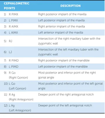
the cephalometric points were digitized twice on Dentofacial Planner Plus (DFP Plus, version 2.0, Toronto, Ontario, Ca) by a single examiner and the digitalization mean was used for cephalometric measurements. The cephalometric points used on the posteroanterior teleradiographs are described on Table 3 and Figure 1.
The transverse growth was calculated by the trans-verse linear distance between the cephalometric points on the right and on the let. Corrections for magniica-tion on transverse linear measurements were necessary before classifying the growth data, because although the posteroanterior teleradiographs had been taken with cephalostat, the radiographic magniication of the gion of the metallic implants is diferent from the re-gion of the acoustic meatus center plane because it is closer to the radiographic ilm, especially compared to the anterior implants on the maxilla. Another reason for correction is that, with the facial growth, maxilla move forward carrying together the metallic implants making them closer to the radiographic ilm. These variations on radiographic magniication were mathematically corrected by a combination of information of lateral and posteroanterior teleradiograph using correction method
recommended by Hsiao et al.15 A reference system,
comprised by Frankfurt’s horizontal plane and a vertical line perpendicular from the Porion, built in each lateral teleradiograph allowed the calculation of distance from the position of the implants mean to the acoustic meatus center plane (Fig 2).
With these measures it was possible to calculate the radiographic magniication on the region of me-tallic implants for each patient based on the formula
described by Hsiao et al:15 Inter-implants real
Table 1 - Characteristics of the studied sample. Table 3 - Cephalometric points digitized on the posteroanterior teleradiograph.
T1 - (beginning of treatment). T2 - (end of treatment with bionator). T3 - (inal evaluation).
Figure 1 - Cephalometric points digitized on the posteroanterior
teleradio-graph. Table 3 identiies each cephalometric point.
Figure 3 - AB: Distance olive; BH: ear rods-implant distance; AC:
focus-ilm distance; DE: inter-implants real distance (posterior inter-implants of the maxilla, anterior inter-implants of the maxilla and posterior inter-implants of the mandible); FG: inter-implants. radiographic distance (Source: Hsiao et al.15).
Figure 2 - Traced line shows the calculation of distance from the position of
implants (mean point between implants) to the acoustic meatus center plane.
Individuals n
T1 T2 T3
Mean (years) ± SD
Mean (years) ± SD
Mean (years) ± SD
Male 9 9.25 ± 1.39 11.08 ± 1.28 16.99 ± 1.62
Female 4 9.55 ± 1.01 11.52 ± 1.7 16.86 ± 2.17
Total 13 9.34 ± 1.25 11.21 ± 1.36 16.95 ± 1.71
Table 2 - Sagittal and vertical angular cephalometric measures.
MEASURES
T1 T2 T3
Mean (years) ± SD Mean (years) ± SD Mean (years) ± SD
SNA 82.92 ± 4.0° 81.53 ± 3.9° 81.26 ± 4.4°
SNB 76.75 ± 3.5° 77.56 ± 3.9° 78.20 ± 4.3°
ANB 6.17 ± 1.9° 3.96 ± 2.3° 3.05 ± 2.6°
SN.GoMe 32.91 ± 5.3° 33.57 ± 5.9° 31.73 ± 6.4°
FMA 23.49 ± 3.8° 23.86 ± 4.4° 22.33 ± 5.4°
SN-ANS-PNS 5.98 ± 2.7° 6.78 ± 4.2° 6.43 ± 3.5°
CEPHALOMETRIC
POINTS DESCRIPTION
1) R PIMX Right posterior implant of the maxilla
2) L PIMX Left posterior implant of the maxilla
3) R AIMX Right anterior implant of the maxilla
4) L AIMX Left anterior implant of the maxilla
5) RJ Intersection of the right maxillary tuber with the zygomatic wall
6) LJ Intersection of the left maxillary tuber with the zygomatic wall
7) R PIMD Right posterior implant of the mandible
8) L PIMD Left posterior implant of the mandible
9) R Go (Right Gonion)
Most posterior and inferior point of the right
gonial angle
10) L Go
(Left Gonion)
Most posterior and inferior point of the left gonial angle
11) R Ag
(Right Antegonion)
Deeper point of the right antegonial notch
12) L Ag
(Left Antegonion)
Deeper point of the left antegonial notch
B B
B
H H
H
A Focus
F
C
G Film B
D H
E
1 2
3 4 5
6
7 8
9 10
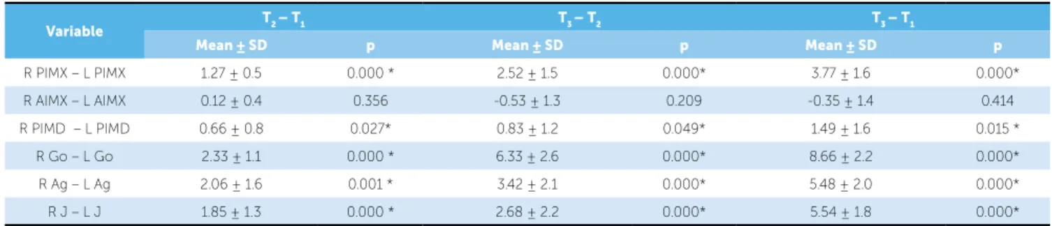
Table 5 - Transverse alterations of the distances between implants and of the maxilla and mandible widths.
* = statistically signiicant values p ≤ 0.05.
Variable T2 – T1 T3 – T2 T3 – T1
Mean ± SD p Mean ± SD p Mean ± SD p
R PIMX – L PIMX 1.27 ± 0.5 0.000 * 2.52 ± 1.5 0.000* 3.77 ± 1.6 0.000*
R AIMX – L AIMX 0.12 ± 0.4 0.356 -0.53 ± 1.3 0.209 -0.35 ± 1.4 0.414
R PIMD – L PIMD 0.66 ± 0.8 0.027* 0.83 ± 1.2 0.049* 1.49 ± 1.6 0.015 *
R Go – L Go 2.33 ± 1.1 0.000 * 6.33 ± 2.6 0.000* 8.66 ± 2.2 0.000*
R Ag – L Ag 2.06 ± 1.6 0.001 * 3.42 ± 2.1 0.000* 5.48 ± 2.0 0.000*
R J – L J 1.85 ± 1.3 0.000 * 2.68 ± 2.2 0.000* 5.54 ± 1.8 0.000*
Table 4 - Means and standard deviation of the maxillary and mandibular
di-mension by period of evaluation.
Variable T1 T2 T3
Mean ± SD Mean ± SD Mean ± SD
R Go – L Go 82.37 ± 4.8 84.71 ± 5.1 91.04 ± 5.5
R Ag – L Ag 75.56 ± 5.5 77.63 ± 5.8 81.05 ± 5.6
R J – L J 57.46 ± 2.0 59.32 ± 2.4 63.01 ± 2.8
The following transverse measurements were per-formed:
» R PIMX - L PIMX: Distance inter-posterior implants of the maxilla.
» R AIMX - L AIMX: Distance inter-anterior implants of the maxilla.
» RJ - LJ: Distance inter-jugal, in relation to the maxilla width.
» R PIMD - L PIMD: Distance inter-posterior implants of the mandible.
» R Go - L Go: Distance inter-gonial, in rela-tion to mandibular width on point Go. » R Ag - L Ag: Distance inter-antegonial, in
relation to mandibular width on point Ag.
Statistical analysis
The mean and standard deviation were calculated for each variable. The different variables presented
normal distribution and the Student’s t test was used
to evaluate the significance of the changes during
evaluation periods (T2–T1, T3–T2, and T3–T1). The
level of significance used was p ≤ 0.05. All calcula-tions were performed with SPSS for Windows (ver-sion 10.0, SPSS Inc., Chicago, Ill).
Method error
To evaluate the error on the localization of cepha-lometric points and digitalization procedures all trac-ings were digitalized again after two weeks by the same examiner. The random error was evaluated us-ing Dahlberg’s formula and the systematic errors were
evaluated using paired t test. The method random
er-ror (Dahlberg’s formula) did not exceed 0,33 mm.
The paired t test did not show statistically significant
systematic error.
RESULTS
Table 4 shows the transverse dimension of the maxilla and mandible on the three periods of evalu-ation. Table 5 shows that there was statistically sig-nificant increase of the maxillary transverse distanc-es on the region of anatomic cephalometric points (Go, Ag and J) and of implants in all evaluated pe-riods, except for the region of anterior implants of the maxilla that did not show statistically significant growth at no time. The lowest gains obtained were on the distance between mandibular implants and the highest were found on the inter-gonial distance.
DISCUSSION
The size of the sample cannot be considered rep-resentative of the population in a statistical sense, on the other side, due to the use of metallic implants, a detailed analysis can provide information on the
facial growth.16 Studies with posteroanterior
radio-graphs present some limitations such as variability on the magniication of the projected transverse
di-mension,9,16,22 problem of standardization of the head
positioning on the cephalostat16,22,25 due to slight up
and down movements of the head and diiculty on
ings agree with other study10 about the posterior move of the maxillary transverse rotation center with aging in function of the immutability of the anterior maxil-lary transverse distance.
Table 6 shows that the annual growth of the poste-rior region of the maxilla was the greatest among the studies that used metallic implants. This result may be associated to the inluence of the facial pattern since the patients with horizontal growth pattern present larger transverse facial dimensions when compared to
other patterns,27 may be due to the fact that the
sam-ple is composed mostly by male that presents facial
widths larger than female9,17,24,25,28 and/or stimulation
of transverse growth by the use of bionator.1,8 During
the treatment with bionator there was an increase of
1.85 mm on the distance RJ - LJ. A previous study19
showed 1.72 mm increase during the same period in Class II division 1 patients and 2.03 mm in Class I patients. This diference may be related to the therapy used because when comparing Class II our results were higher and when compared Class I they were lower, but it must be emphasized that Class II patients present the maxillary transverse dimension smaller
than Class I patients.19 Besides, the remodeling of the
Jugal point during this period was also greater than
the presented by other works.2,9,13 The annual
trans-verse increase between the maxillary implants calcu-lated on the same period was 0.73 mm. In one year of
treatment with Frankel’s appliance, a study8 showed
0.57 mm of increase in patients with age and gender distribution similar to the present study. It was con-cluded that the treatment was capable to increase the basal transverse distances of the maxilla. As the values of annual growth in the present study, were higher it is believed that the bionator also has the capacity to
increase the maxillary bone base,1 although it is not
clinically signiicant. The problem on identification of points is
cor-rected when metallic implants are used, the vari-ability of magnification was individually corrected for the distances between implants in each period of evaluation, but the problems of standardization of the head positioning are impossible to solve
be-cause slight movements of the head are inevitable,14
however, some studies11,20 did not find statistically
significant differences among measures taken with up to 10° of difference.
The study conirmed the increase of bone bases and evidenced that the maxillary growth was greater
than the mandibular.1,10,17 The distance between
pos-terior implants of the maxilla increased more than the distance between the anterior implants conirming
the indings of other studies4,5,10,17 and showing that the
maxillary growth on the posterior region was greater than the anterior besides conirming the existence of transverse growth until the studied age. The mean in-crease of the distance between posterior implants of
the maxilla during all evaluated period, T1-T3, was
of 3.77 mm. Björk and Skieller5 found 3 mm increase
from 10-11 years to 21 years and in a previous study4
they found 2.8 mm from 11 to 19 years of age. The result in this work was a little higher, but considering the standard deviation the values are similar because
they also observed great variability.5 The amount of
maxillary transverse growth between the implants when compared to the increase related to Jugal point
agrees with the present literature4,5 conirming the
median palatine suture as the main site for the maxil-lary transverse growth and, less expressive, the bone apposition in other areas completing the transverse growth (Table 5).
The diferential maxillary transverse increase re-garding the anterior and posterior region implies in a
transverse rotation between the sides.4,5,10,17
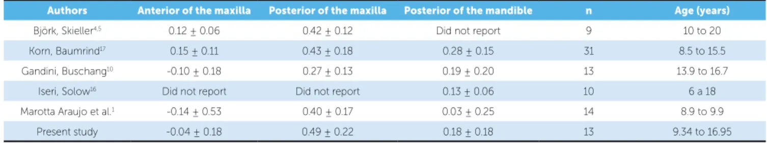
Our ind-Table 6 - Annual changes (mm/year) and standard deviation in transverse growth with metallic implants according to the location.
Authors Anterior of the maxilla Posterior of the maxilla Posterior of the mandible n Age (years)
Björk, Skieller4,5 0.12 ± 0.06 0.42 ± 0.12 Did not report 9 10 to 20
Korn, Baumrind17 0.15 ± 0.11 0.43 ± 0.18 0.28 ± 0.15 31 8.5 to 15.5
Gandini, Buschang10 -0.10 ± 0.18 0.27 ± 0.13 0.19 ± 0.20 13 13.9 to 16.7
Iseri, Solow16 Did not report Did not report 0.13 ± 0.06 10 6 a 18
Marotta Araujo et al.1 -0.14 ± 0.53 0.40 ± 0.17 0.03 ± 0.25 14 8.9 to 9.9
During the bionator therapy, the presented man-dibular basal transverse growth was 0.66 mm.
Anoth-er study,16 on the same period of evaluation, found
0,46 mm and despite not presenting similar sample to the present study, it could be assumed that the bi-onator has the ability of increasing the mandibular bone base, when used appropriately. This
informa-tion was already reported by another study1 that did
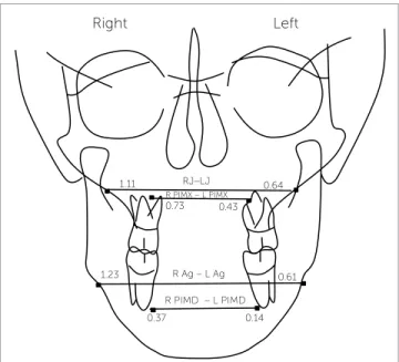
not find statistically significant mandibular transverse increase during one year of treatment with bion-ator, but observed higher value on the treated group. Evaluating the annual changes of growth on the two periods it is observed that on the stage of treatment with bionator the maxillary and mandibular basal transverse growth was 0.73 mm/year and 0.37 mm/ year, respectively. After therapy, the normal growth showed 0.43 mm/year and 0.14 mm/year for maxil-la and mandible showing the stimumaxil-lation of growth with bionator (Figs 4 to 8). The mandibular basal transverse increase is a supposition, but at a dental and dentoalveolar matter it was already identified in
studies with functional appliances.12,14,23,26 The width
of the mandibular implants increased 0.18 mm/year,
value similar to the one found previously10 in older
Class I patients. This result may represent that Class II patients have lower potential of mandibular basal transverse growth even if treated at a young age,
how-ever some authors19 did not identify difference on the
mandible between Class I and II patients.
Ater therapy with bionator, the remodeling on the Jugal point found by the present study was greater than the ones shown by several articles with similar period
of observation.2,9,13,19,27 Diferently from the maxilla,
the contribution of the basal growth on the
mandibu-lar transverse increase is lower than the remodeling10
(Table 5). Regarding the mandibular remodeling, af-ter therapy with bionator, our results were lower than
the ones presented by other works,2,9,19,27 and observing
the increase of the distance R Ag-L Ag during therapy with bionator, the amount of remodeling was identical
to one found in the same period (2 mm),9 but
infe-rior to other works.2,19,27 However the values of annual
growth obtained during the treatment were systemati-cally higher (Fig 4).
The mandibular transverse distance, both on the
re-gion of Gonion and Antegonion, evaluated in T3, is lower
than the presented by Lux et al19 evaluating 15 years old
Figure 5 - Individual annual changes in transverse growth on the region of
posterior implants of the maxilla during bionator therapy.
Figure 6 - Individual annual changes in transverse growth on the region of
posterior implants of the mandible during bionator therapy.
Figure 4 - Annual changes in transverse growth during bionator therapy
(val-ues on the right) and after bionator therapy (val(val-ues on the left).
Right Left
mm/year
1.11 0.64
0.61
0.37 0.14 R PIMD – L PIMD
R PIMX – L PIMX
1.23
0.73 0.43
R Ag – L Ag RJ–LJ
Posterior implants on mandible T2-T1
Patients
mm/year
1.2 1.4
1 0.8 0.6
1 2 3 0.94
-0.05 -0.05
0.05 0.05
0.21 0.45
0.18 0.42
1.29
0.56
5
4 6 7 8 9 10 11 12 13 0.4
0.2
-0.2 0 Posterior implants on maxilla T2-T1
Patients 1,8
1.6 1.4 1.2 1 0.8 0.6
1 2 3 0.32
1.3
0.38 0.31
0.55 0.65
0.34 1.64
0.750.71 0.87 0.94
0.78
5
4 6 7 8 9 10 11 12 13 0.4
Class I or II patients. It was also lower than presented
in other studies.2,13,27 Thus, our results suggest that the
Class II patients, present mandibular transverse growth and dimensions lower that Class I patients, not conirming
the result by Lux et al,19 although the found diferences of
size probably have little clinical meaning once they were not greater than 4 mm. Besides, the gonial region showed wide remodeling during growth and it is the transverse dimension of the lower third of the face that presents greater growth and possibility of morphological variation. On Table 7 it can be noticed the inluence of gender and malocclusion on the mandibular transverse growth since
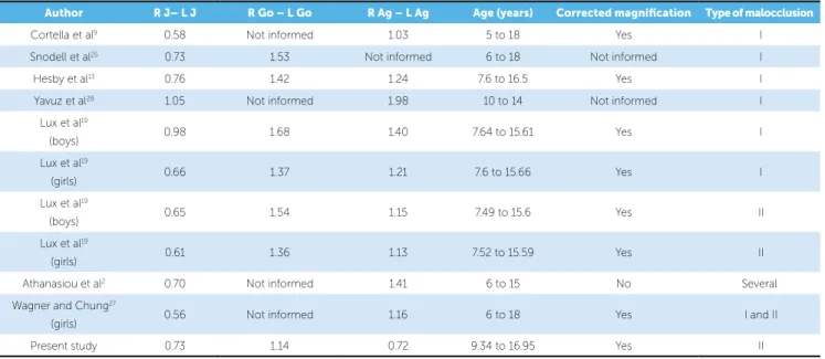
Table 7 - Annual changes (mm/year) in transverse growth of anatomic points according to the location.
Author R J– L J R Go – L Go R Ag – L Ag Age (years) Corrected magnification Type of malocclusion
Cortella et al9 0.58 Not informed 1.03 5 to 18 Yes I
Snodell et al25 0.73 1.53 Not informed 6 to 18 Not informed I
Hesby et al13 0.76 1.42 1.24 7.6 to 16.5 Yes I
Yavuz et al28 1.05 Not informed 1.98 10 to 14 Not informed I
Lux et al19
(boys) 0.98 1.68 1.40 7.64 to 15.61 Yes I
Lux et al19
(girls) 0.66 1.37 1.21 7.6 to 15.66 Yes I
Lux et al19
(boys) 0.65 1.54 1.15 7.49 to 15.6 Yes II
Lux et al19
(girls) 0.61 1.36 1.13 7.52 to 15.59 Yes II
Athanasiou et al2 0.70 Not informed 1.41 6 to 15 No Several
Wagner and Chung27
(girls) 0.56 Not informed 1.16 6 to 18 Yes I and II
Present study 0.73 1.14 0.72 9.34 to 16.95 Yes II
the lowest annual growths are related to studies evaluat-ing female patients and/or with Class II malocclusion. Another aspect is that the comparison of normative val-ues between the studies is not appropriated due to radio-graphic magniication. Some articles do not mention the correction and other do not describe appropriately the used methodology. Due to these problems some
stud-ies9,27 suggest the use of proportion (JJ/AgAg) instead of
normative values to minimize the problem although not solving it because some centers take posteroanterior ra-diographs with Frankfurt’s plane parallel to the ground and others with Frankfurt plane inclined 35° down.
Figure 7 - Individual annual changes in transverse growth on the region of
posterior implants of the maxilla after bionator therapy.
Figure 8 - Individual annual changes in transverse growth on the region of
posterior implants of the mandible after bionator therapy.
mm/year mm/year
Posterior implants on maxilla T3-T2
Patients 1
0.9 0.8 0.7 0.6 0.5 0.4 0.3 0.2 0.1
1 2 3
0.32 0.36 0.32 0.39 0.15 0.16
0.54 0.8
0.89
0.39 0.81
0.1
5
4 6 7 8 9 10 11 12 13 0
Posterior implants on mandible T3-T2
Patients 0.7
0.08 0.05 0.13
-0.14
-0.18 0.47
0.57
0.11 0.17 0.2
0.12 0.6 0.5 0.4 0.3 0.2 0.1 -0.1 -0.2 -0.3
The indings in this study are limited by the size of the sample, bias of the treatment potential and lack of control group. Although the size of the sample is small, the highly signiicant probabilities obtained (p < 0.001) suggest that the changes observed in growth are real. Besides, addition-al studies with larger samples are necessary to provide bet-ter estimates of variation on transverse increase by growth. There is also the possibility of inluence of the treatment subsequent to the bionator on the transverse increase al-though it is hardly likely that conventional ixed appliances have some potential of orthopedic efect.
CONCLUSIONS
1) The maxillary and mandibular bone bases seem to be afected by bionator therapy, during treat-ment, returning to a normal pattern on the post-treatment.
2) The maxillary and mandible remodeling pattern followed the same tendency of transverse growth of metallic implants.
3) With aging, the center of transverse rotation of the maxilla is displaced posteriorly.
1. Marotta Araujo A, Buschang PH, Melo AC. Transverse skeletal base adaptations with Bionator therapy: a pilot implant study. Am J Orthod Dentofacial Orthop. 2004;126(6):666-71.
2. Athanasiou AE, Droschl H, Bosch C. Data and patterns of transverse dentofacial structure of 6- to 15-year-old children: a posteroanterior cephalometric study. Am J Orthod Dentofacial Orthop. 1992;101(5):465-71. 3. Baumrind S, Ben-Bassat Y, Korn EL, Bravo LA, Curry S. Mandibular
remodeling measured on cephalograms: 2. A comparison of information from implant and anatomic best-it superimpositions. Am J Orthod Dentofacial Orthop. 1992;102(3):227-38.
4. Björk A, Skieller V. Growth in width of the maxilla studied by the implant method. Scand J Plast Reconstr Surg. 1974;8(1-2):26-33.
5. Björk A, Skieller V. Growth of the maxilla in three dimensions as revealed radiographically by the implant method. Br J Orthod. 1977;4(2):53-64. 6. Björk A. Facial growth in man, studied with the aid of metallic implants. Acta
Odontol Scand. 1955;13(1):9-34.
7. Björk A. Variations in growth pattern of the human mandible: Longitudinal radiographic study by the implant method. J Dent Res. 1963;42(1):400-11. 8. Brieden CM, Pangrazio-Kulbersh V, Kulbersh R. Maxillary skeletal and
dental change with Frankel appliances: an implant study. Angle Orthod. 1984;54(3):226-32.
9. Cortella S, Shofer FS, Ghafari J. Transverse development of the jaws: Norms for the posteroanterior cephalometric analysis. Am J Orthod Dentofacial Orthop. 1997;112(5):519-22.
10. Gandini LG Jr, Buschang PH. Maxillary and mandibular width changes studied using metallic implants. Am J Orthod Dentofacial Orthop. 2000;117(1):75-80.
11. Ghafari J, Cater PE, Shofer FS. Efect of ilm-object distance on
posteroanterior cephalometric measurements: suggestions for standardized cephalometric methods. Am J Orthod Dentofacial Orthop. 1995;108(1):30-7. 12. Gibbs SL, Hunt NP. Functional appliances and arch width. Br J Orthod.
1992;19(2):117-25.
13. Hesby RM, Marshall SD, Dawson DV, Southard KA, Casko JS, Franciscus RG, et al. Transverse skeletal and dentoalveolar changes during growth. Am J Orthod Dentofacial Orthop. 2006;130(6):721-31.
14. Hime DL, Owen AH 3rd. The stability of the arch-expansion efects of Fränkel appliance therapy. Am J Orthod Dentofacial Orthop. 1990;98(5):437-45. REFERENCES
15. Hsiao TH, Chang HP, Liu KM. A method of magniication correction for posteroanterior radiographic cephalometry. Angle Orthod.
1997;67(2):137-42.
16. Işeri H, Solow B. Change in the width of the mandibular body from 6 to 23 years of age: an implant study. Eur J Orthod. 2000;22(3):229-38. 17. Korn EL, Baumrind S. Transverse development of the human jaws between
the ages of 8.5 and 15.5 years, studied longitudinally with use of implants. J Dent Res. 1990;69(6):1298-306.
18. Leonardi R, Annunziata A, Caltabiano M. Landmark identiication error in posteroanterior cephalometric radiography. A systematic review. Angle Orthod. 2008;78(4):761-5.
19. Lux CJ, Conradt C, Burden D, Komposch G. Dental arch widths and mandibular-maxillary base widths in Class II malocclusions between early mixed and permanent dentitions. Angle Orthod. 2003;73(6):674-85. 20. Major PW, Johnson DE, Hesse KL, Glover KE. Efect of head orientation on
posterior anterior cephalometric landmark identiication. Angle Orthod. 1996;66(1):51-60.
21. Major PW, Johnson DE, Hesse KL, Glover KE. Landmark identiication error in posterior anterior cephalometrics. Angle Orthod. 1994;64(6):447-54. 22. Malkoc S, Sari Z, Usumez S, Koyuturk AE. The efect of head rotation on
cephalometric radiographs. Eur J Orthod. 2005;27(3):315-21. 23. Owen AH. Morphologic changes in the transverse dimension using the
Fränkel appliance. Am J Orthod. 1983;83(3):200-17.
24. Savara BS, Singh IJ. Norms of size and annual increments of seven anatomical measures of maxillae in boys from three to sixteen years of age. Angle Orthod. 1968;38(2):104-20.
25. Snodell SF, Nanda RS, Currier GF. A longitudinal cephalometric study of transverse and vertical craniofacial growth. Am J Orthod Dentofacial Orthop. 1993;104(5):471-83.
26. Vargevik K. Morphologic evidence of muscle inluence on dental arch width. Am J Orthod. 1979;76(1):21-8.
27. Wagner DM, Chung CH. Transverse growth of the maxilla and mandible in untreated girls with low, average, and high MP-SN angles: a longitudinal study. Am J Orthod Dentofacial Orthop. 2005;128(6):716-23.