Printed in Brazil - ©2007 Sociedade Brasileira de Química 0103 - 5053 $6.00+0.00
ArticleArticleArticleArticleArticle
*e-mail: [email protected]
Biotransformation of Digitoxigenin by
Cochliobolus lunatus
Rodrigo M. Pádua,a Alaíde B. Oliveira,a José D. Souza Filho,b Jacqueline A. Takahashi,b Maurício de Abreu e Silvac and Fernão C. Braga*,a
a
Faculdade de Farmácia, Universidade Federal de Minas Gerais, Av. Antônio Carlos, 6627, 31270-010 Belo Horizonte-MG, Brazil
b
Departamento de Química, Universidade Federal de Minas Gerais, Av. Antônio Carlos, 6627, 31270-010 Belo Horizonte-MG, Brazil
c
Faculdade de Farmácia, Universidade Federal de Ouro Preto, Rua Costa Sena, 171, 35400-000 Ouro Preto-MG, Brazil
A reação de biotransformação da digitoxigenina (1) por Cochliobolus lunatusfoi investigada. Foram realizados experimentos com duração de 4 dias, que resultaram no isolamento de quatro produtos, cujas estruturas químicas foram elucidadas como sendo 1β-hidroxidigitoxigenina (2), 7β-hidroxidigitoxigenina (3), 8β-hidroxidigitoxigenina (4) e digitoxigenona (5). A obtenção desses produtos nas condições empregadas nunca foi anteriormente descrita. A produção da substância 4 em uma reação de biotransformação também é inédita.
The biotransformation of digitoxigenin (1) by Cochliobolus lunatuswas investigated. The biotransformation reaction was carried out in a 4-day process, resulting in the isolation of four products, whose structures were elucidated as 1β-hydroxydigitoxigenin (2), 7β-hydroxydigitoxigenin (3), 8β-hydroxydigitoxigenin (4) and digitoxigenone (5). The production of these derivatives under the employed conditions has never been described so far. This is also the first report on the production of compound 4 by a biotransformation reaction.
Keywords: digitoxigenin, Cochliobolus lunatus, 1β-hydroxydigitoxigenin, 7β -hydroxy-digitoxigenin, 8β-hydroxydigitoxigenin, digitoxigenone
Introduction
Enzymes are known to possess a wide substrate tolerance by keeping their exquisite catalytic properties with respect to chemo-, regio- and enantio-selectivity, playing an important role in biotransformations.1 Biotransformation reactions can be accomplished at room temperature and in aqueous medium, presenting itself as a milder alternative to classical chemical reactions,2-4 being employed for the resolution of racemates and to introduce chiral centers in substrates, among other uses.5
Fungi are eukaryotic organisms that possess enzyme systems similar to those of mammalians. They usually present highly flexible metabolism, thus accepting varied sources of carbon and nitrogen. The ecological relations of these organisms include the metabolism of different secondary compounds, a feature sustained by diversified
enzymatic systems, both intra and extra cellular, capable of carrying out numerous reactions.6-8 Such attributes suggest fungi as suitable organisms to perform biotransformation reactions.
The biotransformation of steroidal compounds by fungi has been extensively evaluated, including reactions with cardiac glycosides (Pádua et al.9 and references herein; Table 3 of the present work). Digoxin, a Digitalis
cardenolide, is still the drug of choice for the treatment of congestive heart failure, acting as a selective inhibitor of the Na+,K+ ATPase enzyme. Biotransformation of cardenolides has been investigated either as a strategy to obtain new derivatives or to convert the A-type cardenolides into the corresponding C-type compounds, which have clinical relevance.9
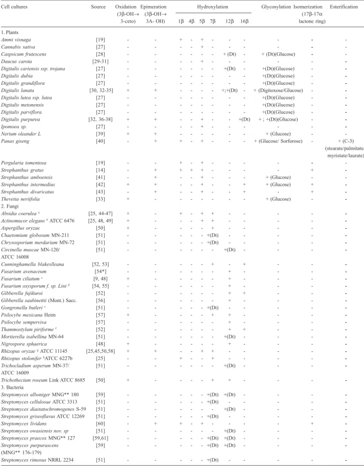
group at C-3 (see Table 3 for references). Analysis of those data indicate that in general plant cell cultures hydroxylize position 5β, whereas the major hydroxylation sites carried out by fungi and bacteria are 7β and 12β. Besides, solely plant cell cultures are capable of performing glycosylation reactions, as indicated by the reviewed literature (Table 3).
Hydroxylation reactions may be seen as a defense mechanism of fungi and are carried out by cytochrome P-450 monooxygenases found in their endoplasmatic reticulum. Difficulties in isolation and stabilization of those enzymes, associated with the necessity of recycling NAD(P)H co-factors, prevent the industrial use of isolated enzymes.7,10 Intact fungus organisms are therefore alternatively employed in biotransformation reactions.6, 11
The fungus Cochliobolus lunatus and its conidial anamorphous form Curvularia lanata are known for their capacity of hydroxylating Δ4-5 steroids.12 The substrate induced 11β-hydroxylation of steroids is a classical biotransformation reaction carried out by this fungus, which tolerates structure variation in substrates. Apart from 11β position (“normal binding”), the major sites for C.
lunatus hydroxylation are 14α and 7α resulting from
inverted and reverse inverted binding of enzyme to steroid, respectively.13
Within this context, the main goal of the present work was to evaluate the biotransformation reaction of the cardenolide digitoxigenin by C. lunatus and to compare the hydroxylation sites with those previously described for other steroids.
Materials and Methods
General
1H NMR, 13C NMR, 1H-1H COSY and HMQC spectra
were recorded on a Bruker DRX-400 spectrometer (1H 400 MHz and 13C 100 MHz) using TMS as internal standard for both nuclei. Chemical shifts (δ) are given in
ppm and J couplings in Hertz (Hz). Optical rotations were measured with Perkin Elmer 341 polarimeter.
Chemicals
Acetonitrile chromatographic grade LiChrosolv and digitoxin were obtained from Merck (Germany). Water was purified using a Milli-Q50 purification system (Millipore, USA). Digitoxigenin, [α]D +197° (CH2Cl2; c.0.14), employed in the biotransformation experiments, was obtained by hydrolysis of digitoxin as previously described by Pádua et al.9
Biotransformation experiments
The filamentous fungus Cochliobolus lunatus
(CCT0271/ NRRL 2178) was obtained from Fundação André Tosello, Coleção de Culturas Tropicais (CCT), Campinas, SP, Brazil (http://www.cct.org.br/sdms.cgi). Before the biotransformation experiment, the fungus culture was incubated in malt agar 2%, pH 6.5 adjusted with 1 mol L-1 NaOH, for 7 days, at room temperature.
A total of 14 erlenmeyer flasks (300 mL), each containing 100 mL of sterile medium (composition: 2.0% glucose, 0.5% peptone, 0.3% yeast extract and 0.5% KH2PO4, pH adjusted to 5.7), was inoculated with fresh fungal suspension. After 24 h incubation, digitoxigenin (1) (10 mg) was dissolved in DMF (1 mL) and added to each flask, following incubation at room temperature (24-26 °C), under stirring (200 rpm) for 4 days. The total amount of digitoxigenin (1) submitted to biotrans-formation was 140 mg. Control experiments containing medium plus substrate (C-1) and medium plus fungus culture (C-2) were carried out in each case. After removing the mycelium by filtration, the biotransformation products were sequentially extracted with chloroform (2 × 100 mL) and chloroform/2-propanol (3:1) (100 mL) in a separator funnel and the solvent was vacuum removed at 50 °C, until residue. The obtained residues were combined (296 mg) and analyzed by TLC (acetone:chloroform: dichlormethane; 50:35:15; Kieselgel 60G; 0.1 mm; 20 × 20 cm, Merck). In sequence, the combined residues were submitted to purification on a Shimadzu HPLC semi-preparative system (Japan) composed of pump SCL-8A and integrator C-R4A. Portions of the combined residues (20 mg) were dissolved in MeOH (1.0 mL) for the injection into the apparatus. An ODS column (250 × 20 mm i.d., Shimadzu, Japan) was employed at room temperature, eluted with 84% aqueous CH3CN / H2O (46:54), at a flow rate of 5.0 mL min-1 and UV
et al.
20 °C: compound 2 [α]D: +56° (c.0.04); 3 [α]D: +278° (c.0.195); 4 [α]D: +162° (c.0.36), 5 [α]D: +228° (c.0.195).
Results and Discussion
TLC analysis of the combined residues from digitoxigenin transformation (1) by C. lunatus, employing Kedde as spray reagent, showed spots with distinct Rf
values of 1 solely for the biotransformation reaction, i.e., no product was observed for controls C1 and C2, as expected.
Chromatographic separation of the combined residues from biotransformation experiments of 1 resulted in the isolation of four compounds, along with the recovery of part of the starting material digitoxigenin (1). The structures of the products were defined based on spectroscopic analysis, using digitoxigenin (1) as model compound, and also by comparison with spectral data reported for the compounds or structurally related cardenolides.
The 1H NMR spectrum of compound 2 showed a complex profile and the only signals easily assigned were those of H-3α (δ 4.13), H-21(δ 5.03 and 4.91), H-22
(δ 5.89), H-18 (δ 0.88) and H-19 (δ 1.09), thus confirming the presence of the C-3 hydroxyl group, the integrity of the α,β unsaturated lactone ring at C-17 and the methyl groups in the steroidal structure. The hydroxylation site was suggested by the paramagnetic shift observed for H-19 (δ 1.09) in comparison to digitoxigenin (1) (δ 0.96), pointing out the hydroxylation position close to C-19 (Table 1). The presence of a broad signal at δ 3.76 was also indicative of an additional hydroxyl group.
The HMQC spectrum obtained for 2 allowed determining 1H/13C one-bond shift correlations of all hydrogen-bearing carbon atoms in the compound. Correlation spots between C-1 / C-2 (δ 29.6 / 27.9) and their corresponding hydrogens H-1α and H-1β (δ 1.49 and 1.49) / H-2α and H-2β (δ 1.53 and 1.53) were clearly observed in the HMQC spectrum of model compound 1. On the other hand, those cross-peaks were absent in the equivalent region of HMQC spectrum of 2, indicating their shift resulting from hydroxylation at C-1 or C-2.
TheCOSY spectrum was helpful for assigning 1H NMR chemical shifts of 2 and also disclosed the precise site of hydroxylation. Hence, the sign of a methine hydrogen at
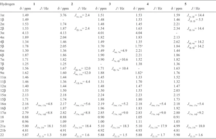
Table 1. 1HNMR assignments of digitoxigenin (1) and its biotransformation products (2-5)
Hydrogen 1 2 3 4 5
δ / ppm J / Hz δ / ppm J / Hz δ / ppm J / Hz δ / ppm J / Hz δ / ppm J / Hz
1α 1.49 3.76 J1α, 2α= 2.4 1.51 1.53 1.59 J1α, 2β= 14.4
1β 1.49 - 1.48 1.53 1.46 J1β, 2β= 5.5
2α 1.53 1.74 1.48 1.45 2.21
2β 1.53 1.87 J2β, 1α= 2.4 1.54 1.63 2.34 J2β, 2α= 14.4
3α 4.13 4.13 4.01 4.04
-4α 1.89 2.04 1.82 1.83 2.13
4β 1.34 1.46 1.49 1.35 2.62 J4β, 4α= 14.2
5β 1.78 2.05 1.70 1.75a 1.84 J
5β, 4β= 14.2
6α 1.30 1.36 1.49 J6α, 7α=4.9 2.21 1.44
6β 1.87 1.86 1.90 2.21 1.86
7α 1.71 1.82 3.90 J7α, 6β=10.6 1.52 1.82
7β 1.25 1.25 - 1.38 1.36
8β 1.56 1.67 J8β, 9α= 12.0 1.71 J8β, 7α= 10.4 - 1.63
9α 1.62 1.60 J9α, 11α=12.0 1.88 1.82a 1.76
11α 1.46 1.44 1.45 1.33 1.52
11β 1.46 1.36 J11β, 9α= 4.4 1.31 1.70 1.32
12α 1.40 1.44 1.48 1.47 1.47
12β 1.53 1.50 1.50 1.53 2.03
15α 2.13 2.18 2.24 2.23 2.15
15β 1.71 1.74 1.88 1.80 1.73
16α 2.16 J16α, 17α=4.8 2.17 J16α, 17α=5.6 2.19 J16α, 17α=5.2 2.18 J16α, 17α=5.4 2.18 J16α, 17α=5.4
16β 1.87 1.87 1.96 1.83 1.92
17α 2.79 J17α, 16β=8.8 2.82 J17α, 16β=8.8 2.85 J17α, 16β=9.0 2.83 J17α, 16β=9.0 2.80 J17α, 16β=9.2
18 0.88 0.88 0.90 1.05 0.91
19 0.96 1.09 1.00 1.11 1.03
21a 5.00 J21a, 21b= 18.1 5.03 J21a, 21b= 18.4 5.10 J21a, 21b= 18.3 5.10 J21a, 21b= 17.9 4.99 J21a, 21b= 18.0
21b 4.81 4.91 4.92 4.93 4.81
δ 3.76 showed cross-peaks with C-2 methylene hydrogens (δ 1.74 and 1.87), whereas C-3 methine proton (δ 4.13) showed correlation with both 2-CH2 (δ 1.74 and 1.87) and 4-CH2 (δ 2.04 and 1.46). These correlations clearly indicated the hydroxylation site at C-1.
Further evidence of the hydroxylation position was given by comparing carbon chemical shifts obtained for compound 2 and those previously reported for 1β-hydroxy-17β-H-digitoxigenin.14 The values showed close correspondence, except for the chemical shifts of C-12, C-16, C-17 and C-18, what was expected result since 2
and 1β-hydroxy-17β-H-digitoxigenin are epimers at C-17. Furthermore, carbon chemical shits obtained for 2
were similar to those of digitoxigenin (1), with the exception of C-1 (δ 74.9), C-2 (δ 33.2), C-3 (δ 69.6), C-5 (δ 32.0) and C-19 (δ 19.6), which presented either paramagnetic or diamagnetic shifts, attributed to α-, β-and γ-effects, resulting from the hydroxylation at C-1.
The stereochemistry of the hydroxylation site was indicated by the vicinal coupling constant of H-1 (J = 2.4 Hz), which pointed out the axial position for the C-1 hydroxyl group. Based on these findings, the structure of compound 2 was defined as 1β-hydroxydigitoxigenin, also named acovenosigenin A.15
Acovenosigenin A and its glycosides have been isolated from different plant species and also as a product of biotransformation reactions.16-19 NMR data previously reported showed good agreement with those of compound
2. As a result of the small amount of 2 isolated in the present work, 13C NMR data were obtained indirectly by HMQC experiment. Therefore, it was not possible to attribute the resonances of non-hydrogenated carbons (Table 2).
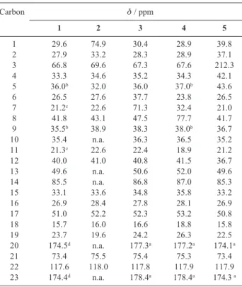
13C NMR spectrum of compound 3 presented 23
signals, disclosed by DEPT-135 experiment as two methyl, nine methylene, seven methine and five non hydrogenated carbons. Compound 3 presents a methine signal at δ 71.3, not found in the spectrum of digitoxigenin (1), indicating that hydroxylation occurred at a methylene group. Besides, a signal with chemical shift typical of C-7 (δ 21.2) or C-11 (δ 21.3) in digitoxigenin (1) was absent in compound
3. Compounds 1 and 3 present equivalent chemical shift values for C-12 (Table 2); hence, it is very unlikely that hydroxylation had occurred at C-11, since β effect would result in diamagnetic shift of C-12.
Therefore, compound 3 is the hydroxylation product of digitoxigenin at C-7, what can be confirmed by the chemical shift (δ 3.90) and coupling constants of H-7 (J = 10.6, 10.4 and 4.9 Hz), consistent with two trans diaxial and one axial-equatorial coupling. Altogether these data allowed identifying compound 3 as 7β-hydroxydigitoxigenin. As
expected, 1H NMR spectrum of 3 was similar to that of digitoxigenin (1), apart from the diamagnetic shifts observed for H-7α (δ 3.90), H-6α (δ 1.49), H-6β (δ 1.90) and H-8β (δ 1.71), resultant from the vicinity of the 7β-hydroxyl group (Table 1). Assignment of these hydrogens was confirmed by data of the HMQC spectrum. Further confirmation of the hydroxylation site at C-7 was furnished by analysis of the COSY spectrum, which showed cross-peaks between H-7α (δ 3.90) both with 6-CH2 (δ 1.90 and 1.49) and C-8 methine proton (δ 1.71). 13C and DEPT-135 NMR spectra of 4 revealed two
methyl, ten methylene, five methine and six non hydrogenated carbons. The signal at δ 77.7 in the spectrum of 1 disappeared in the DEPT-135 spectrum of 4, suggesting the occurrence of hydroxylation at a methine group. Comparison of 13C NMR spectrum obtained for compound 4 and digitoxigenin (1) showed that the signal attributed to C-8 (δ 41.8) in the later, was absent in the first. Consequently, compound 4 was identified as 8β-hydroxydigitoxigenin. As expected, 1H NMR spectra of 4 and digitoxigenin (1) were similar, excluding the signals attributed to H-6α (δ 2.21), H-6β (δ 2.21), H-7α (δ 1.52), H-7β (δ 1.38), H-9α (δ 1.75 or 1.82), H-11α (δ 1.33), H-11β (δ 1.70), H-18 (δ 1.05) and H-19 (δ 1.11), which showed distinct values, due to the influence of the
Table 2. 13CNMR assignments of digitoxigenin (1) and
biotransforma-tion products (2-5)
Carbon δ / ppm
1 2 3 4 5
1 29.6 74.9 30.4 28.9 39.8
2 27.9 33.2 28.3 28.9 37.1
3 66.8 69.6 67.3 67.6 212.3
4 33.3 34.6 35.2 34.3 42.1
5 36.0b 32.0 36.0 37.0b 43.6
6 26.5 27.6 37.7 23.8 26.5
7 21.2c 22.6 71.3 32.4 21.0
8 41.8 43.1 47.5 77.7 41.7
9 35.5b 38.9 38.3 38.0b 36.7
10 35.4 n.a. 36.3 36.5 35.2
11 21.3c 22.6 22.4 18.9 21.2
12 40.0 41.0 40.8 41.5 36.7
13 49.6 n.a. 50.6 52.0 49.6
14 85.5 n.a. 86.8 87.0 85.3
15 33.1 33.6 34.8 35.8 33.2
16 26.9 28.4 27.8 28.1 26.9
17 51.0 52.2 52.3 53.2 50.8
18 15.7 16.0 16.6 18.8 15.8
19 23.7 19.6 24.2 26.3 22.5
20 174.5d n.a. 177.3a 177.2a 174.1a
21 73.4 75.5 75.4 75.3 73.4
22 117.6 118.0 117.8 117.9 117.9
23 174.4d n.a. 178.4a 178.4a 174.3 a
et al.
Table 3. Biotransformation reactions of digitoxigenin and digitoxin (Dt) carried out by cell cultures
Cell cultures Source Oxidation Epimeration Hydroxylation Glycosylation Isomerization Esterification
(3β-OH→ (3β-OH→ (17β-17α
3-ceto) 3A- OH) 1β 4β 5β 7β 12β 16β lactone ring) 1. Plants
Ammi visnaga [19] - - + - + - - - - -
-Cannabis sativa [27] - - - - + - - - - -
-Caspsicum frutescens [28] - - - + (Dt) - + (Dt)(Glucose) -
-Daucus carota [29-31] - - - - + - - -
-Digitalis cariensis ssp. trojana [27] - - - +(Dt) - +(Dt)(Glucose) -
-Digitalis dubia [27] - - - +(Dt)(Glucose) -
-Digitalis grandiflora [27] - - - +(Dt)(Glucose) -
-Digitalis lanata [30, 32-35] + + - - - - +;+(Dt) - + (Digitoxose/Glucose) -
-Digitalis lutea ssp. lutea [27] - - - +(Dt)(Glucose) -
-Digitalis metonensis [27] - - - +(Dt)(Glucose) -
-Digitalis parviflora [27] - - - +(Dt)(Glucose) -
-Digitalis purpurea [32, 36-38] + + - - + - - +(Dt) + ; +(Dt)(Glucose) -
-Ipomoea sp. [27] - - - - + - - -
-Nerium oleanderL [39] + + - - - + (Glucose) -
-Panax giseng [40] - + + - + - - - + (Glucose/ Sorforose) - + (C-3)
(stearate/palmitate/ myristate/laurate)
Pergularia tomentosa [19] - - + - + - - - - -
-Strophanthus gratus [14] - + + + + - - - - +
-Strophanthus amboensis [41] - + - - + - - - + (Glucose) -
-Strophanthus intermedius [42] + + - - + - - + + (Glucose) +
-Strophanthus divaricatus [43] - + - - + - - + - +
-Thevetia neriifolia [33] + - - - + (Glucose) -
-2. Fungi
Absidia coerulea a [25, 44-47] + - + - + + - - - -
-Actinomucor elegans bATCC 6476 [25, 48, 49] + - - - + + - - - -
-Aspergillus oryzae [50] + - - - - + - - - -
-Chaetomium globosum MN-211 [51] - - - +(Dt) - - - -
-Chrysosporium merdarium MN-72 [51] - - - +(Dt) - - - -
-Circinella muscae MN-120/ [51] - - - +(Dt) - - -
-ATCC 16008
Cunninghamella blakeslleana [52, 53] - - - + - + - -
-Fusarium avenaceum [54*] - - - + - - -
-Fusarium ciliatum c [9, 48] + - - - - - + - - -
-Fusarium oxysporum f. sp. Lini d [54, 55] - - - - - - + - - -
-Gibberella fujikuroi [52] - - - + + - -
-Gibberella saubinettii (Mont.) Sacc. [56] - - - + - - -
-Gongronella butleri e [51] - - - - - +(Dt) - - - -
-Psilocybe mexicana Heim [57] + - - - - + + - - -
-Psilocybe semperviva [57] - - - + - - -
-Thammostylum piriforme f [52] - - - - - - + + - -
-Mortierella isabellina MN-64 [51] - - - +(Dt) - - -
-Nigrospora sphaerica [48] + - - - + - - -
-Rhizopus oryzaegATCC 11145 [25,45,50,58] + + - - + + - - - -
-Rhizopus stolonifer hATCC 6227b [25] - - + - - + - - - -
-Trichocladium asperum MN-37/ [51] - - - +(Dt) - - -
-ATCC 16009
Trichothecium roseum Link ATCC 8685 [50] + - - - - + + - - -
-3. Bacteria
Streptomyces alboniger MNG** 180 [59] - - - +(Dt) +(Dt) - - -
-Streptomyces cellulosae ATCC 3313 [51] - - - +(Dt) - - - -
-Streptomyces diastatochromogenes S-59 [51] - - - +(Dt) - - -
-Streptomyces griseoflavus ATCC 12269 [51] - - - +(Dt) - - - -
-Streptomyces lividans [60] - + + - + - - - - +
-Streptomyces owasiensis nov. sp [51] - - - +(Dt) - - -
-Streptomyces praecox MNG** 127 [59,61] - - - +(Dt) +(Dt) - - -
-Streptomyces purpurascens [59] - - - +(Dt) +(Dt) - - -
-(MNG** 176-179)
Streptomyces rimosus NRRL 2234 [51] - - - +(Dt) - - - -
8β-hydroxyl group. Deshielding induced by this group imposed diamagnetic shifts to H-18 and H-19, due to 1,4 effect.20
The COSY spectrum of 4 did not provide additional information on the hydroxylation site, since it occurred in a methine group. On the other hand, comparison of HMQC spectra recorded for 1 and 4 clearly indicated, in the last, the absence of a cross-peak between C-8 (δ 41.8) and H-8β (δ 1.56) found in digitoxigenin (1). Comparison between NMR data assignments carried out for compound
4 and literature records for its glycoside21 hasconfirmed the structure of 4 as 8β-hydroxydigitoxigenin, also named cerdollagenin.
The major differences between 13C NMR spectra of compound 5 and digitoxigenin (1) were the presence of a carbonyl carbon signal (δ 212.3) and the absence of the C-3 carbinol carbon signal (δ 66.8) in the first. In total, these data strongly suggest that compound 5 is digitoxigenone, a product from digitoxigenin oxidation at C-3. Aside from the diamagnetic shifts observed for C-1 (δ 39.8), C-2 (δ 37.1), C-4 (δ 42.1) and C-5 (δ 43.6) in compound 5, as a result from deshielding effect of the carbonyl at C-3, 13C NMR chemical shifts observed for 5
Figure 1. Semi-preparative RP-HPLC chromatogram obtained for the
residue from digitoxigenin biotransformation. Identified peaks: 1 = digi-toxigenin; 2 = 1β-hydroxydigitoxigenin; 3 = 7β-hydroxydigitoxigenin; 4 = 8β-hydroxydigitoxigenin; 5 = digitoxigenone. Chromatographic con-ditions: see Materials and Methods.
Retention time / min
Detec
tor
re
s
p
ons
e/
mA.U
Figure 2. Chemical structures of biotransformation products from digitoxigenin (1) by Cochliobolus lunatus: 1β-hydroxydigitoxigenin (2), 7β
-hydroxydigitoxigenin (3), 8β-hydroxidigitoxigenin (4) and digitoxigenone (5). O
O H
OH CH3
CH3
O
H
H
H
O
O
OH CH3
CH3
O
H
H
H
O
O H
OH CH3
CH3
O
H
H
H
OH O
O H
OH CH3
CH3
O
OH
H
H
O
O H
OH CH3
CH3
O
H
H
H OH
1 2
5
3 4
1 2
3 4
5
6 7
8 9 10
11 12
13
14 15 16 17 18
19
20 21
et al.
and digitoxigenin were similar. Assignments of the above mentioned carbons were attested by HMQC spectrum, whereas COSY data allowed confirming the attribution of some hydrogens. Hence, H-2β (δ 2.34 J = 14.4, 14.4 and 5.5 Hz) showed correlation spots with 1-CH2 (δ 1.59 and 1.46) and H-2α (δ 2.21), while H-4β (δ 2.62 J = 14.2, and 14.2 Hz) showed cross-peaks with 5-CH (δ 1.84) and H-4α (δ 2.13). Definitive confirmation of the structure of
5 was given by comparison with NMR data previously reported for digitoxigenone.22
The biotransformation of digitoxigenin by C. lunatus
is here reported for the first time and it afforded hydroxylated products at positions 1β, 7β and 8β. These hydroxylation sites are distinct from those previously described for Δ4-5 steroids in reactions with the same fungus, that usually occur at the positions 11β, 14α and 7α.7,23,24 Such differences may be explained by distinct enzyme/ substrate interactions, arising from the uncommon configuration of digitoxigenin steroidal frame (cis, trans,
cis), in comparison to other steroids, or due to the presence of the α,β-unsaturated lactone ring at C-17. Such hypotheses consider that the same hydroxylases do participate in reactions with digitoxigenin and other steroids. In this sense, Nozaki et al.25 improved the 7β hydroxylation of digitoxigenin by Absidia coerulea, Rhizopus oryzae and
Rhizopus stolonifer after pre-incubation with progesterone
and deoxycorticosterone. Such result demonstrates that monooxygenases induced by Δ4-5 steroids are also capable of hydroxylating digitoxigenin.
Hydroxylation of digitoxigenin at positions 1β and 7β, as well as oxidation of its hydroxyl group at C-3, have been previously reported for plant cell cultures and fungi (Table 3). It should be stressed, however, that this is the first report on compounds 2, 3 and 5 as biotransformation products of digitoxigenin by C. lunatus. Hydroxylation at 1β-position is of special interest considering that some glycosides of 1β-hydroxydigitoxigenin have been reported to exhibit potent in vitro activity against ovarian adenocarcinoma and lung carcinoma.15, 16 Hence, such reaction may be employed for the future production of new bioactive cardenolide derivatives.
The 8β-hydroxylation of digitoxigenin employing a cell culture, the fungus C. lunatus, is here described for the first time (Table 3). Some cardiac glycosides hydroxylated at 8β-position have been isolated from the plant species Nerium oleander, Cerbera manghas and
Cerbera odalamm.21 The 8β-hydroxydigitoxigenin
obtained in the present work is clear evidence that C.
lunatus hydroxylases involved in the reaction are not
affected by the steric hindrance of the 14β-OH group. Therefore, it is feasible to obtain a product with two vicinal
hydroxyls at 8β and 14β-positions. Such reaction may present several synthetic applications in the future and can also lead to new bioactive cardenolides and steroid derivatives.
As a future perspective, the conditions for this biotransformation reaction have to be optimized to overcome the obstacles and to allow its application in large scale: the low aqueous solubility of digitoxigenin, which results in limited substrate accessibility to the biocatalyst, and toxicity of both substrate and product against fungus culture.26 The use of surfactants and water-miscible or immiscible solvents is suggested by several authors as a strategy to diminish these difficulties.26
Acknowledgments
This research was supported by grant CDS1203/98 from FAPEMIG (Brazil). CNPq (Brazil) is also acknowledged for research (F. C. B.) and graduate (R. M. P.) fellowships.
References
1. Faber, K.; Pure Appl. Chem. 1997, 69, 1613.
2. Leuenberger, H.G.W.; Pure Appl. Chem. 1990, 62, 753. 3. Mahato, S.B.; Garai, S.; Steroids1997, 62, 332.
4. Fernandes, P.; Cruz, A.; Angelova, B.; Pinheiro, H.M.; Cabral, J.M.S.; Enzyme Microb. Technol. 2003, 32, 688.
5. Jeromin, G.E.; Bertau, M. Bioorganikum; Jeromin, G. E.; Bertau, M.; eds.; Wiley-VCH: Dresden, 2005, ch. 1. 6. van den Brink, H. (J.) M.; van Gorcom, R.F.M.; van den Hondel,
C.A.M.J.J.; Punt, P.J.; Fungal Genet. Biol. 1998, 23, 1. 7. Vitas, M.; Smith, K.E.; Plavec, J.; Kesselmeier, J.; Pajiè, T.;
Ferlan, A.; Žigon, D.; Kelly, S.L.; Komel, R.; Chemosphere
1999, 38, 853.
8.Žnidaršič, P.; Vitas, M.; Komel, R.; Pavko, A.; Physiol. Mol. Plant. Pathol. 1999, 55, 251.
9. Pádua, R.M.; Oliveira, A.B.; Souza Filho, J.D.; Vieira, G.J.; Takahashi, J.A.; Braga, F.C.; J. Braz. Chem. Soc. 2005, 16, 614.
10. Bernhardt, R.; J. Biotechnol.2006, 124, 128.
11. Omura, T.; Biochem. Biophys. Res. Commun. 1999, 266, 690. 12. Vitas, M.; Smith, K.; Rozman, D.; Komel, R.; J. Steroid.
Biochem. Mol. Biol.1994, 49, 87.
13. Holland, H.L.; Lakshmaiah, G.; Ruddock, P.L.; Steroids1998,
63, 484.
14. Furuya, T.; Kawaguchi, K.; Hirotani, M.; Phytochemistry1988,
27, 2129.
16. Kitanaka, S.; Takido, M.; Mizoue, K.; Nakaike, S.; Chem. Pharm. Bull. (Tokyo)1996, 44, 615.
17. Hanna, A.G.; Elmagal, M.H.A.; Hassan, A.Z.; Duddeck, H.; Simon, A.; Kovács, J.; Tóth, G.; Magn. Reson. Chem. 1998,
36, 936.
18. Ueda, J.; Tezuka, Y.; Banskota, A.H.; Tran, Q.L.; Tran, Q.K.; Saiki, I.; Kadota, S.; J. Nat. Prod.2003, 66, 1427.
19. El Olemy, M.M.; Elhag, H.; El Domiaty, M.; Sattar, E.A.; Al Azizi, M.M.; Al Said, M.S.; Saudi Pharm. J.1994, 2, 76. 20. Kirk, D.N.; Toms, H.C.; Douglas, C.; White, K.A.; Smith, K.E.;
Latif, S.; Hubbard, R.W.P.; J. Chem. Soc., Perkin Trans. 21990, 1567.
21. Abe, F.; Yamauchi, T.; Phytochemistry1992, 31, 2459. 22. Habermehel, G. G.; Hammann, P. E.; Magn. Reson. Chem.1985,
23, 959.
23. Undisz, K.; Groh, H.; Stopsack, H.; Hörhold-Schubert, C.; J. Steroid. Biochem. Mol. Biol.1992, 43, 543.
24. Vitas, M.; Pajiè, T.; Kelly, S.L.; Komel, R.; J. Steroid. Biochem. Mol. Biol.1997, 63, 345.
25. Nozaki, Y.; Masuo, E.; Satoh, D.; Agr. Biol. Chem. (Tokyo)1962,
26, 398.
26. Mahato, S.B.; Majumdar, I.; Phytochemistry1993,34, 883. 27. Reinhard, E.; Alfermann, A.W.; Adv. Biochem. Eng. 1980, 16,
49.
28. Rao, S. R.; Tripathi, U.; Ravishankar, G. A.; Biocatal. Biotransform. 2002, 20, 137.
29. Jones, A.; Veliky, I.A.; Ozubko, R.S.; Lloydia1978, 41, 476. 30. Stohs, S.J.; Adv. Biochem. Eng. 1980, 16, 85.
31. Jones, A.; Veliky, I.A.; Eur. J. Appl. Microbiol. Biotechnol.1981,
13, 84.
32 Stohs, S.J.; Rosenberg, H.; Lloydia1975, 38, 181. 33. Döller, P.C.; Reinhard, E.; Planta Med.1979, 37, 277. 34. Kreis, W.; Hoelz, H.; May, U.; Reinhard, E.; Plant. Cell., Tiss.
Organ Cult.1990, 20, 191.
35. Theurer, C.; Kreis, W.; Reinhard, E.; Planta Med.1998, 64, 705.
36. Furuya, T.; Hirotani, M.; Shinohara, T.; Chem. Pharm. Bull. (Tokyo)1970, 18, 1080.
37. Alfermann, A.W.; Boy, H.M.; Döller, P.C.; Hagedorn, W.; Heins, M.; Wahl, J.; Reinhard, E. In Plant Tissue Culture and its Biotechnological Applications; Barz, W.; Reinhard, E.; Zenk, M. H.; eds., Springer: Berlin, 1977, ch. 5.
38. Hirotani, M.; Furuya, T.; Phytochemistry1980, 19, 531.
39. Paper, D.H.; Franz, G.; Plant Cell Rep. 1990, 8, 651. 40. Kawaguchi, K.; Watanabe, T.; Hirotani, M.; Furuya, T.;
Phytochemistry1996, 42, 667.
41. Kawaguchi, K.; Hirotani, M.; Furuya, T.; Phytochemistry1988,
27, 3475.
42. Kawaguchi, K.; Hirotani, M.; Furuya, T.; Phytochemistry1989,
28, 1093.
43. Kawaguchi, K.; Hirotani, M.; Furuya, T.; Phytochemistry1991,
30, 1503.
44. Ishii, H.; Nozaki, Y.; Okumura, T.; Satoh, D.; Yakugaku Zasshi
1961, 81, 1051. (CA 55:144366)
45. Nozaki, Y.; Okumura, T.; Agr. Biol. Chem. (Tokyo)1961, 25, 515.
46. Ishii, H.; Nozaki, Y.; Okumura, T.; Satoh, D.; Chem. Pharm. Bull. (Tokyo)1963, 11, 156.
47. Okumura, T.; Nozaki, Y.; Satoh, D.; Chem. Pharm. Bull. (Tokyo)
1964, 12, 1143.
48. Nozaki, Y.; Agr. Biol. Chem. (Tokyo)1961, 25, 461. 49. Nozaki, Y.; Agr. Biol. Chem. (Tokyo)1961, 25, 559. 50. Juhasz, G.; Tamm, Ch.; Helv. Chim. Acta1961, 44, 1063. 51. Nozaki, Y.; Mayama, M.; Akaki, K.; Satoh, D.; Agr. Biol. Chem.
(Tokyo)1965, 29, 783.
52. Nawa, H.; Uchibaysahi, M.; Kamiya, T.; Yamano, T.; Arai, H.; Abe, M., Nature1959; 184, 469.
53. Okada, M.; Hasunuma, M.; Yakugaku Zasshi1966, 86, 67. (CA 64:68072)
54. Tamm, Ch.; Gubler, A.; Helv. Chim. Acta1959, 42, 239. 55. Gubler, A.; Tamm, Ch.; Helv. Chim. Acta1958, 41, 297. 56. Okada, M.; Yamada, A.; Ishidate, M.; Chem. Pharm. Bull.
(Tokyo)1960, 8, 530.
57. Weiss-Berg, E.; Tamm, Ch.; Helv. Chim. Acta1963, 46, 2435. 58. Satoh, D.; Eitaro, M.; Nozaki, Y.; Japanese Patent 43029583
1968. (CA 70:86300)
59. Albrecht, K.; Szeleczky, Z.; Kişs, J.; Trinn, M.; Tömörkeny, E.; Balogh, T.; Ila, L.; Nagy, L.; Tölgyesi, L.; UK Patent GB2 034 353A1979. (CA 93:43934)
60. Dahiya, J.S.; Indian J. Microbiol1991, 31, 251.
61. Natonek, M.; Albrecht, K.; Szeleczky, Z.; Szarka, E.; Szentirmai, A.; Udvardy-Nagy, M.; Trinn, M.; Nagy, L.; Budai, M.; Kişs, J.; Trompler, A.; Balogh, T.; Tölgyesi, L.; Ila, L.; UK Patent GB2 023 144A1979. (CA 93:68657)