DOI: 10.1590/0004-282X20130217
ARTICLE
Gamma knife surgery for pineal region tumors:
an alternative strategy for negative pathology
Cirurgia com
gamma knife
como tratamento experimental para tumores da região da
pineal: uma estratégia alternativa quando os dados anatomopatológicos são negativos
Peng Wang1,2, Qing Mao1, Wei Wang1, Liang-Xue Zhou1, Yan-Hui Liu1
1Department of Neurosurgery, West China Hospital, Sichuan University, Chengdu, China; 2Department of Neurosurgery, The Fifth People’s Hospital of Chengdu, Chengdu, China.
Correspondence: Yan-Hui Liu; Department of Neurosurgery, West China Hospital, Sichuan University, 37# Guoxue Alley, Chengdu, 610041 China; E-mail: [email protected]
Conflict of interest: There is no conflict of interest to declare.
Received 28 June 2013, received in final form 20 October 2013. Accepted 28 October 2013. ABSTRACT
Objective: Pineal region tumors (PRTs) are uncommon, and treatments vary among neoplasm types. The authors report their experience with gamma knife surgery (GKS) as an initial treatment in a series of PRT patients with unclear pathological diagnoses. Method: Seventeen PRT patients with negative pathology who underwent GKS were retrospectively studied. Nine patients had further whole-brain and spinal cord radiotherapy and chemotherapy 6–9 months after GKS. Results: Sixteen of 17 cases were followed up over a mean of 33.3 months. The total response rate was 75%, and the control rate was 81.3%. No obvious neurological deficits or complications were attributable to GKS. Conclusion: The findings indicate that GKS may be an alternative strategy in selected PRT patients who have negative pathological diagnoses, and that good outcomes and quality of life can be obtained with few complications.
Keywords: gamma knife surgery, pineal region tumors, radiosurgery. RESUMO
Tumores da região da pineal (TRP) são pouco frequentes e as propostas de tratamento são bastante variadas. Os autores relatam sua expe-riência em cirurgias com uso gamma knife (CGK) como tratamento experimental inicial em séries de TRP que não têm diagnóstico anatomo-patológico ou nos quais o diagnóstico não ficou claro. Foram estudados retrospectivamente 17 pacientes com TRP nestas condições e que foram submetidos a CGK. Destes, 9 pacientes foram submetidos posteriormente a radioterapia de todo o encéfalo e medula espinhal entre 6 e 9 meses depois da CGK. Dezesseis dos 17 pacientes foram acompanhados por um período médio de 33,3 meses. A taxa total de resposta nos pacientes foi de 75% e a taxa dos controles, 81,3%. Não houve nenhum déficit neurológico evidente que pudesse ser atribuído à CGK. A CGK como tratamento experimental pode ser uma estratégia alternativa no grupo específico de pacientes com TRP em que não há diagnós-tico anatomopatológico, podendo ser obtida uma boa qualidade de vida com poucas complicações para esse grupo de pacientes.
Palavras-chave: cirurgia com gamma knife, tumors da região da pineal, radiocirurgia.
Pineal region tumors (PRTs) account for 1–3% of all pri-mary brain tumors1. hey have very complex pathological
types, including germ cell tumors, pineal parenchymal tu
-mors, gliomas, meningiomas, ependymomas, lymphomas,
neuronal tumors, and metastases2,3. Treatment strategies for
these tumors vary, and many experts suggest that PRT mana
gement strategies depend on an accurate histologic diagno
-sis. Radiotherapy and chemotherapy are usually suggested for radiosensitive or malignant tumors, while surgical resections
are recommended for benign tumors4-9.
However, it is not possible to obtain accurate pathologi cal diagnoses in a subset of PRT patients, either because open biopsy (craniotomy) is high risk or because stereotactic bio p sy carries a certain rate of diagnostic failure. Although suf
-icient specimen and total or partial tumor resection are op
-tions in open biopsy, the surgery is high risk and has a 5–20%
morbi dity (major or minor) rate4-6, even with advancements
in microsurgical techniques. In addition, the operation re
-quires extensive technical skills and experience10. Stereotactic
yields only a small pathology specimen, which can be chal
-lenging for even experienced neuropathologists11. Moreover,
for some tumors with a combination of benign and malig
-nant components, it is hard to make an accurate pathological
diagnosis with a limited amount of biopsy material2,3. here
is also a risk of hemorrhage because of the nature of tumor
vasculature and the density of vascular structures in the pi
-neal region12. Recently, neuroendoscopy has been used in the
initial management of PRTs, as this minimally invasive tech
-nique can obtain more tumor tissue during biopsy. However, 6–25% of patients had a negative pathological diagnosis7-9,
and some experts still argue that this procedure provides
limited tissue samples13.
In patients with a negative pathological diagnosis, radio therapy could be a viable alternative treatment because some PRTs are radiosensitive14. his is especially true in
Asia15,16, where radiosensitive PRTs account for a higher per -centage17,18. Kanamori and colleagues16 showed that 32 of 41
PRT patients treated with neoadjuvant chemotherapy and radiation therapy without histological veriication achieved an excellent response.
Gamma knife surgery (GKS) is a stereotactic radiation
treatment with better safety and fewer associated complica
-tions; it avoids adjacent brain tissues but delivers a high dose of radiation to the target lesion. Here, we report our expe rience using GKS for pathologynegative PRT patients.
METHOD
We retrospectively reviewed data from 17 PRTs patients
treated with GKS who had no or unclear pathological diagno
-ses in our department from January 2005 to December 2009 (Table 1). he mean age of the patients was 25.5 years (1265 years), and the male:female ratio was 15:2. All patients had preoperative threedimensional cranial magnetic resonance imaging (MRI) and underwent laboratory tests for the tumor
markers alpha fetoprotein (AFP) and beta chorionic gonado
-trophin (βHCG). he average tumor diameter was 2.20 cm
(range, 1.1–3.6 cm), and 88.2% of tumors were <3 cm. A Leksell stereotactic frame (Elekta Instruments AB, Stockholm, Sweden) was used for all patients, and images
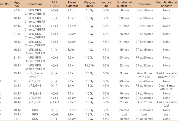
Table 1. Patient summary*.
Case No. Age,
sex Treatment
AFP, β-HCG†
Mean diameter
Marginal dose
Isodose line
Duration of
clinical FU Response
Complications/ or death
1 27, M VPS, GKS,
Chemo, WBSRT
(-), (-) 2.1 cm 13 Gy 50% 56 mos CR at 56 mos None
2 16, M VPS, GKS,
Chemo, WBSRT
(-), (+) 1.8 cm 13 Gy 50% 30 mos PR at 30 mos None
3 12, M VPS, GKS,
Chemo, WBSRT
(-), (-) 1.1 cm 13 Gy 50% 61 mos CR at 61 mos None
4 21, M VPS, GKS,
Chemo, WBSRT
(-), (-) 2.5 cm 13 Gy 50% 25 mos CR at 25 mos None
5 20, M VPS, GKS,
Chemo, WBSRT
(-), (-) 3.6 cm 13 Gy 45% 64 mos CR at 64 mos None
6 16, M VPS, GKS,
Chemo, WBSRT
(-), (+) 3.0 cm 14 Gy 45% 15 mos CR at 15 mos None
7 31, M VPS, GKS,
Chemo, WBSRT
(-), (-) 1.3 cm 15 Gy 50% 60 mos PR at 60 mos None
8 18, M VPS, GKS,
Chemo, WBSRT
(-), (-) 1.6 cm 14.5 Gy 50% 32 mos CR at 32 mos None
9 45, M GKS, Chemo,
WBSRT
(-), (+) 2.3 cm 15 Gy 50% 9 mos PG at 9 mos
with VSS
Died 9 mos after GKS with ISS
10 65, F VPS, GKS (-), (+) 2.2 cm 13 Gy 50% 43 mos CR at 43 mos None
11 13, M VPS, GKS (+), (-) 2.4 cm 13 Gy 45% 18 mos PG at 18 mos Died 18 mos
after GKS
12 54, M VPS, GKS (-), (-) 1.5 cm 15 Gy 50% 16 mos CR at 16 mos None
13 34, M VPS, GKS (-), (-) 1.6 cm 14 Gy 50% 39 mos CR at 39 mos None
14 16, M VPS, GKS (+), (+) 2.0 cm 13 Gy 50% 2 mos PG at 2 mos Died 2 mos after
GKS
15 18, M GKS (+), (-) 3.1 cm 15 Gy 50% 35 mos PR at 35 mos None
16 13, M GKS (-), (-) 2.9 cm 13 Gy 45% Lost Lost Lost
17 14, F GKS (-), (+) 2.4 cm 14 Gy 45% 28 mos NC at 28 mos None
*GKS: gamma knife surgery; VPS: ventriculoperitoneal shunt; Chemo: chemotherapy; WBSRT: whole-brain and spinal cord radiotherapy; AFP: alpha fetoprotein;
β-HCG: beta chorionic gonadotrophin; CR: complete response; PR: partial response; MR: minor response; NC: no change; PG: progression; VSS: ventricular and spinal spread; (-): negative; (+): positive.
for dose planning were obtained with a 1.5Tesla supercon
-ducting magnetic resonance scanner (Siemens, Erlanger, Germany). Dose planning was carried out with Leksell Gamma Plan (Elekta Instruments AB). he average marginal dose was 13.7 Gy (range, 12–15 Gy), and the average isodose line was 48.5% (range, 45–50%). Ventriculoperitoneal shunt
(VPS) was employed in patients with high intracranial pres
-sure before GKS.
Clinical followup was by means of patient interview ( face to face or telephone) and regular MRI examination eve ry 36 months. he efects of GKS were evaluated based on clinical manifestations and radiological changes. MRI ima ges were analyzed by two independent experienced radio logists according to a ivegrade system, including complete
response (CR), partial response (PR), minor response (MR),
no change (NC), and progression (PG). Total response rate was deined as the percentages of CR and PR, and control
rate was the ratio of CR, PR, MR, and NC to total19. Karnofsky
performance status (KPS) was assessed at admission and 6 months after GKS, and these two scores were compared to investigate the inluence of GKS on quality of life.
RESULTS
Sixteen of 17 cases (94.1%) were followed up over a mean
of 33.3 months (range, 264 months), and one was lost to fol
-lowup. hirteen patients (81.3%) were alive at the inal fol
-lowup (Table 1). CR was achieved in nine cases (56.3%), PR in three cases (18.8%), NC in one case (6.3%), and PG in three cases (18.8%); no patients showed MR. he total response rate was 75%, and the control rate was 81.3% (Table 2). VPS
was performed in 13 cases (76.5%), as high intracranial pres
-sure is the main symptom in patients with PRTs. No extra nervous system metastases were found.
Serum testing for tumor markers (AFP and β-HCG)
was negative in nine (52.9%) cases, and either one or both were positive in eight cases (47.1%). he relationship bet ween tumor markers and response revealed that patients
with negative tumor markers had a tendency to achieve a CR regar dless of whether they were treated with GKS plus WBSRT and chemotherapy or GKS only (Table 3).
Nine patients had further wholebrain and spinal cord ra
-diotherapy (WBSRT) and chemotherapy (Chemo) 69 months
after GKS. he whole brain and spinal cord were fractional
-ly irradiated with doses from 34203600 cGy. he remaining seven patients (excluding the lost patient) did not undergo
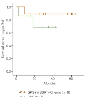
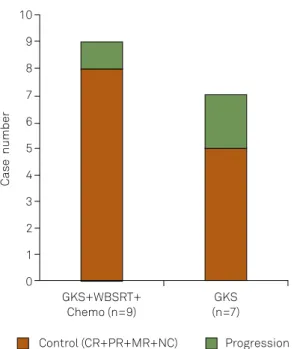
further treatment after GKS. here was no dife rence in sur
-vival (p=0.382) (Figure 1) or control rate (71.4% vs. 88.9%, p=0.375) (Figure 2) between the patients treated with GKS and those with GKS followed by WBSRT and Chemo.
hree patients died. One died 2 months after GKS due to disease progression. he second died 9 months after
Table 2. Tumor control*.
Response No. of cases Percentage
CR 9 56.3%
PR 3 18.8%
MR 0 0.0%
NC 1 6.3%
PG 3 18.8%
Total response rate 12 75.0%
Control rate 13 81.3%
Total no. of cases 16 100%
* One case was excluded due to follow-up loss.
CR: complete response; PR: partial response; MR: minor response; NC: no change; PG: progression.
Table 3. Tumor markers and response*.
Reponses
AFP/β-HCG in serum
GKS, Chemo, WBSRT GKS
-/- +/-, -/+, +/+ -/- +/-, -/+, +/+
CR 5 1 2 1
PR 1 1 0 1
MR 0 0 0 0
NC 0 0 0 1
PG 0 1 0 2
No. of cases 6 3 2 5
* One case was excluded due to follow-up loss.
CR: complete response; PR: partial response; MR: minor response; NC: no change; PG: progression; GKS: gamma knife surgery; WBSRT: whole-brain and spinal cord radiotherapy; AFP: alpha fetoprotein; β-HCG: beta chorionic gonadotrophin.
1.0
0.8
0.6
0.4
0.2
0.0
0 20 40 60 Months
GKS+WBSRT+Chemo (n=9) GKS (n=7)
Survival per
centag
es (%)
GKS: gamma knife surgery; Chemo: chemotherapy; WBSRT: whole-brain and spinal cord radiotherapy.
radiosurgery with ventricular and spinal spread. he third died 18 months following multiple recurrences; however, the MRI of this patient taken 3 months after GKS revealed a sa tisfactory decrease in tumor size.
he mean KPS at 6 months after GKS (excluding those who died or were lost to followup) was 94.6, compared to 63.8 when they were irst admitted to hospital. No obvious neurological deicits or complications, such as brain edema or cerebral radiation necrosis, were attributable to GKS.
Selected cases
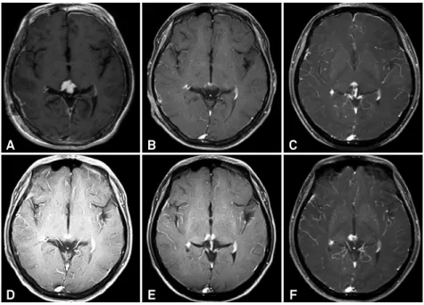
Case 1
A 27yearold man complained of a 1month history of headache and vomiting. Cranial MRI revealed a lesion with
enhancement in the pineal region (Figure 3). VPS was per
-formed, followed by GKS with a marginal dose of 13 Gy and a 50% isodose line. Four months later, the patient underwent WBSRT with a dose of 3600 cGy, as well as Chemo. Followup conirmed a CR for more than 56 months.
Case 2
An 18yearold boy complained of headache that had las ted for 10 days. MRI showed an enhanced lesion in the pineal region (Figure 4). he patient only underwent GKS, and the
tumor reduced 3 months later. However, a small enhanced le
-sion was still present on MRI 6 months after GKS, but did not show any obvious changes at later followup assessments.
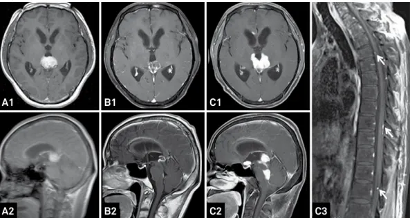
Case 3
A 45yearold man complained of headache for 15 days. MRI revealed an enhanced lesion in the pineal region (Fi gure 5A), and GKS was performed followed by WBSRT at a dose of 3420 cGy, as well as Chemo with temozolamide 3 months later. At 4 months after GKS there was a minor
change in the lesion, with tumor seeding to the fourth ven
-tricle (Figure 5B). he tumor showed PG at 7 months with ventricular and spinal spread (Figure 5C), and the patient died 9 months after GKS.
DISCUSSION
Compared to conventional irradiation, GKS is a better
treatment tool for PRT patients. It can greatly protect adja
-cent brain tissue while directing a high dose to the tumor.
Kobayashi and colleagues19 reported on their use of stereo
-tactic gamma radiosurgery for 30 pineal and related tumors,
among which 3 cases without histological diagnosis were ini
-tially treated with GKS. A CR was obtained in 1 case and PRs in 2 during a mean followup period of 23.3 months. In the present research of 17 cases, 9 cases had a CR, and 3 cases obtained a PR over a mean followup period of 33.3 months. he total response rate was 75%, and the control rate was
81.3%. hirteen patients (81.3%) were alive at the end of fol
-lowup. hese outcomes were analogous to a previous report
in which pathological diagnoses were proven19,20.
Good quality of life was also seen in the present patients.
Many studies have reported that good outcomes were ob
-tained with fewer complications, although more invasive diag nostic treatments were applied13,1925. We recorded the
KPS of patients at admission and 6 months after GKS (ex
-cluding those who died or were lost to followup). he mean KPS at the 6month followup was 94.6, compared to the KPS of 63.8 when patients were irst admitted to the hospital. We did not ind any evidence of obvious neurological deicits or
complications, such as brain edema or cerebral radiation ne
-crosis. Most of our patients were able to return to school or their previous job 1–3 months after treatment.
One of the factors underlying outcome is the radiosensi
-tive nature of some PRTs, such as germinomas, which have 5year survival rates of roughly 90% with radiotherapy alone1.
hese tumors account for a higher rate of PRTs in Asia, and
usually occur in males17. he male:female ratio in our patients
was 15:2, which suggests that radiosensitive PRTs may have accounted for the majority of cases. According to this cha racteristic and preoperative imaging evaluations, we selected
midrange doses for GKS compared to previous studies20,23.
Another factor was the minimal invasiveness of GKS.
Conventional radiation therapy for PRTs usually produces se
-quelae, such as cognitive deicits, endocrinopathies, secon
dary malignancies, growth arrest, and marrow suppression26.
10
9
8
7
6
5
4
3
2
1
0
Case number
GKS+WBSRT+ Chemo (n=9)
GKS (n=7)
Control (CR+PR+MR+NC) Progression
GKS: gamma knife surgery; Chemo: chemotherapy; WBSRT: whole-brain and spinal cord radiotherapy; CR: complete response; PR: partial response; MR: minor response; NC: no change.
A1 B1 C1 D1 E1
A2 B2 C2 D2 E2
GKS: gamma knife surgery; Chemo: chemotherapy; WBSRT: whole-brain and spinal cord radiotherapy; CR: complete response; MRIs: magnetic resonance images; VPS: ventriculoperitoneal shunt.
Figure 3. Follow-up MRIs of a 27-year-old patient treated with VPS, GKS, Chemo, and WBSRT. A lesion with enhancement (A1, A2) was found in the pineal region before treatment and decreased 4 months later (B1, B2). MRIs at 23 months (C1, C2), 38 months (D1, D2), and 56 months (E1, E2) showed a CR.
A
D
B
E
C
F
MRIs: magnetic resonance images; GKS: gamma knife surgery.
Figure 4. Follow-up MRIs of an 18-year-old male patient treated with GKS only. An enhanced lesion (A) was found in the pineal region and decreased 3 months after GKS (B). However, a small enhanced lesion was found at 6 months (C) and persisted at 12 months (D), 24 months (E), and 35 months (F).
he risk for these morbidities is particularly high in young children and in patients with a long life expectancy1. GKS can
greatly protect adjacent brain tissue when delivering large singlefraction radiation doses to a focal area. In the publi
shed research on PRTs treated with GKS13, 1925, obvious neu
-rological deicit and complications were not typically attribu table to GKS. A similar result was also observed in our study.
Tumor markers, such as AFP and β-HCG, are useful
prog-nostic factors, and all patients should undergo routine tests14.
We found that CR was most likely to be obtained in patients
with negative markers regardless of whether they were trea ted with GKS only or GKS followed by WBSRT and Chemo. Our indings indicate that patients with positive markers are more likely to have malignant tumors, and those with both
negative AFP and βHCG results may have the best outcomes
following radiosurgery.
It is important to note that some PRTs have a cer
-tain rate of ventricular and spinal spreading. For example, 2–37% of germinomas have distant metastases after appa
A1 B1 C1
A2 B2 C2 C3
GKS: gamma knife surgery; Chemo: chemotherapy; MRIs: magnetic resonance images; WBSRT: whole-brain and spinal cord radiotherapy.
Figure 5. Follow-up MRIs of a 45-year-old male patient treated with GKS, Chemo, and WBSRT. An enhanced lesion (A1, A2) was found in the pineal region and slightly 4 months after treatment (B1, B2). MRIs taken at 7 months (C1-C3) revealed that the tumor had spread to the saddle area and fourth ventricle, as well as the spinal cord (C3, white arrow).
alone, as it treats a limited irradiation region. Hence, GKS
followed by WBSRT and Chemo is theoretically the better choice for PRT patients with no or unclear pathological
diagnoses. Endo and colleagues21 reported that combined
radiotherapy using GKS is efective for pineal germinoma and reduces the cost of treatment by shortening hospita
lization. Hasegawa and colleagues13 pointed out that mul
-timodality therapy, inclu ding stereotactic radiosurgery, fractionated radiotherapy, and Chemo, is required for more aggressive pineal parenchymal tumors. One of our patients whose enhanced lesion disappeared at 3 months after GKS died of multiple recurrences at 18 months, which
might be attributable to GKS monothe rapy without adjunct WBSRT and chemotherapy. However, a diference in survi val and control rates between GKS only and GKS followed
by WBSRT plus Chemo was not observed in this retrospec
-tive study. his may be because of the small sample size. For further studies, it remains an open question whether every
patient with a negative pathological diagnosis should rou
-tinely receive WBSRT and Chemo after initial GKS.
In conclusion, GKS may be an alternative strategy in a
subset of PRT patients who have negative pathological diag
-noses, and good quality of life can be obtained with a low risk
of complications.
References
1. Blakeley JO, Grossman SA. Management of pineal region tumors. Curr Treat Options Oncol 2006;7:505-516.
2. Hirato J, Nakazato Y. Pathology of pineal region tumors. J Neurooncol 2001;54:239-249.
3. Fevre-Montange M, Vasiljevic A, Champier J, Jouvet A. Histopathology of tumors of the pineal region. Future Oncol 2010;6:791-809.
4. Konovalov AN, Pitskhelauri DI. Principles of treatment of the pineal region tumors. Surg Neurol 2003;59:250-268.
5. Kang JK, Jeun SS, Hong YK, et al. Experience with pineal region tumors. Childs Nerv Syst 1998;14:63-68.
6. Shin HJ, Cho BK, Jung HW, Wang KC. Pediatric pineal tumors: need for a direct surgical approach and complications of the occipital transtentorial approach. Childs Nerv Syst 1998;14:174-178.
7. Yamini B, Refai D, Rubin CM, Frim DM. Initial endoscopic management of pineal region tumors and associated hydrocephalus: clinical series and literature review. J Neurosurg 2004;100:437-441.
8. Pople IK, Athanasiou TC, Sandeman DR, Coakham HB. The role of endoscopic biopsy and third ventriculostomy in the management of pineal region tumours. Br J Neurosurg 2001;15:305-311.
9. Ferrer E, Santamarta D, Garcia-Fructuoso G, Caral L, Rumia J. Neuroendoscopic management of pineal region tumours. Acta Neurochir (Wien) 1997;139:12-21.
10. Radovanovic I, Dizdarevic K, de Tribolet N, Masic T, Muminagic S. Pineal region tumors--neurosurgical review. Med Arh 2009;63:171-173.
11. Edwards MS, Hudgins RJ, Wilson CB, Levin VA, Wara WM. Pineal region tumors in children. J Neurosurg 1988;68:689-697.
12. Field M, Witham TF, Flickinger JC, Kondziolka D, Lunsford LD. Comprehensive assessment of hemorrhage risks and outcomes after stereotactic brain biopsy. J Neurosurg 2001;94:545-551.
14. Bruce JN, Ogden AT. Surgical strategies for treating patients with pineal region tumors. J Neurooncol 2004;69:221-236.
15. Choi JU, Kim DS, Chung SS, Kim TS. Treatment of germ cell tumors in the pineal region. Childs Nerv Syst 1998;14:41-48.
16. Kanamori M, Kumabe T, Tominaga T. Is histological diagnosis necessary to start treatment for germ cell tumours in the pineal region? J Clin Neurosci 2008;15:978-987.
17. Nomura K. Epidemiology of germ cell tumors in Asia of pineal region tumor. J Neurooncol 2001;54:211-217.
18. Shibui S, Nomura K. Statistical analysis of pineal tumors based on the data of Brain Tumor Registry of Japan. Prog Neurol Surg 2009;23:1-11.
19. Kobayashi T, Kida Y, Mori Y. Stereotactic gamma radiosurgery for pineal and related tumors. J Neurooncol 2001;54:301-309.
20. Amendola BE, Wolf A, Coy SR, Amendola MA, Eber D. Pineal
tumors: analysis of treatment results in 20 patients. J Neurosurg 2005;102:S175-S179.
21. Endo H, Kumabe T, Jokura H, Tominaga T. Stereotactic radiosurgery followed by whole ventricular irradiation for primary intracranial
germinoma of the pineal region. Minim Invasive Neurosurg 2005;48:186-190.
22. Lekovic GP, Gonzalez LF, Shetter AG, Porter RW, et al. Role of Gamma Knife surgery in the management of pineal region tumors. Neurosurg Focus 2007;23:E12.
23. Mori Y, Kobayashi T, Hasegawa T, Yoshida K, Kida Y. Stereotactic radiosurgery for pineal and related tumors. Prog Neurol Surg 2009;23:106-118.
24. Kano H, Niranjan A, Kondziolka D, Flickinger JC, Lunsford D. Role of stereotactic radiosurgery in the management of pineal parenchymal tumors. Prog Neurol Surg 2009;23:44-58.
25. Reyns N, Hayashi M, Chinot O, et al. The role of Gamma Knife radiosurgery in the treatment of pineal parenchymal tumours. Acta Neurochir (Wien) 2006;148:5-11.