Article
Printed in Brazil - ©2017 Sociedade Brasileira de Química0103 - 5053 $6.00+0.00*e-mail: [email protected]
Sesquiterpenoids from
Nectandra megapotamica
(Lauraceae)
Carolina Q. Oliveira, Liziane B. Morandini, Marcelo Pedroso, Alexandre T. Neto, Ubiratan F. Silva, Marco A. Mostardeiro, Ionara I. Dalcol and Ademir F. Morel*
Departamento de Química (NPPN), Universidade Federal de Santa Maria, 97105-900 Santa Maria-RS, Brazil
Five sesquiterpenoid oxides, named nectandrene A, B, C, D, and E, were isolated from the essential oil of the leaves of Nectandra megapotamica. Their structures were elucidated by spectroscopic analysis, and the relative configurations were proposed by their nuclear Overhauser effect (NOESY) spectrum. Three of these isolated compounds displayed significant antimicrobial activity; the compound most active had minimal inhibitory concentration (MIC) values between 3.12 and 25.0 µg mL-1 against some tested bacteria, and antifungal activity with MIC values between 12.5 and 25.0 µg mL-1.
Keywords: Nectandra megapotamica, Lauraceae, sesquiterpenoid spectroscopic analysis, antimicrobial activity
Introduction
The Lauraceae is a large pantropical family mainly composed of trees and shrubs and comprised of about 53 genera and ca. 3000 species.1 It is a family with distribution concentrated in tropical and subtropical rain forests of Asia and the Americas,2 and Brazil itself houses 22 genera and about 43 species.3,4 The genera Anibal, Nectandra, and Ocotea present the greatest number of species of economic importance.5 The Lauraceae family comprises many species rich in essential oils that can be found in the leaves and the wood itself such as Anibal roseodora, Ocotea pretiosa and Cinnamomum cassia, among others.6 The Nectandra Rol. ex Rottb is among the most important genera of the family in number of species. This genus is restricted to tropical and subtropical America and it is represented by 114 species recognized to date being 43 native to Brazil.3,4 In addition to the essential oils, this genus is characterized by the presence of benzoquinolinic,7 aporfinic and indole alkaloids,8 lignans9 and neolignans.10 Essential oils of this genus are rarely studied. The literature has reported studies of the volatile oils of N. rigida (Kunth) Nees, whose main components are
α-phellandrene and β-phellandrene,11 while N. augustifolia Nees contains p-menta-1(7),8-diene and α-terpinolene.12 The species N. salicina C. K. Allen contains in its essential oil α-pinene, β-caryophyllene, viridiflorene, as well as
the sesquiterpene atractylone and germacrene D.13 The essential oil of the fruits of N. shady (Kunth) Metz present cadinol, germacrene B and spathulenol.14 The species
wounds being caused by fungi and bacteria, we decided to study the antimicrobial activities of the purified compounds of the essential oil of N. megapotamica collected in the city of Santa Maria-RS, utilizing microdilution methods.19
Experimental
General experimental procedures
Optical rotations were taken on a PerkinElmer 341 digital polarimeter. Low resolution electron impact mass spectrometry (EIMS) were recorded on a Varian 3800 operating in the ionization potential mode at 70 eV. 1H and 13C NMR (nuclear magnetic resonance) spectra were recorded at 400.1/100.6 MHz on a Bruker DPX-400 spectrometer using CDCl3 as solvent, and tetramethylsilane (TMS) as internal standard. Thin layer chromatography (TLC) was performed on a pre-coated TLC plate (Merck, silica 60 F-254), by spraying with 10% H2SO4/EtOH, followed by heating.
Plantmaterial
The aerial parts of N. megapotamica were collected in 2010 (in spring), in the city of Santa Maria (Rio Grande do Sul State, Brazil) from the same population. Specimens were identified by Prof Thais do Canto-Dorow(Department of Biology, University of Santa Maria, RS, Brazil). Voucher specimens (SMDB 14502) were deposited at the Herbarium of the University of Santa Maria, RS, Brazil.
Essential oil isolation and chemical analysis
The essential oil was submitted to gas chromatograph (GC) analysis in a Varian 3800 Gas Chromatograph equipped with a capillary fused silica column (25 m × 0.25 mm; film thickness 0.2 µm) coated with HP5-MS. Fresh plant leaves (100 g) were subjected to hydrodistillation using a modified Clevenger-type Apparatus. After removal of the solvent, the yield of the crude oil (d 0.8701 g mL-1; η 1.5485; [α]D25 –45.9 (c 0.05, CH2Cl2)) was 0.81%. The GC conditions used were: carrier gas H2 (1 mL min-1); injector split/splitless 220 °C; flame ionization detector (FID) detector 280 °C; column temperature 50 to 250 °C at 4 °C min-1. GC-MS analyses were performed on a Shimadzu QP-2010, equipped with an INNOWAX (PEG) cross-linked capillary column (60 m × 0.32 mm; film thickness 0.25 µm). The GC-MS conditions used were: carrier gas He (1 mL min-1); injector split/splitless 250 °C; detector 200 oC, and column temperature 35 to 310 °C at 3 °C min-1. The retention index (RI) values were determined relative to the retention times
(RT) of a series of C8-C30n-alkanes with linear interpolation on the HP5-MS column.
Isolationof compounds 1-5
The essential oil (300 mg) of leaves of N. megapotamica was subjected to silica gel chromatography (30 g). Elution with n-hexane-ethyl acetate (100:0, 97:3, 95:5 and 90:10) furnished 48 fractions (1-48, 20 mL each). Fractions 20-24 eluted with n-hexane-ethyl acetate (95:5) gave a mixture of compounds (GC) as colorless oil. Fractions 20-14 were combined (90.0 mg) and submitted to preparative TLC (n-hexane:ethyl acetate 95:5) affording two fractions, a major (55 mg) and a minor fraction (30 mg). The major fraction was submitted to preparative TLC (n-hexane:ethyl acetate 95:5 containing a drop of trifluoroacetic anhydride-TFAA, two elutions) to yield 1 (20.0 mg) and 2 (18.5 mg). The minor and less polar fraction (30 mg) consisting of two components (63:37 ratio determined by GC) was subjected to a preparative TLC (n-hexane:ethyl acetate 95:5, two elutions) providing two fractions: the less polar fraction (20 mg) was rich (89:11 ratio determined by GC) with the major constituent (3), and a more polar fraction (10 mg) was rich (90:10 ratio determined by GC) with the minority constituent (4). Fraction 30-38 (70 mg), containing two components, was subjected to preparative TLC (silica gel, n-hexane-acetone 90:10 two elutions) to give 5 (25 mg).
Nectandrene A (1)
Colorless oil; [α]D25 –33 (c 0.02, CHCl3); Rf 0.55 (n-hexane:AcOEt 96:4); 1H NMR (400.1 MHz, CDCl
3) and 13C NMR (100.6 MHz, CDCl
3) see Tables 1 and 2; EIMS 70 eV m/z 220 [M]+, 109 (100%); HRMS m/z, calcd. for C15H25O [M + H]+: 221.1905; found: 221.1942.
Nectandrene B (2)
Colorless oil; [α]D25 –98 (c 0.02, CHCl3); Rf 0.58 (n-hexane:AcOEt 96:4); 1H NMR (400.1 MHz, CDCl
3) and 13C NMR (100.6 MHz, CDCl
3) see Tables 1 and 2; EIMS 70 eV m/z 220 [M]+, 107 (100%); HRMS m/z, calcd. for C15H25O [M + H]+: 221.1905; found: 221.1927.
Nectandrene C (3)
Colorless oil; Rf 0.65 (n-hexane:AcOEt 96:4); 1H NMR (400.1 MHz, CDCl3) and 13C NMR (100.6 MHz, CDCl3) see Tables 1 and 2; EIMS 70 eV m/z 220 [M]+.
Nectandrene D (4)
Nectandrene E(5)
Colorless oil; [α]D25 –85 (c 0.03, CHCl3); Rf 0.35 (n-hexane:AcOEt 95:5); 1H NMR (400.1 MHz, CDCl
3) and 13C NMR (100.6 MHz, CDCl
3) see Tables 1 and 2; HRME [M + H – H2O]+ at m/z 221.1973, [M + H]+ at m/z 239.2021, [M + H + Na]+ at m/z 261.1884.
Antimicrobialassays
The antibacterial activities were assayed using the broth micro dilution method.19 A collection of eight microorganisms was used, including five Gram-positive bacteria: Staphylococcus aureus (ATCC 6538p), Staphylococcus epidermidis (ATCC 12228), Bacillus subtillis (ATCC 6633), Staphylococcus saprophyticus (ATCC 15305) and Streptococcus pyogenes ( AT C C 1 9 6 1 5 ) ; t h r e e Gram-negative bacteria: Shigella sonnei (ATCC 25931), Escherichia coli (ATCC 25792), and Pseudomonas a e r u g i n o s a ( AT C C 2 7 8 5 3 ) a n d f o u r y e a s t s : Saccharomyces cerevisiae (ATCC 2601), Candida albicans (ATCC 10231), Candida tropicalis (ATCC 18803) and Cryptococcus neoformans (ATCC 28952). Standard strains of microorganisms were obtained from American Type Culture Collection (ATCC), and standard antibiotics chloramphenicol and nystatin were used in order to control the sensitivity of the microbial test. The minimal inhibitory concentration (MIC) was determined on 96 well culture plates by a micro dilution method using a microorganism suspension at a density of 105 CFU mL-1 with Casein Soy Broth incubated for 24 h at 37 °C for bacteria, and Sabouraud Broth incubated for 72 h at 25 °C for yeasts. The cultures that did not present growth were used to inoculate plates of solid medium (Muller Hinton Agar and Sabouraud Agar) in order to determine the minimal lethal concentration (MBC for bacteria and MFC for fungi).
Proper blanks were assayed simultaneously and samples were tested in triplicate.
Results and Discussion
Chemical investigation of the oil
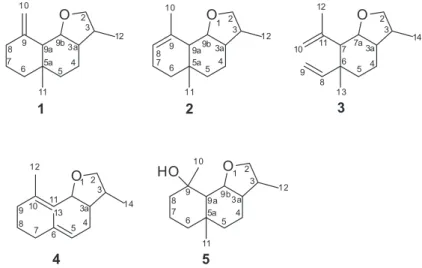
The essential oil yields of the leaves of N. megapotamica were mainly composed (> 90%) of sesquiterpenoids (Figures S34, S35 and 36, Supplementary Information section). The major components of the oil showed retention index RI consistent with compounds known in literature,20 but with small differences in their mass fragmentation. This result led us to attempt to isolate them by chromatographic methods and to identify them through usual NMR and mass spectrometry (MS) techniques. Thus, five components of the oil were isolated (Table S1, Supplementary Information section) and identified as new sesquiterpenoids containing an unusual tetrahydrofuran ring (Figure 1).
Compounds 1 and 2 were initially identified as a single oil component because the chromatogram appeared in only one broad peak, which RI was 1705. The mass spectrum showed a molecular ion of 220 amu. Comparison of experimentally obtained data with literature did not identify any known compound. With the purpose of isolating the pure component, the oil (300 mg) was chromatographed over silica gel. Fractions 20-24 (n-hexane:EtOAc, 90:10), comprising the main component of the oil and a minor component, were subjected to preparative TLC separation. Thus, the desired fraction (RI 1705) was obtained. Analysis of the separated fraction by GC showed a split signal into two components, suggesting two compounds with very close retention rates. NMR analysis of this fraction revealed the presence of 30 carbons, suggesting two sesquiterpenes with very close structures and even molecular ion. After repeated
attempts to separate the two components by preparative TLC, success was obtained when the liquid phase (hexane:ethyl acetate, 94:6) was used with a drop of trifluoroacetic acid. Thus, the two components were obtained by preparative TLC in pure form and in small amounts, but enough for structural analysis and for the determination of its antimicrobial activity. The NMR spectra of both compounds separately show exactly the same peaks observed in the mixture, therefore no structural alteration by addition of TFAA in the solvent. The EIMS of 1 gave a [M]+ peak at m/z 220. Its high resolution (HRME) spectra (Figure S1, Supplementary Information section) displayed a prominent [M + H]+ at
m/z 221.1942 (calculated for C15H25O, m/z 221.1905). Its 1H NMR spectrum (Figure S2, Supplementary Information
section) determined in CDCl3 (Table 1)indicated the presence of two methyl groups, one singlet at dH 0.73 (CH3-11), and a doublet at dH 0.99 (CH3-12), four methine hydrogens at
dH 1.79 (d, 1H, J 10.4 Hz, H-9a), 1.89 (m, 1H, H-3a), 2.25 (m, 1H, H-3) and 4.27 (dd, 1H, J 7.2, 10.4 Hz, H-9b), seven methylene at dH1.20/1.29 (m, 2H, H-6), 1.35/1.32 (m, 2H, H-5), 1.65/1.87 (m, 2H, H-4), 1.58 (m, 2H, H-7), 2.0/2.34 (m, 2H, H-8), 3.29 (dd, 1H, J 8.5, 8.2 Hz, H-2), 4.04 (dd,
1H, J 8.5, 7.8 Hz, H-2’) and 4.77/4.92 (d, 2H, J 1.5 Hz, H-10). The signal at dH 4.27 (1H) and at dH 3.28/4.04 (2H) suggested one methine and two methylene hydrogens bearing oxygen. The absence of hydroxyl groups in the molecule (IR spectrum) suggests ether oxygen joining both carbons (–CH–O–CH2–). Signals at 4.77/4.92 suggest a methylene group of a double bond. The 13C NMR spectra (Table 2) for the hydrogen bonded carbons obtained from the DEPT (distortionless enhancement by polarisation transfer) and HETCOR (heteronuclear shift correlation experiments) (Figures S3-S5 Supplementary Information section) showed the presence of four methine, seven methylene, two methyl and two quaternary carbons, which indicated a sesquiterpene, consistent with the molecular formula C15H24O. COLOC (correlation through long-range coupling) correlations between C-2, C-3, C-3a/CH3-12; C-3,-3a, C-9a, C-9b/H2-2, supported a tetrahydrofuran ring in this structure.
All spectroscopic data indicated that 2 was a closely related analog of 1. Its EIMS gave a [M]+ peak at m/z 220. In contrast to 1, 1H NMR spectrum of 2 (Table 1, Figure S6, Supplementary Information section) shows, instead of signals of two olefinic metilenic hidrogens (H2-10),
Table 1. 1H NMR spectroscopic data of compounds 1-5 in CDCl 3a
Proton position 1
b 2b 3b 4c 5b
(mult., J ) / Hz (mult., J ) / Hz (mult., J ) / Hz (mult., J ) / Hz (mult., J ) / Hz
2, CH2 4.04, dd (8.5, 7.8)
3.29, dd (8.5, 8.2)
4.02, dd (8.5, 8.2) 3.29, dd (8.5; 8.0)
4.03, dd (8.0, 8.4) 3.30, dd (8.4, 8.8)
3.96, dd (7.8, 7.5) 3.18, dd (8.4, 7.5)
4.13, dd (8.8, 8.0) 3.30, dd (8.8, 9.2)
3, CH 2.25, m 2.20, m 2.26, m 1.85, m 2.33, m
3a, CH 1.89, m 1.98, m 1.86, m 1.72, m 1.77, m
4, CH2 1.87, m
1.65, m
1.89, m 1.65, m
1.86, m 1.81, m
1.93, m 1.44, m
1.75, m 1.68, m
5, CH2 (1-3, 5), CH (4) 1.35/1.32, m 1.34/1.30, m 1.52/1.21, m 4.92, brs 1.36/1.35, m
6, CH2 1.29/1.20, m 1.26/1.22, m 1.28/1.26, m
7, CH2 (1, 2, 4, 5), CH (3) 1.58, m 2.05/1.74, m 1.90, d (10.7) 2.20/1.89, m 1.35/1.18, m
7a, CH – – 4.28, dd (10.7, 7.2)
8, CH2 (1, 4, 5), CH (2, 3) 2.34/2.0, m 5.34, brs 5.76, dd (17.4, 10.9) 2.10, m 1.78/1.42, m
9, CH (3), CH2 (4) – – 4.88, dd (10.9, 1.2)/4.86, dd
(17.4, 1.2)
2.47/1.74, m
9a, CH 1.79, d (10.4) 1.90, m 1.42, d (11.2)
9b, CH 4.27, dd (10.4, 7.2) 4.05, m 4.45, dd (11.2, 7.2)
10, CH2 (1, 3)
CH3 (2, 5)
4.92, d (1.5) 4.77, d (1.5)
1.82, brs 4.99, brs 4.71, brs
1.30, s
11, CH (4), CH3 (1, 2, 5) 0.73, brs 0.79, brs 4.85, d (10.2) 0.84, brs
11a, CH 4.54, dd (10.2, 7.5)
12, CH3 0.99, d (6.4) 1.00, d (6.5, 3.0) 1.76, brs 1.43, brs 1.0, d (6.5)
13, CH3 – – 0.97, brs 1.50, brs
14, CH3 – – 1.05, d (6.8) 1.06, d (7.2)
resonances of a methyl group at dH 1.82 (s, Me-10), and one methine at dH 5.34 (H-8). A striking difference between the 13C NMR spectra of 1 and 2 (Table 2) is the absence of the olefinic methylene carbon in 2, the appearance of a new methyl group at d 23.2 (CH3-10), and a new methine carbon at d 122.1 (C-8). The detailed analysis of their 13C NMR, H-H correlation spectroscopy (COSY), DEPT, and HETCOR spectroscopic data (Figures S7-S10, Supplementary Information section) supported the proposed structures as 3,5a-dimethyl-9-methylene dodecahydronafto[1,2-b] furane, named nectandrene A (1), and as 3,5a ,9-trimethyl-4,5,5a,6,7,9a,9b-decahydronafto[1,2-b]furane, named nectandrene B (2). A diastereoisomer of this compound {3a-epi-2, [α]D25 +10.3 (CHCl3)} has previously been obtained by organic synthesis.21
On account of the possibility of 1 and 2 being artifacts due to the extraction method or that one becomes the other by heating, we performed an extraction of the essential oil with cold n-hexane. Gas chromatography of the hexane extract showed the presence of the same fraction obtained from hydrodistillation. This fraction was purified by preparative TLC and when analyzed by carbon NMR presented the same mixture as observed previously. Therefore, both compounds are natural sesquiterpenoids and not artifacts.
From the same preparative plate, a less polar fraction
composed of two main components (63 and 37%) was isolated. In an attempt to isolate the major component (RI 1547) by PTLC, a concentrated mixture of the main component (89:11) was obtained, which led us to determine the structure of the major component, therefore its optical rotation was not measured. The mass spectrum showed a molecular ion of 220 amu for both components (Figure S11, Supplementary Information section), which in combination with the 1H NMR and 13C NMR spectroscopic data (Figures S12 and S13, Supplementary Information section), suggested that both compounds have the same molecular formula C15H24O. The 1H NMR spectrum (Table 1) of the major component (3) displayed three methyl groups: two tertiary methyl at dH 1.76 (s, 3H, H-12), and 0.97 (s, 3H, H-13), and a secondary methyl at dH1.05 (d, 3H, J 6.8 Hz, H-14). Moreover, the spectrum showed an oxygenated methine at dH 4.28 (dd, 1H, J 10.7, 7.2 Hz, H-7a), two oxygenated methylene protons at dH 4.03 (dd, 1H, J 8.4, 8.0 Hz, H-2), and dH 3.30 (dd, 1H, J 8.4, 8.8 Hz, H-2’). Unlike 1 and 2, this compound has in its structure two pairs of methylene groups, suggesting the opening of a six-membered ring (ring A). Two of these geminal hydrogens appear at dH 4.88 (dd, 1H, J 10.9, 1.2 Hz, H-9), and at dH 4.86 (dd, 1H, J 17.4, 1.2 Hz, H-9). Both show coupling in the COSY spectrum (Figure S14, Supplementary
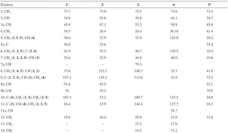
Table 2.13C NMR spectroscopic data of compounds 1-5a
Position 1b 2b 3c 4c 5b
2, CH2 73.7 73.0 73.5 73.6 73.4
3, CH 34.6 35.6 34.8 44.1 34.7
3a, CH 45.0 47.1 53.3 50.9 45.6
4, CH2 19.5 20.4 20.4 38.18 41.4
5, CH2 (1-3, 5); CH (4) 36.6 37.9 33.9 125.8 39.2
5a, C 36.8 33.6 35.4
6, CH2 (1, 2, 5); C (3, 4) 41.9 35.3 40.7 139.5 19.4
7, CH2 (1, 2, 4, 5); CH (3) 23.6 22.9 44.8 40.0 19.6
7a, CH – – 79.3
8, CH2 (1, 4, 5); CH (2, 3) 37.6 122.1 148.7 25.7 41.8
9, C (1, 2, 5); CH (3); CH2 (4) 147.1 135.3 113.6 32.9 72.3
9a, CH 51.6 45.9 52.1
9b, CH 76 79.2 79.9
10, C (4); CH2 (1, 3);CH3(2, 5) 107.3 23.2 109.7 133.5 24.0
11, C (3); CH (4); CH3 (1, 2, 5) 16.4 15.9 144.4 127.7 18.1
11a, CH 76.7
12, CH3 15.6 16.4 25.0 21.0 15.8
13, CH3 – – 17.2 17.0
14, CH3 – – 15.5 17.2
Information section) with a vicinal olefinic proton at dH 5.76 (dd, 1H, J 17.4, 10.9 Hz, H-8). The other two germinal protons appear at dH 4.99 (sl, 1H, H-10), and at dH 4.71 (sl, 1H, H-10). The remaining methine protons absorb at
dH1.90 (d, 1H, J 10.7 Hz, H-7), dH 1.86 (m, 1H, H-3a), and dH 2.26 (m, 1H, H-3). The methylene protons H-4 and H-5 appear at dH 1.86/1.81 and1.52/1.21, respectively, as multiplets. The 13C NMR spectrum of 3 (Table 2) contained resonances for all 15 carbons, while a DEPT, heteronuclear multiple quantum coherence (HMQC) (Figures S15 and S16, Supplementary Information section), heteronuclear multiple-bond correlation (HMBC) and nuclear Overhauser effect spectroscopy (NOESY) experiments (Figures 2, S17 and S18, Supplementary Information section) revealed the presence and the position of five methylenes (among them two geminal olefinic and one bonded to oxygen), three methyls, five methines (among them one bonded to an oxygenated carbon), and two quaternary carbons. These data led us to conclude the structure of 3 to be 3,6-dimethyl-7-(prop-1-en-2-yl)-6-vinyloctahydrobenzofuran, named nectandrene C (3).
From the same PTLC, we obtained a fraction with lower retention rates, yet very close to 3. Analysis of this fraction by GC presented a greater concentration of the other component (4, IK 1549) of the mixture but still contaminated with 3 (90:10). Although being isolated in a small amount, it was enough for structural determination. Its 1H and 13C NMR spectra (Figures S19 and S20, Supplementary Information section) showed that the structure 4 consists of two quaternary olefinic, three methyl, five methylene (DEPT), and five methine carbons. The major difference between the two compounds is in the absence of the four vinyl methylene hydrogens in 4. The 1H and 13C NMR spectra (Tables 1 and 2) of 4 indicated the presence of two olefinic H/C, one at dH/C 4.85 (J 10.2 Hz)/127.7 (CH-11) identified by presenting an intersection with H-11a in the COSY spectrum, and at
dH/C 4.92 (br s)/125.8 (CH-5), three methyl groups at dH/C
1.06 (d, J 7.2 Hz)/17.2 (CH3-14), 1.43 (s)/21.0 (CH3-12) and 1.50(s)/17.0 (CH3-13), five methylenes at dH/C 2.10 (m)/25.7 (CH2-8), 1.93/1.44 (m)/ 38.18 (CH2-4), 2.47/1.74 (m)/32.9 (CH2-9), 2.20/1.89 (m)/40.0 (CH2-7), and one bearing a hydroxyl group at 3.96 (dd, J 7.8, 7.5 Hz); 3.18 (dd, J 8.4, 7.5 Hz) /73.6 (CH2-2), and two quaternary olefinic carbons at dC 133.5 (C-10) and 139.5 (C-6). HMBC correlations between CH3-14/C-2/C-3/C-3a, and between H2 -2/C-2/C-3/C-11a, supported a tetrahydrofuran ring in this structure. The detailed analysis of their H-H-COSY, DEPT, HMQC (Figures S21-23, Supplementary Information section), HMBC and NOESY spectra (Figures 2, S24 and 25, Supplementary Information section), led to the determination that this compound is a new sesquiterpenoid (5E,10E)-3,6,10-trimethyl-2,3,3a,4,7,8,9,11a-octahydro cyclodeca[b]furan, named nectandrene D (4).
Fraction 30-38 (70 mg), containing two components, was subjected to preparative TLC (silica gel, n-hexane:acetone 90:10, twice) to give 5 (25 mg). Compound 5 (RI = 1817) displayed in the HRME spectrum a prominent [M + H – H2O]+ at m/z 221.1973, a molecular peak at [M + H]+ at m/z 239.2021 (Figure S26, Supplementary Information section), and a prominent [M + H + Na]+ at m/z 261.1884, which in combination with the 13C NMR spectroscopic data, was assigned a molecular formula of C15H26O2. The 1H NMR spectrum (Table 1) of compound 5 (Figure S27, Supplementary Information section) displayed three methyl groups: two tertiary methyl at dH 0.84 (s, CH3-11), and 1.30 (s, CH3-10), and a secondary methyl at dH1.0 (d, 3H, J 6.5 Hz, CH3-12). Moreover, the spectrum showed an oxygenated methine at dH 4.45 (dd, 1H, J 11.2, 7.2 Hz, H-9b), two oxygenated methylene protons at dH 4.13 (t, 1H, J 8.8, 8.0 Hz, H-2), and dH 3.30 (dd, 1H, J 8.8, 9.2 Hz, H-2’). The remaining methine protons absorbed at dH2.33 (m, 1H, H-3), 1.77 (m, 1H, H-3a), and dH 1.42 (d, 1H, J 11.2 Hz, H-9a). The methylene protons H-4, H-5, H-6, H-7, and H-8 appeared at
dH 1.75/1.68, 1.36./1.35, 1.28/1.26, 1.35/1.18, and 1.78/1.42, respectively, as multiplets.
The 13C NMR spectrum of 5 (Table2, Figure S28, Supplementary Information section) contained resonances for all 15 carbons, while a DEPT, HMQC, and HMBC experiments revealed the presence and the position of six methylenes at dC 73.4 (C-2), 41.8 (C-8), 41.4 (C-4), 39.2 (C-5), 19.6 (C-7),and19.4 (C-6), three methyls at dC 15.8 (C-12), 18.1 (C-11), and 23.9 (C-10), four methines at
dC 34.7 (C-3),45.6 (C-3a), 52.1 (C-9a) and 79.9 (C-9b, bonded to an oxygen), one quaternary carbon at dC 35.4 (C-5a), and one dehydrogenated carbon at dC 72.3 (C-9, bonded to an hydroxyl group). This compound differs from 1 and 2 since it does not present a double bond in its structure, but rather a tertiary OH group. This was supported by the methyl singlet atdH 1.30 (s, CH3-10) and the quaternary carbon at dC 72.3 (C-9). Assignments of the H/C chemical shift (Table 2) were made possible by the combination of the H-H COSY, DEPT, HMQC, and HMBC experiments (Figures S29-S32, Supplementary Information section). The relative stereochemistry configuration of C-3, C-3a, C-5a, C-9, C-9a and C9b was determined from 2D NOESY experiments (Figure S33, Supplementary Information section) through NOE cross-peaks between H-9b and H-3a, Me-10 and Me-11 (Figure 2) suggesting that these substituents are on the same face. H-9a also shows
a correlation with H-3, suggesting that both hydrogen’s are co facial. These data suggest anti-junction between rings A and B and a cis-junction between the rings B and C of the structure 5.
These data led us to conclude the structure to be a new sesquiterpene (3R*,3aR*,5aR*,9R*,9aS*,9bS*)-3,5a ,9-trimethyldodecahydronaphtho[1,2-b]furan-9-ol, named nectandrene E (5).
The relative configuration at C-3a, C-5a C-9a, and C- 9b position in compounds 1 and 2 was assigned as 3R*, 3aR*, 5aR*, 9R*, 9aS* based on 1H NMR coupling constant values, and comparing the data with compound 5. Similarly, compounds 3 and 4 had their relative stereochemistry determined as 3R*, 3aR*, 6R*, 7S*, 7aS*and3R*, 3aR*, 5aR*, 11aS*, respectively.
Biosyntheticconsiderations
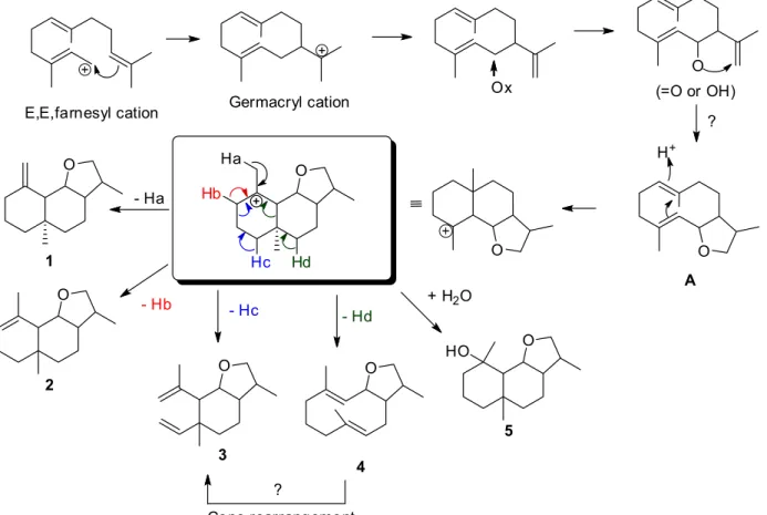
Biosynthetic considerations led to the hypothesis that compound A (not isolated) is a key intermediate in the biosynthesis of 1-5 (Figure 3). In addition, it can be assumed that the germacryl cation is the precursor of eudesmanes, elemanes, guaianes and germacranes. Further biosynthetic experiments are needed to substantiate the
Table 3. Antimicrobial activity (µg mL-1) of compounds 1, 2 and 5
Bacteriaa
Compound
1 2 5 Antibioticb
MIC / (µg mL-1)
MBC / (µg mL-1)
MIC / (µg mL-1)
MBC / (µg mL-1)
MIC / (µg mL-1)
MBC / (µg mL-1)
MIC/ (µg mL-1)
Staphylococcus aureus 6.25 100 3.12 100 25.0 100 3.12
Staphylococcus epidermidis 25.0 200 12.5 > 200 nt nt 3.12
Bacillus subtilis 25.0 200 12.5 200 25.0 100 3.12
Staphylococcus saprophyticus 25.0 200 12.5 200 nt nt 3.12
Streptococcus pyogenes 12.5 200 12.5 200 nt nt 3.12
Escherichia coli 25.0 200 12.5 200 25.0 100 3.12
Shiguella sonnei 25.0 200 12.5 200 25.0 25.0 3.12
Pseudomonas aeruginosa 25.0 200 12.5 > 200 25.0 25.0 3.12
Yeastsa MIC MFC MIC MFC MIC MFC MIC
Candida albicans 25.0 100 12.5 25.0 > 100 nt 6.25
Candida tropicalis 25.0 100 25.0 25.0 25.0 50.0 6.25
Cryptococcus neoformans 25.0 100 12.5 25.0 25.0 25.0 10.3
Saccharomyces cerivisiae 25.0 200 12.5 25.0 25.0 25.0 5.15
aATCC (American Type Culture Collection); bcloramphenicol for bacteria and nystatin for fungi; nt: not tested; MIC: minimal inhibitory concentration;
MBC and MFC: minimal lethal concentration for bacteria and for fungi, respectively.
above biogenetic proposition, particularly the formation of the tetrahydrofuran ring. Moreover, because of the possibility of a Cope rearrangement due to the extraction method,22,23 compound 3 could be an artifact of the compound 4.
Antibacterialactivity of compounds 1, 2 and 5
The antimicrobial activity of the isolated pure compounds
1, 2 and 5 was evaluated by the broth micro dilution method in order to determine the minimum inhibitory concentration (MIC) and the minimum lethal concentration (MBC or MFC). As shown in Table 3, the isolated compounds 1, 2 and 5 showed promising antibacterial (3.12 to 25.0 µg mL-1) and antifungal (12.5 to 25.0 µg mL-1) activities against some of the tested strains, compared with chloramphenicol for bacteria (3.12 µg mL-1) and nystatin for fungi (5.15-10.3 µg mL-1). Exception was compound 5 which proved to be inactive against C. albicans with a MIC > 100 µg mL-1. As demonstrated in Table 3, compounds
1 and 2 showed bacteriostatic activity (MIC) against the analyzed strains of bacteria, whereas for the tested fungi, both substances exhibited fungiostatic (MIC) and fungicidal (MFC) activity. Comparing the activity of both compounds, except for Pseudomonasaeruginosa (twice more active) and Staphylococcus pyogenes (same activity), compound 2, with the endo double bond, was twice more active against
the other bacteria than its isomer 1 with the exo double bond. The best result was observed for compound 2, which showed excellent activity against Staphylococcus aureus (MIC = 3.12 µg mL-1), compared to cloramphenicol (MIC = 3.12 µg mL-1). The same behavior can be observed for the fungi. Compound 2 was twice more active against Candida albicans, Sacharomyes cerevisae, and against Cryptococcus neoformans than 1. Both displayed the same activity against Candida troppicalis. Compound 5 was less effective against Staphylocous aureus, Bacillus subtilis and Escherichia coli (MIC/MBC = 25/100 µg mL-1) compared with compounds 1 and 2, but it showed to be bacteriostatic and bactericidal against Shiguella sonnei and Pseudomonas aeruginosa (MIC/MBC = 25/25 µg mL-1). Furthermore, compound 5 showed antifungal activity against Candida tropicalis (MIC/MFC = 25/50 µg mL-1),
Cryptococcus neoformans (MIC/MFC = 25/25 µg mL-1) and Saccharomyces cerivisiae (MIC/MFC = 25/25 µg mL-1). Compounds 3 and 4 were not analyzed because they are not pure compounds.
Conclusions
them, compounds 1, 2 and 5 were tested against a series of Gram (+/−) bacteria and fungi, showing promising results.
Supplementary Information
Supplementary data are available free of charge at http://jbcs.sbq.org.br as PDF file.
Acknowledgments
The authors thank CNPq (Conselho Nacional de Desenvolvimento Científico e Tecnológico) for financial support for this work. We are indebted to Prof E. M. Flores (UFSM) for the HRMS.
References
1. Rohwer, J. G.; Kubitzki, K.; Bot. Acta1993, 106, 88. 2. Souza, V. C.; Lorenzi, H.; Botânica Sistemática:Guia Ilustrado
para Identificação das Famílias de Angiospermas da Flora Brasileira, Baseado em APG II, 2a ed.; Instituto Plantarum de Estudos da Flora Ltda: Nova Odessa, SP, 2008.
3. Baitello, J. B.; ActaBot. Bras.2001, 15, 445.
4. Baitello, J. B.; Lorea-Hernández, F. G. L.; Moraes, P. L. R.; Esteves, R.; Marcovino, J. R. In Flora Fanerogâmica do Estado de São Paulo, vol. 3; Wanderley, M. G. L.; Shepherd, G. J.; Giulietti, A. M.; Melhem T. S., eds.; Fapesp-RiMa: São Paulo, 2003, p. 149.
5. Marques, C.; Floresta e Ambiente2001, 8, 195.
6. Craveiro, A. A.; Fernandes, A. G.; Andrade, C. H. S.; Matos, F. J. A.; Alencar, J. W.; Machado, M. I. L.; Óleos Essenciais de Plantas do Nordeste; UFC: Fortaleza, 1981.
7. Böhlke, M.; Guinaudeau, H.; Angerhofer, C. K.; Wongpanich, V.; Soejarto, D. D.; Farnsworth, N. R.; J. Nat. Prod.1996, 59, 576.
8. dos Santos Filho, D.; Gilbert, B.; Phytochemistry1975, 14, 821. 9. da Silva Filho, A. A.; Andrade e Silva, M. L.; Carvalho, J. C.;
Bastos, J. K.; J. Pharm. Pharmacol.2004, 56, 1179.
10. Braz Filho, R.; Figliuolo, R.; Gottlieb, O. R.;Phytochemistry
1980, 19, 659.
11. Morais, A. A.; Mourão, J. C.; Gottlieb, O. R.; Koketsu, M.; Moura, L. L.; da Silva, M. L.; Marx, M. C.; Mendes, P. H.; Magalhaes, M. T.; An. Acad. Bras. Cienc.1972, 44, 320. 12. Torres, A. M.; Riciardi, G. A. L.; Agrelo de Nassif, A. E.;
Ricciardi, A. A.; Dellacassa, E.; Univ. Nac. Nordeste Comum. Cient. Technol.2005, E-013.
13. Cicció, J. F.; Chaverri,C.; Díaz,C.; Quim. Nova2009, 32, 417. 14. Valley, P. S. M.; Scora C. A. R. W.; Calif.Avocado Soc. Yearb.
1999, 83, 163.
15. Garcez, F. R.; Garcez, W. S.; Hamerski, L.; Miguita. C. H.;
Quim. Nova2009, 32, 407.
16. da Silva Filho, A. A.; Costa, E. S.; Cunha, W. R.; Silva, M. L. A.; Dhammika Nanayakkara, N. P.; Bastos, J. K.; Phytother. Res.2008, 22, 1307.
17. Apel, M. A.; Lima, M. E. L.; Souza, A.; Cordeiro, I.; Young, M. C. M.; Sobral, M. E. G.; Suffredini, I. B.; Moreno, P. R. H.;
Pharmacologyonline2006, 3, 376.
18. Romoff, P.; Ferreira, M. J. P.; Padilla, R.; Toyama, D. O.; Fávero, O. A.; Lago, J. H. G.; Quim. Nova2010, 33, 1119.
19. National Committee for Clinical Laboratory Standards (NCCL);
Reference Method for Broth Dilution Antifungal Susceptibility Testing of Yeast: Approved Standard, CLSI document M27-A2, National Committee for Clinical Laboratory Standards: Wayne, PA, 2008.
20. Adams, R. P.; Identification of Essential Oil Components by Gas Chromatography/Quadrupole Mass Spectroscopy; Allured Publishing Corporation: Illinois, USA, 2001.
21. Cardona, L.; Garcia, B.; Giménez, E.; Pedro, J. R.; Tetrahedron
1992, 48, 851.
22. König, W. A.; Bülow, N.; Phytochemistry2000, 55, 141. 23. Adio, A. M.; Tetrahedron2009, 65, 1533.
Submitted: December 15, 2015 Published online: May 6, 2016