Daniela Mesquita Moutinho
Degree in Molecular and Cellular Biology
Human ceruloplasmin and neurotransmitters:
complex stabilization and crystallization
Dissertation to obtain a
Master Degree in Molecular Genetics and Biomedicine at Faculty of Sciences and Technology,
Universidade Nova de Lisboa
Supervisor:
Isabel Bento, Ph.D., ITQB-UNL
Co-supervisor: José Paulo Sampaio, Ph.D., FCT-UNL
Júri
Presidente: Doutora Margarida Casal Ribeiro Castro Caldas Braga Arguente: Doutor Colin Edward McVey
Vogal: Doutora Isabel Maria Travassos de Almeida de Jesus Bento
Daniela Mesquita Moutinho
Degree in Molecular and Cellular Biology
Human ceruloplasmin and neurotransmitters:
complex stabilization and crystallization
Dissertation to obtain a
Master Degree in Molecular Genetics and Biomedicine at Faculty of Sciences and Technology,
Universidade Nova de Lisboa
Supervisor
Isabel Bento, Ph.D., ITQB-UNL
Co-supervisor: José Paulo Sampaio, Ph.D., FCT-UNL
Júri
Presidente: Doutora Margarida Casal Ribeiro Castro Caldas Braga Arguente: Doutor Colin Edward McVey
Vogal: Doutora Isabel Maria Travassos de Almeida de Jesus Bento
“Human ceruloplasmin and neurotransmitters: complex stabilization and crystallization”
© Daniela Mesquita Moutinho, FCT/UNL, FCT
“Just because something doesn't do what you planned it to do doesn't mean it's useless.”
A
gradecimentos
Acknowledgments
A realização deste trabalho marca o final de mais uma etapa na minha vida e só pode ocor-rer devido à confiança, apoio, incentivo, dedicação e conhecimento de várias pessoas a quem não poderia deixar de expressar a minha gratidão e apreço.
A todos aqueles que me acompanharam ao longo deste ano na Unidade de Cristalografia Macromolecular do Instituto de Tecnologia Química e Biológica da Universidade Nova de Lisboa e que de alguma forma contribuíram para a realização deste trabalho e que me levam a querer prosseguir e alargar os meus horizontes nesta área.
Agradeço em especial,
A Professora Doutora Maria Arménia Carrondo por me ter recebido na Unidade de Cristalo-grafia Macromolecular e pela sua disponibilidade.
À Doutora Isabel Bento por me ter possibilitado trabalhar num projecto que me causou grande interesse e me pode ensinar tanto e por poder fazer parte do Laboratório de Genómica Estrutural. Pela sua orientação e entusiasmo. Por todo o seu apoio e crítica.
Ao Doutor Pedro Matias e ao Doutor Tiago Bandeiras por me terem dado a possibilidade de realizar este trabalho ao mesmo tempo que trabalhei com eles e fiz parte da Unidade de Proteómi-ca do Instituto de Biologia Experimental e TecnológiProteómi-ca. Por todo o seu apoio, entusiasmo, com-preensão, preocupação, interesse e ajuda.
Aos meus colegas da Unidade de Proteómica do iBET e do Laboratório de Cristalografia Aplicada à Medicina e à Industria do ITQB pelo seu apoio e compreensão. À Cristiana Sousa pela cumplicidade, partilha de momentos e boa disposição! Ao Micael Freitas, Margarida Silva e Sara Silva pela sua ajuda, apoio e partilha de conhecimentos.
Aos meus colegas do Laboratório de Genómica Estrutural Ana Teresa Gonçalves, Bruno Correia, Sara Brandão e à Doutora Ana Maria Gonçalves por toda a ajuda e apoio partilhado.
Às minhas colegas e amigas do Laboratório de Biologia Estrutural Ana Rita Silva e Patrícia Borges por todos os momentos de afecto e apoio sempre que necessitei. Pelo incentivo e partilha de ideias.
A todos os meus amigos que estiveram presentes durante esta etapa e que de uma forma ou de outra me ajudaram a conseguir ultrapassá-la com sucesso.
Aos meus pais e irmã por sempre me apoiarem em todas as decisões da minha vida. Pela sua orientação, compreensão, amor e sacrifício constantes para me ajudarem no meu sucesso.
A
bstract
Human ceruloplasmin (hCp) is the molecular linker between the copper and iron metabolism and its importance in the homeostasis of human body has been implied in some neurological dis-eases. This plasma cuproenzyme has ferroxidase activity, oxidizing Fe2+ to Fe3+ and incorporating it into apotransferrin. hCp also has aminoxidase activity regulating the levels of amine stress hor-mones in the bloodstream and brain. Thus, it is thought to have an important role in neurodegener-ative diseases such as Alzheimer’s or Parkinson’s. To know more about the role of cerulopalsmin on the oxidation of neurotransmitters and on brain homeostasis it is essential to know which protein residues are implied in the binding and stabilization of these neurotransmitters. The primary source of structural information for protein-ligand complexes is X-ray crystallography. This is the most suc-cessful method to determine macromolecular 3D structures but has some limitations as obtaining good diffracting protein crystals.
In this study several attempts were made to achieve better hCp diffracting crystals and crys-tals of hcp in complex with dopamine, L-dopa, epinephrine or serotonin in order to further deter-mine its tridimensional structure.
To improve hCp stabilization and solubility, differential scanning fluorimetry and dynamic light scattering were used in a search for a better buffer for crystallization. For hCp crystallization the vapour-diffusion technique was used in combination with several other methods. Commercial crystallization screens, crystal seeding, additives, crosslinking were the several methods used to improve crystal diffraction.
Co-crystallization of hCp with neurotransmitters was performed with no success. Soaking of hCp crystals with the neurotransmitters was performed in an attempt to get crystals of the hCp-neurotransmitter complexes. All crystals were sent for analysis at European Synchrotron Radiation Facility (ESRF) and structural data will be further processed.
R
esumo
A ceruloplasmina humana (hCp) é a ligação molecular entre o metabolismo do cobre e do ferro sendo de extrema importância para a homeostase corporal. Esta cuproenzima plasmática tem actividade de ferroxidase, oxidando Fe2+ a Fe3+ e incorporando-o na apotransferina. A hCp regula os níveis de hormonas na corrente sanguínea e no cérebro. Esta actividade de aminoxidase é muito importante podendo estar implicada em doenças neurodegenerativas como Alzheimer e Parkinson. Para de saber mais sobre o papel da ceruloplasmina na oxidação de neurotrans-missores e na homeostase cerebral é necessário conhecer a natureza desta interacção e quais os resíduos proteicos envolvidos na ligação e estabilização destes complexos. A cristalografia de raios-X é a principal fonte de informação sobre complexos de proteína-ligandos. Apesar deste ser o método mais bem sucedido na determinação de estruturas 3D, tem algumas limitações, como a obtenção de cristais de proteína com bom poder de difracção.
Neste estudo, foram feitas várias tentativas de melhorar cristais de hCp, já previamente descritos, e de obter cristais de hCp combinados com dopamina, L-dopa, epinefrina e serotonina de forma a poder obter a sua estrutura tridimensional.
A solução tamponante onde é armazenada a proteína é muito importante para a sua estabi-lização e solubilidade. Para melhorar estes aspectos da hCp utilizaram-se técnicas de fluorometria de varrimento diferencial (DSF) e varrimento de luz dinâmica (DLS). Para a cristalização da hCp usou-se o método de difusão de vapor combinado com outros métodos. Usaram-se screens de cristalização comerciais, semearam-se cristais, usaram-se aditivos e fez-se crosslinking para ten-tar melhorar os cristais.
A co-cristalização da ceruloplasmina com neurotransmissores não obteve sucesso e assim usaram-se cristais de hCp e mergulharam-se em neurotransmissores para tentar obter os cristais dos complexos. Todos os cristais foram enviados para análise no European Synchrotron Radiation Facility (ESRF) e os dados estruturais serão depois processados.
C
ontents
Index of Figures ….……….……XVII
Index of Tables …….……….…..XIX
Abbreviations……….………..XXI
1. INTRODUCTION...1
1.1. Copper and Iron Metabolism ...1
1.1.1. Copper metabolism and homeostasis ...2
1.1.2. Iron metabolism and homeostasis ...4
1.2. Cooper and Iron Disorders...7
1.2.2. Wilson’s disease ...8
1.2.3. Menkes’ diseases ...9
1.2.4. Hemochromatosis ...9
1.2.5. Aceruloplasminemia ...9
1.3. Ceruloplasmin ... 10
1.3.1. Gene structure and expression ... 10
1.3.2. Metabolism ... 11
1.3.3. Role and function ... 12
1.3.4. Functional mechanism ... 12
1.3.5. Tridimensional structure ... 13
1.3.5.1. Copper binding sites ... 15
1.3.5.1.1. Mononuclear type I copper centres ... 15
1.3.5.1.2. Trinuclear copper centre ... 16
1.3.5.2. Iron binding sites ... 17
1.4. Ceruloplasmin and neurodegeneration ... 19
1.4.1. Ceruloplasmin and Alzheimer’s and Parkinson’s diseases... 19
1.4.2. Ceruloplasmin and neurotransmitters ... 20
1.5. X-ray crystallography ... 21
1.5.2. X-ray diffraction analysis ... 23
1.5.3. The phase problem ... 24
1.5.3.1. Multiple Isomorphous replacement ... 24
1.5.3.2. Molecular replacement ... 25
1.6. Objective ... 25
2. METHODOLOGIES... 27
2.1. Materials ... 27
2.2. Methods ... 28
2.2.1. Human ceruloplasmin purification and quantification ... 28
2.2.2. Differential scanning fluorimetry (Thermofluor) ... 29
2.2.2.1. Addition of CuSO4, FeCl3 or CoCl2 ... 29
2.2.2.2. Optimum solubility screening ... 29
2.2.2.2.1. Buffer screen ... 29
2.2.2.2.2. Dynamic light scattering (DLS) ... 29
2.2.3. Human ceruloplasmin crystallization ... 30
2.2.3.1. Crystallization screens ... 30
2.2.3.2. Scale up conditions ... 31
2.2.3.3. Crystallization assess ... 32
2.2.3.4. Additive screen ... 36
2.2.3.5. Seed Screen ... 36
2.2.3.6. Crosslinking experiments ... 36
2.2.3.7. Co-crystallizations ... 36
2.2.3.7.1. hCp with CuSO4, FeCl2 and CoCl2 ... 36
2.2.3.7.2. hCp with serotonin, epinephrine, dopamine and L-dopa ... 36
2.2.3.8. Soaking experiments ... 37
2.2.4. Crystals cryoprotection ... 38
3. RESULTS ... 39
3.1. Ceruloplasmin purification... 39
3.2. Ceruloplasmin stability and solubility evaluation ... 41
3.2.1. Differential Scanning Fluorometry ... 42
3.3. Human ceruloplasmin crystallization ... 49
3.3.1. Crystallization conditions improvement ... 49
3.3.1.1. Co-crystallizations: FeCl3, CuSO4, CoCl3 ... 51
3.3.1.2. Additive screen ... 52
3.3.1.3. Seed Screen ... 54
3.3.2. Crystallization screenings ... 54
3.3.2.1. Commercial screens: the hCp challenge... 54
3.3.2.2. Scaled up drops ... 56
3.3.2.3. Optiscreen ... 57
3.4. Ceruloplasmin and neurotransmitters crystallization ... 58
3.4.1. Co-crystallization ... 58
3.4.2. Soaking experiments ... 58
3.5. Crystals cryoprotection ... 60
3.6. Crystals analysis ... 60
4. DISCUSSION... 61
4.1. Ceruloplasmin purification, stability and solubility for crystallization ... 62
4.2. Ceruloplasmin crystallization ... 63
4.3. Ceruloplasmin and neurotransmitters crystallization ... 64
4.4. Crystallography data analysis ... 65
5. CONCLUSIONS & FUTURE PERSPECTIVES ... 67
6. REFERENCES ... 71
I
ndex of Figures
Chapter 1: INTRODUCTION
Figure 1.1 | Haber-Weiss cicle. Fenton and Hiber-Weiss reactions. ...1
Figure 1.2 | Model of copper metabolism in humans). ...2
Figure 1.3 | Model of copper metabolism in hepatocytes ...3
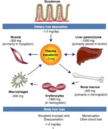
Figure 1.4 | Iron distribution in the adult human body ...5
Figure 1.5 | Iron absorption in intestinal epithelium. ...6
Figure 1.6 | Cellular iron metabolism model.. ...7
Figure 1.7 | A view of the human ceruloplasmin X-ray crystallography structure ... 14
Figure 1.8 | hCp cupredoxin-like domain 1.. ... 15
Figure 1.9 | Schematic diagram of hCp structure ... 15
Figure 1.10 | Copper centers in hCp ... 16
Figure 1.11 | Trinuclear copper centre ... 17
Figure 1.12 | Cation binding to ceruloplasmin by Fe2+ ... 17
Figure 1.13 | Oxidation reactions and products of biogenic amines. ... 20
Figure 1.14 | How X-rays interact with the atoms in a crystal... 21
Figure 1.15 | Schematic illustration of a typical crystallization phase diagram. ... 22
Figure 1.16 | Vapour-diffusion crystallization methods. ... 23
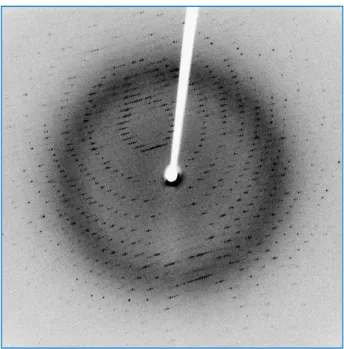
Figure 1.17 | An X-ray diffraction pattern of a crystallized enzyme. ... 24
Chapter 3: RESULTS Figure 3.1 | Work strategy diagram. ... 40
Figure 3.2 | Ceruloplasmin final size exclusion purification step. ... 41
Figure 3.3 | Normalized graphics of ligand-dependent hCp stabilization measured by DSF ... 44
Figure 3.4 | Melting temperatures for hCp in the presence of buffers listed in table 2.4... 45
Figure 3.5 | Melting temperatures for hCp in the presence of buffers listed in table 2.4 and 1 mM CuSO4.. ... 46
Figure 3.6 | Normalized graphics of best buffers for hCp stabilization measured by DSF. ... 47
Figure 3.7 | hCp blue crystals using the hanging-drop vapour diffusion method at 293K... 49
Figure 3.8 | hCp blue crystals using the hanging-drop vapour diffusion method at 293K... 50
Figure 3.10 | hCp co-crystals using the hanging-drop vapour diffusion method at 293K ... 51
Figure 3.11 | hCp blue crystals grown in some screen conditions... 56
Figure 3.12 | hCp blue crystals grown in a screen condition. ... 56
Figure 3.13 | Scaled up hit conditions and hCp blue crystals ... 57
Figure 3.14 | hCp blue crystals grown using the optiscreen at 293K. ... 57
Figure 3.15 | hCp crystals soaked with neurotransmitters. ... 59
I
ndex of Tables
Chapter 1: INTRODUCTION
Table 1.2 | Mammal cuproenzymes. ...8
Table 1.3 | hCp sequence features. ... 18
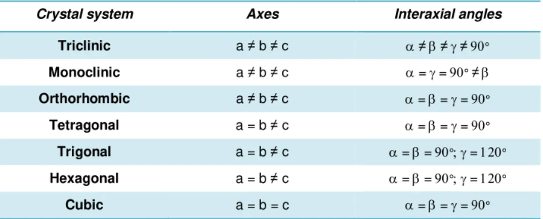
Table 1.4 | Crystal systems.. ... 23
Chapter 2: METHODOLOGIES Table 2.1 | Reagents and manufacturers ... 27
Table 2.2 | Kits, crystallization screens and manufacturers ... 27
Table 2.3 | Equipments, materials and manufacturers ... 28
Table 2.4 | Optimum solubility screen buffer (JBS solubility kit). ... 30
Table 2.5 | Crystallization screens and respective conditions used for hCP crystallization. ... 31
Table 2.6 | Crystallization condition improvement. ... 32
Table 2.7 | Crystallization condition modification and improvement. ... 34
Table 2.8 | Crystallization condition modification and improvement. ... 35
Table 2.9 | Ceruloplasmin and neurotransmitters incubations. ... 37
Table 2.10 | Crystallization screens for hCp and neurotransmitters co-crystallization. ... 37
Table 2.11 | hCp soaking experiments with neurotransmitters... 38
Chapter 3: RESULTS Table 3.1 | Metal-dependent hCp stabilization and melting temperatures. ... 43
Table 3.2 | Dymanic light scattering results for hCp-buffer assays at 20ºC. ... 48
Table 3.3 | Additive screen results on optimized crystallization condition at 293K... 52
Table 3.4 | Crystallization screen hit conditions... 55
A
bbreviations
Å – Angstrom aa – amino acid Ab – albumin
AD – Alzhemer’s disease
ADA – N-(2-Acetamido)-iminodiacetic acid APP – amyloid precursor protein
APS – ammonium persulphate ATOX1 – antioxidante protein 1 ATP – adenosine triphosphate
CAPS – 3-(cyclohexylamino)-1-propanesulfonic acid Ccs – copper chaperone for SOD
cDNA – complementary desoxiribonucleic acid CHES – 2-(cyclohexylamino)ethanesulfonic acid CNS – central nervous system
Co – cobalt
CO – carbon monoxide COX – cytochrome c oxidase
COX17 – cytochrome c oxidase copper chaperone
Ctr1 – Cu transporter 1 Cu – copper
Dcytb – duodenal cytochrome b reductase
DMT1 – divalent metal transporter 1 DNA – desoxiribonucleic acid DLS – dynamic light scattering
DSF – differential scanning fluorometry
EPPS – 4-(2-Hydroxyethyl)-1-piperazinepropanesulfonic acid EPR – electron paramagnetic resonance
GPI – glycosylphosphatidylinositol GTS – glutathione
HAH1 – human ATOX1 homologue hCp – human ceruloplasmin HFE – hemochromatosis gene HO – heme oxygenase IRE – iron responsive element
IREG1 – iron-regulated transporter 1 or ferroportin 1 IRP – iron regulatory protein
K – Kelvin degree kDa – kilo Dalton
LDL – low density protein
L-dopa – 3,4-dihydroxy-L-phenylalanine LIP – labile iron pool
MES – 2-(N-morpholino)ethane sulfonic acid MPD – m-phenylenediamine
MNK – Menkes ATPase
MOPS – 3-(N-morpholino) propanesulfonic acid MT – metallothioein
NADH – nicotinamide adenine dinucleotide PAGE – polyacrylamide gel electrophoresis PCR – polymerase chain reaction
PD – Parkinson’s disease PEG – polyethyleneglycol
PIPPS – piperazine-N,n'-Bis (3-propanesulfonic acid SOD – superoxide dismutase
Tc – transcuprein
TCEP – tris-(2-carboxyethyl) phosphine hydrochloride Tf – transferrin
UTR - untranslated region WND – Wilson ATPase
SDS – sodium dodecyl sulphate
1
. I
NTRODUCTION
Iron (Fe) and copper (Cu) are essential nutrients and its excess or deficit cause cell function fail leading to cell death. There is a metabolic linkage between these nutrients. Copper deficiency generates cellular iron deficiency, which in humans results in diminished work capacity, reduced intellectual capacity, diminished growth, alterations in bone mineralization, and diminished immune response which can be associated to several diseases and syndromes (Fox, 2003). In humans, the molecular linker between Fe and Cu was identified as being the plasma multi-copper oxidase ceruloplasmin (hCp).
1.1. Copper and Iron Metabolism
Copper and iron belong to the sub-family of transition elements that also includes zinc, nickel, cobalt, manganese and chromium. Cu has two oxidation states, cuprous (Cu1+) and cupric (Cu2+). Cu2+ is soluble, whereas Cu1+ solubility is in the sub-micromolar range. In biological systems, Cu is found mainly in the Cu2+ form, since in the presence of oxygen or other electron acceptors Cu1+ is readily oxidized to Cu2+. Cu oxidation is reversible as Cu2+ can accept an electron from strong reductants such as ascorbate and reduced glutathione (Galhardi et al., 2004).
Iron exists in two stable oxidative states, ferrous (Fe2+) and ferric (Fe3+). In aqueous media, as plasma, Fe2+ is spontaneously oxidized by molecular oxygen to Fe3+ to form Fe(OH)3. The maximal solubility of Fe in an oxidative environment such as extracellular fluids is limited by the product solu-bility constant of Fe(OH)3. At pH 7.0 the maximal solubility of Fe3+ is much lower than the maximal solubility of Fe2+. Thus, because of the low solubility of Fe in the presence of oxygen, over time or-ganisms have been forced to evolve proteins that are able to bind Fe3+ and keep it thermodynami-cally stable and, at the same time, kinetithermodynami-cally available for biological processes. In vertebrates, Fe3+ binds to the plasma protein transferrin (Tf), which has two Fe3+ binding sites, and is delivered where needed (Aisen and Listowsky, 1980).
The essentiality of iron and copper resides in their capacity to participate in one-electron ex-change reactions, but the same property that makes them essential also generates free radicals that can be deleterious to cells. Thus, a concerted regulation of cellular copper and iron levels is crucial (Arredondo and Nunez, 2005). Fe2+ e Cu1+ transform through Fenton reaction, hydrogen peroxide (H2O2), a weak oxidant, into hydroxyl radical (OH.-), one of the most strong reactive species found in nature (Messerschmidt, 1989). This process was first described in 1894 by H.J.H. Fenton. In cellular reductive environment, Fe3+ or Cu2+ are reduced to Fe2+ or Cu1+ (Haber-Weiss reaction) generating a vicious cycle of OH.- production (figure 1.1).
Figure 1.1 | Haber-Weisscicle. Fenton and Hiber-Weiss reactions.
Fenton reaction: Fe2+ + H2O2 Fe 3+
+ OH- + .OH
Haber-Weiss reaction: Fe3+ + O2
Fe2+ + O2
Fe3+ + H2O Fe2+ + H+ + .OH
Fe3+ + H2O2 Fe 2+
Copper is a cofactor for numerous enzymes and plays an important role in central nervous system development; low concentrations of copper may result in incomplete development, whereas excess copper maybe injurious. Copper is required for the function of over 30 proteins, some critical for the metabolism, including superoxide dismutase (SOD), ceruloplasmin (hCp) or cytochrome c oxidase (COX). SOD converts superoxide anions to hydrogen peroxide for further dismutation by catalase. Ceruloplasmin oxidizes Fe2+, prior to Tf binding, and its absence does not produce marked changes in copper metabolism but leads to a gradual accumulation of Fe in liver and other tissues (Harris et al., 1995; Miyajima, 2003).Cumetabolismisalteredininflammation, infection, and cancer. Iron is required as a cofactor for several essential biochemical activities, such as oxygen transport, energy metabolism and DNA synthesis. The biological functions of iron are based on its flexible coordination and its favorable redox potential which allows its association with proteins, oxy-gen binding, electron transfer and catalysis (Aisen et al., 2001). Iron is present in numerous essen-tial proteins, such as the heme-containing proteins, electron transport chain and microsomal electron transport proteins, iron–sulfur proteins and enzymes such as ribonucleotide reductase, and tyrosine hydroxylase. Animal studies have revealed the Fe has relevant roles in neuronal and immune func-tions (Beard et al., 2003). A poor diet on iron leads to irreversible alterafunc-tions of brain function related to insufficient myelination and defective establishment of the dopaminergic tracts (Agarwal, 2001; Beard et al., 2003). The immune response is also affected and macrophages exhibit reduced bacte-ricidal activity (Hallquist et al., 1992).
1.1.1. Copper metabolism and homeostasis
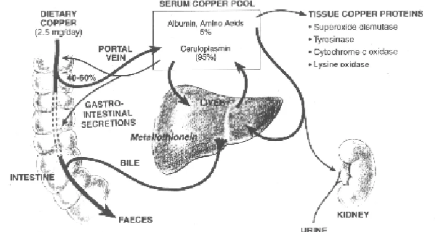
Cooper metabolism it still not fully understood in humans. An efficient Cu absorption varies depending upon the diet. Infancy represents one of the most critical periods in life in terms of Cu requirements because rapid growth increases Cu demands, whereas diets based on milk provide low amounts of the element (Lonnerdal, 1996). 40-50% Cu is absorbed by the small intestine, and transported to liver bound to specialized proteins, to be then distributed as a cofactor in cuproproteins. Excess Cu is disposed through bile and secreted into intestine in faeces (figure 1.2).
Cu regulation seems to be very thigh. Body Cu levels are the result of a balance between Cu absorption and Cu excretion by the bile but although important advances have been made in the understanding of Cu excretion, the mechanisms that govern intestinal Cu absorption remain largely a mystery. In humans, Cu is absorbed primarily by the small intestine however the mechanism by which Cu enters the cells of the intestinal mucosa and crosses into the interstitial fluid and blood is not well understood. Once in blood stream, Cu binds to two proteins, albumin (Ab) and transcuprein (Tc). In hepatocytes Cu binds ceruloplasmin during its synthesis in the trans Golgi network.
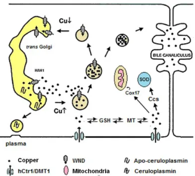
Cell Cu homeostasis, is maintained by two homologous cation transporting P-type ATPases, Menkes (MNK) and Wilson ATPases (WND) (Bull et al., 1993; Mercer et al., 1993; Tanzi et al., 1993). They have similar structures and parallel roles in regulating the Cu status of cells and tissues and in supplying Cu to secreted cuproproteins. In most cells, MNK is responsible for excreting Cu when levels become high. In hepatocytes, this role is carried out by WND, disposing excess Cu through the bile. Both proteins supply Cu to the enzymes that are secreted by the cell by pumping the metal ion into the trans Golgi network, where the metal is incorporated into the apo-ceruloplasmine. The regulation of Cu efflux by the cell is achieved by copper-induced relocalization from the trans Golgi network to the plasma membrane in the case of MNK or to an intracellular ve-sicular compartment in the case of WND (figure 1.3).
Ctr1 (Cu transporter 1), or hCtr1 in humans, promotes intestinal cells copper absorption and is also thought to facilitate the diffusion of Cu across the brush border membrane, even at low Cu con-centrations (Moller et al., 2000).Another likely Cu transporter in the brush border is DMT1(Divalent Metal Transporter 1), already described as a transporter of Fe2+, Cd2+ e Mn2+ (Arredondo et al., 2003). Several other Cu chaperone were identified. These specialized proteins carry Cu to specific intracellular sites and enzymes, preventing the presence of free Cu ions in cell. The Cu chaperone HAH1/ATOX1 (Antioxidant protein 1 homolog) carries Cu ions to the trans Golgi network where de-liver it to the P-type ATPase present there (WND in hepatocytes or MNK in enterocytes) (Hamza et al., 1999; Hung et al., 1998). Another chaperone is Ccs (Copper chaperone for SOD1) which deliv-ers Cu to the SOD in cytoplasm. Cox17 (cytochrome c oxidase copper chaperone) takes Cu to the mitochondria, where it is required for cytochrome c oxidase. Glutathione (GTS) could also play the role of Cu chaperone delivering it to cytosolic metallothioneins (MT) or to hCtr1 in the plasma mem-brane, but this pathway is not fully understood and remains to be further explored.
Body copper homeostasis is the result of a balance between Cu absorption and Cu excretion by the bile (Linder and Hazegh-Azam, 1996). In the enterocytes, Cu is absorbed by Cu transporters and kept in the cytosol, binding to MTs. Cu accumulation in the hepatic or neuronal tissue results in copper intoxication and cell damage. When there is copper deficiency the iron uptake and transport within the body is affected, and iron tends to accumulate in several tissues. Thus, copper deficiency results in an anemia similar to that observed in iron deficiency.
P-type ATPases, MNK and WND are very important in Cu homeostasis and its impaired re-sults in a severe phenotype. Wilson’s disease, associated with WND ATPase impair, leads to copper accumulation in hepatic cells. Cu is not excreted into the bile nor is incorporated into hCp, causing chronic cirrhosis during childhood. Menkes’ disease, associated with fail of MNK ATPase, is caused by deficient Cu absorption in the intestine leading to severe fail of several organs and tissues (mus-cle, kidney, lungs), bone anomalies and neuronal disorders.
Cu metabolism is also altered in inflammation, infection, and cancer, and Cu and hCp levels rise, in contrast to Fe levels, which decline in serum during infection and inflammation. Synthesis and secretion of hCp by hepatocytes is stimulated by interleukin-1 and -6 (Linder and Hazegh-Azam, 1996). Copper is essential for an efficient immune response, being necessary to the production of interleukin-2 by activated lymphocytic cells (Percival, 1998). In cancer, plasma hCp is positively cor-related with disease stage and malignant tumors have concentrations of Cu that are often higher than those of their tissue of origin (Eagon et al., 1999).
1.1.2. Iron metabolism and homeostasis
only 1 - 2 mg is absorbed. Iron body distribution is described in figure 1.4.
A significant fraction of cellular iron is associated with proteins in the form of heme, a prosthet-ic group composed of protoporphyrin IX and a Fe atom. The Fe insertion into protoporphyrin IX is catalyzed by ferrochelatase in the mitochondria. Heme is then exported to the cytosol for incorpora-tion into hemoproteins. Heme degradaincorpora-tion is catalyzed by the microsomal heme oxygenase (HO) and the free Fe2+ is reutilized (Ryter and Tyrrell, 2000). This reaction also produces carbon monox-ide (CO) and biliverdin, which is further converted into the antioxidant bilirubin. The most comum mammalian hemoproteins are hemoglobin and myoglobin, oxygen carriers in the erythrocytes or muscle cells, respectively. Other hemoproteins are oxygenases, peroxidases or nitric oxide syn-thases. Heme may also act in electron transfer reactions (such as in cytochromes a, b or c), as a substrate activator (in cytochrome oxidase, cytochrome P450, catalase) or as a nitric oxide sensor (in guanylate cyclase) (Papanikolaou and Pantopoulos, 2005).
The most abundant forms of non-heme iron in metaloproteins are iron-sulfer clusters, such as 2Fe-2S, 3Fe-4S or 4Fe-4S, that may be involved in electron transfer (e.g. the Rieske proteins in complex III of the respiratory chain), transcriptional regulation, structural stabilization or catalysis (Beinert et al., 1997). Other forms if catalytically active, protein-associated iron may include iron-oxo clusters (e.g. ribonuleotide reductase) or mononuclear iron centers (e.g. lipoxygenase and cycloxygenase).
Two thirds from absorbed iron derives from heme and the remaining fraction is inorganic. Fe absorption occurs in the apical membrane if duodenal enterocytes, but the pathway for heme uptake is not fully characterized. The mechanism for inorganic iron transport is better established. The low pH of the gastric effluent dissolves ingested inorganic iron and facilitates its enzymatic reduction to Fe2+ by the ferrireductase Dcytb (
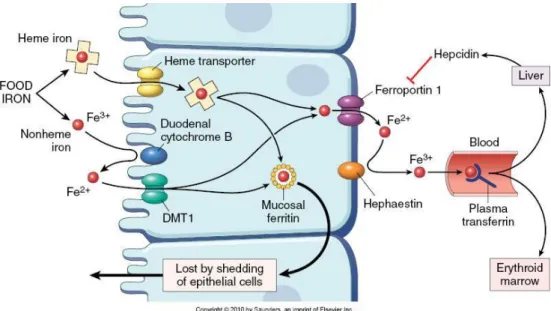
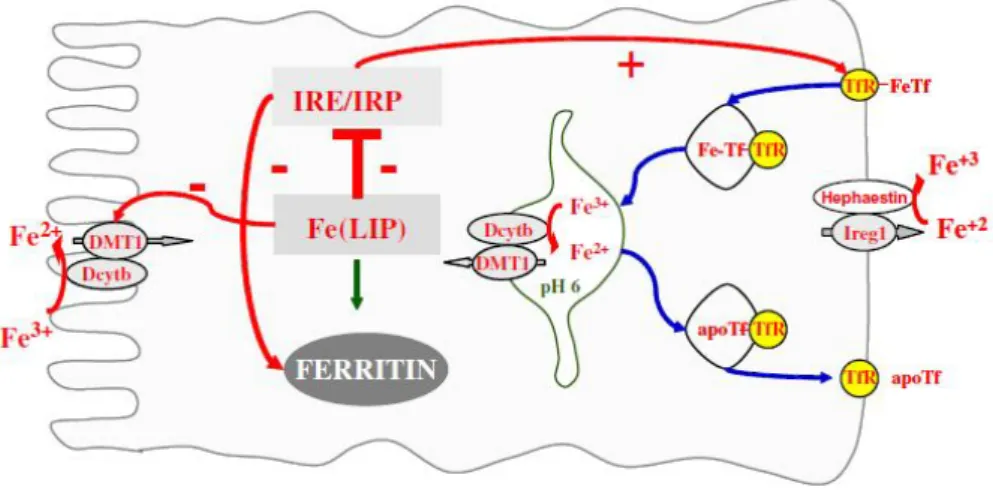
Duodenal citochrome b reductase) (McKie et al., 2001). Fe2+ is then transported across the membrane by DMT1 (Divalent Metal Transporter 1), also capable of transporting Mn2+, Cu2+, Co2+, Zn2+ or Cd2+ (Fleming et al., 1997). In the enterocytes, heme iron is liberated by HO and follows the fate of inorganic iron, it is either stored in ferritin or transported across the membrane by ferroportin 1 (also known as IREG 1)(Donovan et al., 2000), converted to Fe3+ by membrane ferroxidase hephaestin and transferred to plasma transferrin (Tf) (Figure 1.5). hCp shares homology with hephaestin and it may also act as a ferroxidase facilitating iron exporta-tion from enterocytes to Tf(Hellman and Gitlin, 2002).
Figure 1.5 | Iron absorption in intestinal epithelium. Representation of iron uptake by enterocytes.
Cell iron homeostasis is shown in figure 1.6. When in plasma Fe-Tf binds to its specific recep-tor on the cell surface, TfR1 (transferrin receptor 1) and undergoes endocytosis being transported to the endosome (Ponka et al., 1998). Fe3+ is then released from Tf by acidification of the endosome to pH 5.5, reduced to Fe2+ by Dcytb and transported across the endosomal membrane by DMT1 to cytosol. Fe is used for incorporation into iron-containing proteins and the excess is stored into ferri-tin. The mechanisms of intracellular iron transport to organelles, such as mitochondria, are still not fully understood. It seems that there is a ‘labile iron pool’ (LIP) in the cytosol, which remains bound to chelates, such as citrate, ATP, AMP or pyrophosphate (Kakhlon and Cabantchik, 2002; Petrat et al., 2002; Wang and Pantopoulos, 2011).
Figure 1.6| Cellular iron metabolism model. TfR1 and ferritin are essential for the control of cellular iron homeostasis and their expression is coordinately and reciprocally controlled in response to iron levels by a mechanism involving mRNA-protein interactions (Hentze et al., 2004; Pantopoulos, 2004). TfR1 and ferritin mRNA contain ‘iron responsive elements’ (IRE) in their 3’ or 5’ UTRs. These motifs of about 30 nucleotides fold and form a loop providing a binding site for ‘iron regulatory proteins’ (IRP), which are activated in iron-starved cells. The IRE-IRP interactions stabilize TfR1 mRNA, which contains five IRE copies, increasing TfR1 expression levels and the iron-starved cells acquire more iron from Tf. The IRE-IRP interaction in ferri-tin mRNAs inhibits their translation and iron-starved cells do not express ferriferri-tin, which under these condi-tions is not necessary. In iron-replete cells IRPs are inactive leading to TfR1 mRNA degradation and ferritin mRNA translation. This inhibits iron uptake from transferring and storage and detoxification of excess intra-cellular iron (Papanikolaou and Pantopoulos, 2005; Wang and Pantopoulos, 2011).
1.2. Cooper and Iron Disorders
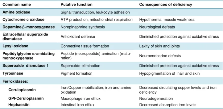
Table 1.1 | Mammal cuproenzymes.
(Adapted from (de Bie et al., 2007; Prohaska, 2011; Vashchenko and Macgillivray, 2013))
Common name Putative function Consequences of deficiency
Amine oxidase Signal transduction, leukocyte adhesion
Cytochrome c oxidase ATP production, mitochondrial respiration Hypothermia, muscle weakness Dopamine--monoxygenase Norepinephrine synthesis Neurological defeats
Extracellular superoxide
dismutase Antioxidant defense Diminished protection against oxidative stress Lysyl oxidase Connective tissue formation Laxity of skin and joints
Peptidylglycine -amidating
monooxygenase Peptide (neuropeptide) amination (matu-ration) Neuroendocrine defects
Superoxide dismutase 1 Superoxide elimination Diminished protection against oxidative stress
Tyrosinase Pigment formation Hypopigmentation of hair and skin
Ferroxidases:
Ceruloplasmin Iron/Copper mobilization; iron and amine oxidation Decreased circulating copper levels and iron deficiency
GPI-Ceruloplasmin Macrophage iron efflux Neurodegeneration
Hephaestin Intestinal iron efflux Decreased absorption iron levels
Iron is a component of several metalloproteins having a crucial role in essential metabolic pathways. Its deficit or excess can lead to complications involving organs failure, including neurolog-ical alterations, and eventually death. Different neuropathologies are involved in iron metabolism. Iron deficit may lead to cognitive failure, learning incapability and motor deterioration (Sadrzadeh and Saffari, 2004). Brain iron accumulation has been associated with Alzheimer’s disease, Parkin-son disease and other neurological disorders. Iron is responsible for the oxygen reactive species formation leading to cell death, since iron excess is related to toxicity (Halliwell, 1992; Halliwell and Gutteridge, 1985). There are also some iron homeostasis alterations that are hereditary, as is the case of hemochromatosis associated with HFE gene and its protein, which forms a complex with TfR on cell membrane in order to lower the affinity of the receptor to Tf.
1.2.2. Wilson’s disease
1.2.3. Menkes’ diseases
Menkes’ disease (OMIM#309400) is an infantile neurogenetic disorder caused by mutations in an X-linked gene, ATP7A or MNK, first described in 1962 as a copper deficiency (Menkes et al., 1962). MNK mutations result in diminished activity and thus, weak copper intestinal absorption caus-ing systemic body copper deficit. MNK gene is expressed in muscle, kidney, lung, brain but not in the liver. MNK plays a significant role in the development and maintenance of the central nervous system, which can be seen by the marked behavioral, neurological, and developmental abnormali-ties observed in Menkes’ disease patients. Children born with this genetic alteration show develop-ment delay, bone anomalies, weak and disordered hair growth, neurological alterations and multiple organs failure. These symptoms reflect the lack of activity of several cuproproteins, including hCp, until death by 3 years of age, depending upon the disease severity. Nowadays, common treatment relies on an early diagnostic and regular copper salt injections (Tumer and Moller, 2010).
1.2.4. Hemochromatosis
Hemochromatosis (HFE; OMIM#235200) is an autosomal recessive disorder and the most prominent iron-related disease in which pathologic symptoms arise from an excessive uptake of dietary iron and its deposition in many organs including the brain. The gene involved in hemochro-matosis is known as HFE and is located at chromosome 6p21.3. The function of HFE protein is to complex with the transferrin receptor on the cell membrane and to lower the affinity of the receptor for Tf (Feder et al., 1998). The homozygous mutation of the C282Y HFE is responsible for the se-vere type of this disorder (Feder et al., 1997). The iron deposition is observed in patients mainly after 40 years old, in the liver, heart, skin and pituitary (Connor, 2003). It is suggested that mutations in the hemochromatosis gene HFE that can result in increased iron accumulation in the brain might be a risk factor for AD and Parkinsonism associated with hemochromatosis (Dekker et al., 2003; Moalem et al., 2000).
1.2.5. Aceruloplasminemia
brain, which is what distinguish aceruloplasminemia from other iron disorders, such as HFE, sug-gesting an important role for hCp in brain iron homeostasis.
1.3. Ceruloplasmin
Human ceruloplasmin is a 132kDa α2-globulin from human blood serum that binds six copper ions. It contains ~95% of serum copper (Harris et al., 1998) and was first described in 1944 by Holmberg (Holmberg, 1944). hCp was latter characterized in 1948 as a cuproprotein (Holmberg and Laurell, 1948; Mittal et al., 2003). In 1952, Scheinberg and Gitlin observed a decreased concentra-tion of plasma hCp in patients with Wilson’s disease (Aldred et al., 1987; Scheinberg and Gitlin, 1952). Now it is known that is not hCp concentration, but its enzymatic activity which drops as the protein is synthesized in liver predominantly in the apo-form, as there is no copper incorporation by WND.
Soon after, the hCp was described as being related to neurodegenerative processes in hu-mans. In 1966, Friden demonstrated that hCp is a ferroxidase suggesting a role for ceruloplasmin in iron homeostasis (Osaki et al., 1966). In the 1970’s indirect indications concerning possible involve-ment of hCp in oxidation of epinephrine and serotonin in the CNS (Barrass and Coult, 1972; Barrass et al., 1972; Barrass et al., 1973; Barrass et al., 1974) were described and the protein was detected in the brain (Linder and Moor, 1977). Later the details of its synthesis in glial cells were described (Klomp and Gitlin, 1996; Mollgard et al., 1988; Patel and David, 1997; Patel et al., 2000).
In 1984, Putnam determined the complete amino acid sequence of hCp, revealing the single-chain structure of this molecule (Takahashi et al., 1984). Isolation and characterization of hCp cDNA confirmed the amino acid sequence obtained by protein chemistry and demonstrated abundant ex-pression of the ceruloplasmin gene in the liver (Koschinsky et al., 1986; Yang et al., 1986). hCp was identified as an essential protein in 1995 with the identification of patients with aceruloplasminemia (Harris et al., 1995).
It is thought that hCp plays an important role in the metabolism and development of nervous tissue (Friedenberg and Brighton, 1966), as its deficiency, its impaired function, or the failure of cop-per metabolism can lead to severe neurodegenerative diseases as Parkinson’s disease, Alzheimer’s disease or aceruloplasminemia (Castellani et al., 1999; Kaneko et al., 2002b; Loeffler et al., 1996).
1.3.1. Gene structure and expression
(Patel and David, 1997). hCp is an acute phase reactant and the serum concentration increases during processes of inflammation, infection or trauma as a result of increased gene transcription in hepatocytes mediated by the inflammatory cytokines (Gitlin, 1988).
Serum hCp is predominantly synthesized in the liver, but extrahepatic gene expression has been described in brain, spleen, lung and testis (Aldred et al., 1987; Fleming and Gitlin, 1990; Klomp et al., 1996). In the CNS, hCp is expressed in neurons and astroglial cells, e.g. cerebral microvascular network involving dopaminergic neurons in substantia nigra (Klomp and Gitlin, 1996; Kuhlow et al., 2003; Loeffler et al., 2001). In astrocytes, and Sertoli cells, hCp is membrane an-chored by glycosylphosphatidylinositol (GPI), which is caused by alternative splicing of exons 19 and 20 in hCp gene (Fortna et al., 1999; Mittal et al., 2003; Patel and David, 1997; Patel et al., 2000; Salzer et al., 1998). As a result, the latest 5 C-terminal aa found in serum hCp are replaced by a 30 aa stretch in GPI-hCp. This isoform seem to have an important role in iron oxidation and mobilization across blood brain barrier, which in cooperation with the iron exporter protein IREG1 provides the Fe efflux from astrocytes (Klibanoff et al., 1966; Trader and Frieden, 1966). The plasma hCp cannot cross the blood brain barrier. The insufficiency of hCp synthesis or its decreased activity in the cells of the CNS is regarded as one of the mechanisms underlying the development of a number of neu-rodegenerative disorders (Connor et al., 1993; Kuhlow et al., 2003; Qian and Wang, 1998; Snaedal et al., 1998; Torsdottir et al., 2001; Torsdottir et al., 1999). Both hCp isoforms are synthesized with six atoms of copper incorporated during biosynthesis in the Golgi complex (Harris, 2003; Holmberg and Laurell, 1948; Miyajima et al., 2003). Serum hCp has a half-life of 5.5 days with little or no ex-change of hCp-bound copper after synthesis. Although copper has no effect on the rate of synthesis or secretion of hCp, failure to incorporate Cu during synthesis results in apo-hCp, an unstable pro-tein with no ferroxidase activity (Kaneko et al., 2002b; Miyajima et al., 2002a; Perez-Aguilar, 2002). This apoprotein is rapidly catabolized in 5 hours (Nittis and Gitlin, 2002).
1.3.2. Metabolism
As referred, hCp is implicated in Cu and Fe metabolism. Fe is absorbed by epithelial cells in duodena and released into blood stream (Harris, 2002). hCp is involved in iron transport across the cell membrane. This protein oxidizes ferrous iron and incorporates the ferric form into apotransferrin (Kaneko et al., 2002a; Miyajima et al., 2002b). It is known that, at physiological pH, Fe2+ is autox-idized to Fe3+ with no need for a specific catalyst (Hellman et al., 2002). However, this non-enzymatic oxidation has a high probability of trigger reactions of Fenton and/or Haber-Weiss chem-istry with the production of oxygen radicals. The role of hCp might be an efficient control of the level of ferrous iron oxidation without the production of hydrogen peroxide as an end product.
copper stored into hepatocytes can generate hydroxyl radicals caused by the redox cycle between Cu1+ e Cu2+ which may cause damage in DNA, proteins, lipids and cell intoxication, leading to cir-rhosis. Cu can also accumulate in the brain causing neurodegeneration with hyperkinesis, dysarthria and dementia.
1.3.3. Role and function
Ceruloplasmin is a metalloenzyme belonging to the iron (II) oxygen oxidoreductase class, EC 1.16.3.1,and to the multicopper oxidases family, also known as blue oxidases.
In vitro studies showed that hCp is capable of catalyze the oxidation of different substrates: i) Fe2+, the substrate with the higher Vm and lower Km;
ii) a group of bifunctional amines and phenols (including biogenic amines, epinephrine and serotonin (5-hydroxy-indole family) and phenothiazines) which do not depend on iron for its activity;
iii) pseudo-substrates including several reducing agents capable of reducing Fe3+ or partially oxidize the intermediaries in ii) (Frieden and Hsieh, 1976b).
So, hCp has ferroxidase activity, oxidizing Fe2+ to Fe3+ and incorporating it into apotransferrin (Osaki et al., 1966, 1971) and has an essential role in iron metabolism (Attieh et al., 1999; Harris et al., 1995; Lahey et al., 1952; Vulpe et al., 1999). It is also an acute phase reactant having antioxi-dant properties and being capable of remove H2O2 (Giurgea et al., 2005; Gutteridge, 1978). Howev-er, studies show that this protein can also act as a prooxidant promoting LDL (low density protein) oxidation (Mukhopadhyay et al., 1996; Mukhopadhyay et al., 1997). It is known that chloride inhibits hCp ferroxidase activity at pH 5.5, but it is thought that at neutral pH chloride might enhance the oxidase activity of hCp and this enzyme might even exhibits an important role in the oxidation of several substrates present in the serum plasma (Musci et al., 1995). A group showed that such ani-ons as chloride or azide increase the aminoxidase activity of hCp and that this effect is observed with non iron substrates, while Fe2+ oxidation remains unaffected (Musci et al., 1999).
Human ceruloplasmin functions include:
a) Ferroxidase activity, eliminating free iron in the plasma, protecting blood and lipidic mem-branes from peroxidative damage (antioxidant), and/or mobilize iron from cells for transporting via Tf;
b) Aminoxidase activity for controlling the levels of biogenic amines in the plasma, spine cord and interstitial fluids;
c) Copper transport for metal distribution for extra hepatic tissues.
1.3.4. Functional mechanism
a) Type I: a single Cu2+ coordinated by 2 histdine residues and a cysteine residue, in which thiolate ion act as an electron donor, and a variable axial ligand, in a trigonal planar structure. Those proteins with type I copper center show high redox potentials (>250mV) and a strong absorption at 600nm, due to S Cu charge transfer conferring a blue color and the name ‘blue oxidases’. This copper center type has a detectable EPR (electron paramagnetic resonance) spectrum (Sakurai and Kataoka, 2007; Yoshida et al., 1995).
b) Type II: a single Cu2+ coordinated by N/NO in a tetragonal square-planar configuration. EPR parameters are detectable but those ions have no specific absorption and no blue color.
c) Typo III: two antiferromagnetically (i.e. spin pairing) coupled copper ions (Cu2+-Cu2+), each one coordinated by 3 histidine residues. EPR parameters are not detectable but those ions absorb strongly at 330nm.
Generally, multicopper oxidases are promiscuous with regards to their reducing substrates and are capable of performing various functions in different species. These oxidases catalyze the four-electron reduction of dioxygen to two molecules of water, with the concomitant reduction of one electron from the substrate. The type I Cu2+ is the primary electron acceptor. This electron cannot be passed directly to the type III center before type II Cu2+ is reduced, because the type III center acts as a 2 electrons cooperative acceptor. The reduction of the two type III Cu2+ occurs by an intramolecular transferof the 2 electrons from type I and type II reduced centers, and then to the oxygen moiety (Malmstrom, 1982). The aa sequences responsible for the copper binding are highly conserved between the multicopper oxidases, but the substrates, number of type I copper centers and the intramolecular mechanisms for electronic transfer vary between proteins (Bento et al., 2007). As exemples of this family members there is the lacase, the ascorbate oxidase, Fet3 and hephaestin, a hCp homologue needed to the iron efflux from placenta and enterocytes (Marques et al., 2012; Vashchenko and Macgillivray, 2013).
Ceruloplasmin, the only blue copper oxidase found in humans, has 3 type I cooper centers, which conferee a intense blue color to this blue oxidase, and a type II Cu2+ coordinated by 2 nitrate atoms close to the 2 antiferromagnetically coupled type III Cu2+. The type II and type III copper ions form a trinuclear Cu2+ cluster where the oxygen binds during the catalytic cycle and gets reduced to water (Miyajima et al., 1998). The hCp X-ray tridimensional structure confirmed the presence of this trinuclear cluster and the identity of the residues involved in the cooper binding and stabilization (Bento et al., 2007; Jeong and David, 2003).
1.3.5. Tridimensional structure
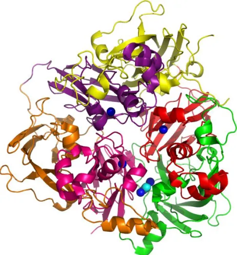
Later, the overall organization of ceruloplasmin and the nature of the 6 copper binding sites was determined by X-ray crystallography at a resolution of 3.1 Å (Zaitseva et al., 1996) and more recently at 2.8 Å (Bento et al., 2007) (figure 1.7 and table 1.2). The six cupredoxin-like domains are arranged in triangular array and each of them comprises a -barrel. Cupredoxins are single domain β-barrel proteins that bind a mononuclear type I copper redox site and are involved in inter-molecular electron transfer reactions (figure 1.8) (Roberts et al., 2002). A -barrelis a large beta-sheetthat twists and coils to form a closed structure in which the first strand is hydrogen bonded to the last. -strands in -barrels are typically arranged in anantiparallelway.
hCp has five disulphide bridges located in domains 1–5 and serve to anchor the last strand of each domain. The C-terminal strand in domain 6 is not anchored to domain 6 through a disulphide bridge. Ceruloplasmin is readily cleaved into three fragments with molecular weights of 67, 50 and 19 kDa respectively. The 19-kDa fragment corresponds almost exactly to domain 6 (Takahashi et al., 1983; Zaitseva et al., 1996). hCp shows heterogeneity with respect to its carbohydrate moieties, but has at least 3 glycan chains ending in sialic acid (Bento et al., 2007; Endo et al., 1982).
Figure 1.7 | A view of the human ceruloplasmin X-ray crystallography structure. Domains 1, 2, 3, 4, 5 and 6 in green, red, yellow, purple, orange and pink, respectively, and the locations of the metal binding sites for Cu2+ as blue spheres and O
Figure 1.8 | hCp cupredoxin-like domain 1. The trinuclear copper cluster (Cu2+ as orange spheres) and the glucan chain at N119 residue are shown (Bento et al., 2007).
1.3.5.1. Copper binding sites
As expected, three copper atoms bind in domains 2, 4 and 6 as mononuclear centers, and the remaining 3 form a trinuclear cluster between domains 1 and 6 (figure 1.9). The disposition if the trinuclear cluster and the nearest mononuclear copper, that in domain 6 is closely similar to that found in the subunit of ascorbate oxidase (Messerschmidt et al., 1989), might support the hCp oxi-dase function.
Figure 1.9 | Schematic diagram of hCp structure. Domains 1-6 and (●) copper centers
(Zaitseva et al., 1996).
1.3.5.1.1. Mononuclear type I copper centres
time. This could accomplish a more effective transfer of four electrons to the dioxygen molecule from the reducing substrate. The exact role of copper ion in domain 2 is yet not known. The type I Cu site in domain 2 differs from the others in lacking an axial methionine being replaced by an aliphatic leucine residue (Bento et al., 2007; Salzer et al., 1998). Metal soaking or organic substrate-soaking experiments showed no reducing substrates binding to domain 2 (Bento et al., 2007). Some studies showed that the type I copper in domain 2 remains permanently reduced, at least in part due to a high reduction potential, and since this site cannot be oxidized it is not catalytically relevant and is likely a vestige of gene duplication (Patel and David, 1997; Patel et al., 2000).
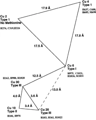
Figure 1.10 | Copper centers in hCp. Separation of the copper atoms in the trinuclear centre, the distance of this cluster from the nearest mononuclear copper in domain 6, and the separation between the mononu-clear copper atoms. Copper binding histidine residues are represented in the diagram (Zaitseva et al., 1996).
1.3.5.1.2. Trinuclear copper centre
Figure 1.11 | Trinuclear copper centre. Located between domains 1 and 6 with a water molecule attached to the type II copper and a diatomic species, assumed to be dioxygen, placed within the type III cluster (Bento et al., 2007).
1.3.5.2. Iron binding sites
Structural data indicates that the iron-binding sites seem to be in domains 4 and 6 of the hCp structure, which seems to be consistent with earlier biochemical data suggesting two iron-binding sites in the enzyme (Frieden and Hsieh, 1976a). The access to these sites is limited to relatively small substrates by three large protuberances at the top of the hCp molecule (Bento et al., 2007). It is not yet fully understood and there is no published structural data but, both Fe2+ and Fe3+ seem to bind to sites near the outside of the protein. The Fe2+ releases an electron to the nearest mononu-clear copper and then translocate to the other site with E597 and E935 in domains 4 and 6, respec-tively, playing key roles in the translocation process (figure 1.12). At this site the ferric iron would be available for collection, for example, by serum transferrin (Bento et al., 2007).
Figure 1.12 | Cation binding to ceruloplasmin by Fe2+. The red electron density is negative indicating loss
Table 1.2 | hCp sequence features (Bento et al., 2007; Lindley et al., 1997; Zaitseva et al., 1996).
Feature Positions (bp) Length (bp) Description
Molecule processing
Signal peptide 1 – 19 19 (Takahashi et al., 1984)
Ceruloplasmin chain 20 - 1065 1046
Domains
1 20 – 211 191
2 212 – 359 147
3 360 – 572 212
4 573 – 722 149
5 723 – 903 180
6 904 – 1065 161
Sites
Metal binding H120 1 Type 2 Copper 1 – Domain 1
Metal binding H122 1 Type 3 Copper 2 – Domain 1
Metal binding H180 1 Type 3 Copper 2 – Domain 1
Metal binding H182 1 Type 3 Copper 3 – Domain 1
Metal binding H295 1 Type 1 Copper 4 – Domain 2
Metal binding C338 1 Type 1 Copper 4 – Domain 2
Metal binding H343 1 Type 1 Copper 4 – Domain 2
Metal binding H656 1 Type 1 Copper 5 – Domain 4
Metal binding C699 1 Type 1 Copper 5 – Domain 4
Metal binding H704 1 Type 1 Copper 5 – Domain 4
Metal binding M709 1 Type 1 Copper 5 – Domain 4
Metal binding H994 1 Type 1 Copper 6 – Domain 6
Metal binding H997 1 Type 2 Copper 1 – Domain 6
Metal binding H999 1 Type 3 Copper 3 – Domain 6
Metal binding H1039 1 Type 3 Copper 3 – Domain 6
Metal binding M1040 1 Type 1 Copper 6 – Domain 6
Metal binding H1041 1 Type 3 Copper 2 – Domain 6
Metal binding H1045 1 Type 1 Copper 6 – Domain 6
Metal binding M1050 1 Type 1 Copper 6 – Domain 6
Amino acid modifications
Glycosylation 138 1 N-linked
Glycosylation 358 1 N-linked
Glycosylation 397 1 N-linked
Glycosylation 588 1 N-linked
Glycosylation 762 1 N-linked
Glycosylation 926 1 N-linked
Disulfide bond 174 ↔ 200
Disulfide bond 276 ↔ 357
Disulfide bond 534 ↔ 560
Disulfide bond 637 ↔ 718
1.4. Ceruloplasmin and neurodegeneration
Both GPI-anchored and secreted hCp enable IREG 1 to conduct the efflux of iron from cells of the CNS (Jeong and David, 2003). All these notions tend to indicate the important role of Cp in iron metabolism and this concept has been strengthened by some recent findings, including the one described for severe disorders of iron metabolism in aceruloplasminemia. It has been suggested that hCp catalyzed oxidation of biogenic amines, such as epinephrine, norepinephrine and serotonin, may be of importance in regulating the level of these stress hormones in the bloodstream and even-tually in those areas of the brain where they act as neurotransmitters. Thus, hCp by its effect on the lifetime of biogenic amines in the bloodstream could play an important role in the regulation of brain chemistry necessary for mental function. It is also important to understand if hCp is able to enhance the oxidation of 6-hydroxydopamine (Medda et al., 1996), an intermediate product on the way to the formation of dopamine-melanin (Chapman et al., 1970). Oxidation of 6-hydroxydopamine by hCp does not result in H2O2 production. It seems likely that Cp might be the enzyme that controls or par-ticipates in controlling catecholamine oxidation in the brain and that its deficiency in cerebral struc-tures underlies Parkinson’s disease and perhaps other neurodegenerative syndromes (Floris et al., 2000). Enhanced oxidation of dopamine by hCp has been invoked to explain the lower dopamine levels found in Parkinson’s disease.
1.4.1. Ceruloplasmin and Alzheimer’s and Parkinson’s diseases
Parkinson’s disease (PD; OMIM#168600) is a common progressive neurodegenerative motor pathology first described by James Parkinson in 1817. It is characterized by shaking,rigidity, slow-ness of movement, difficulty with walking and loss of equilibrium. Later,thinkingand behavioral problems may arise, withdementiacommonly occurring in the advanced stages of the disease (Gaggelli et al., 2006). This phenotype is associated dopaminergic neurons death in the substantia nigra and accumulation of Lewy bodies associated with mutations in -synnuclein, a protein which is expressed in CNS with uncertain function (Rasia et al., 2005). This protein binds iron and copper and aggregates leading to the production of ROS and cell death at substantia nigra which causes motor dysfunction (Rasia et al., 2005; Sadrzadeh and Saffari, 2004). In PD patients there is loss of ferritin and iron is increased in the brain and hCp activity is lowered (Jin et al., 2011). It is possible that hCp variants may be associated with iron accumulation in the substantia nigra in PD patients. The oxidative activity of hCp is involved in the oxidation of catecholamines, as dopamine and epi-nephrine, in neurons. The resulting product is neuromelanin which binds iron keeping the levels of Fe2+ low in the midbrain. In PD the dying neurons release neuromelanin with is digested by microglia and the iron is released increasing the oxidative stress. hCp might have an important role in protect-ing neurons and preventprotect-ing tissue damage and PD progression (Kristinsson et al., 2012). On the other hand enhanced oxidation of dopamine by hCp has been claimed to explain the lower dopa-mine levels found in Parkinson’s disease.
1.4.2. Ceruloplasmin and neurotransmitters
As described before, biogenic amines oxidation by hCp seems to have an important role in hormones and neurotransmitters levels regulation in brain and bloodstream. hCp can use as sub-strates epinephrine, norepinephrine, dopamine, dopa and serotonin (figure 1.13). Zaitsev and col-leagues showed that these substrates seem to bind at the same place in the hCp molecule, in do-main 6, 9.7 Å distant from the type I copper site (Zaitsev et al., 1999). Apparently, residue D1025 is one of the ligands which bind the labile copper together with H940, E935 and E272. It is proposed that the amine binds to any labile copper, an electron is transferred to the domain 6 copper causing oxidation of the substrate, and eventual reduction of oxygen at the trinuclear copper site.
1.5. X-ray crystallography
The aim of X-ray crystallography is to obtain a three dimensional molecular structure from a crystal. A purified sample at high concentration is crystallized and then the crystals are exposed to an X-ray beam. The resulting diffraction patterns are then processed, initially to yield information about the crystal packing symmetry and the size of the repeating unit that forms the crystal. This is obtained from the pattern of the diffraction spots. The intensities of the spots can be used to deter-mine the “structure factors” from which a map of the electron density can be calculated. Several methods can be used to improve the quality of the map until it is of sufficient clarity to permit the building of the molecular structure using the protein sequence. The resulting structure is then refined to fit the map more accurately and to adopt a thermodynamically favored conformation.
The x-rays were first discovered in 1895 by Wilhelm Roentgen and in 1912, Max von Laue, Walter Friedrich and Paul Knipping showed that X-rays are diffracted by crystals forming a pattern (Eckert, 2012). In 1914, William Henry Bragg and is son William Lawrence Bragg analyzed a crystal structure by means of X-rays, showing that the scattering of X-rays could be represented as a reflec-tion by successive planes of atoms within a crystal, which can be used to determine relative posi-tions of atoms within a crystal, and so the molecular structure (Bragg’s law).
X-ray crystallography is the most powerful method to obtain a macromolecular structure. Structural crystallography is based on the scattering of X-rays by the electrons in the molecules of the analyzed sample. The interference of the scattered waves can be constructive or destructive and is called diffraction. For diffraction to be observed, the wavelength () of radiation must be about equal to the distances between the atoms (0 – 5Å; 1Å = 0.1 nm) which corresponds to the X-rays wavelength. The pattern of diffracted X-rays is useful to obtain the orientation of atoms in space. This pattern can only be achieved if there is repetition of the sample macromolecule in order to am-plify and enhance the scattering of the X-radiation. This is why crystals are needed. A crystal is a solid with regular 3D ordering. The regular repeat and high similarity of the structural motifs forming the individual unit cell allows the enhancement of the scattering of X-radiation in selected direction and extinguish completely in others (figure 1.14).
Figure 1.14 | How X-rays interact with the atoms in acrystal (from wikimedia).
1.5.1. Protein crystallization
solu-tion of the sample at high concentrasolu-tion and induce it to come out of solusolu-tion in an ordered way to form a crystal (Gernert et al., 1988). However, if it happens too fast precipitation will occur. Unfortu-nately, some proteins are apparently not crystallizable. Some variables must be taken in account when trying to crystallize a protein: the precipitant, its concentration, the buffer, its pH, the tempera-ture, the protein concentration, the inclusion of additives and the crystallization technique. The inter-action of multiple variables and resulting solubility with respect to crystallization outcomes are repre-sented using a phase diagram (figure 1.15). The two dimensional space diagram is a representation of the phase of a protein regarding its concentrations and the crystallizing agent that can be the pH, the buffer, the temperature, etc. The diagram can be divided into zones defined as undersaturated, saturated, metastable, labile, and precipitation. Undersaturated solutions will remain in a liquid phase. Saturation is a solution state where a crystal, if present, would remain in equilibrium with the surrounding solution. If a crystal is in a clear metastable solution (supersaturated), the protein would add to the surface of the growing crystal until the solution reached saturation. The two zones most relevant to crystallization are the metastable and labile zones. The labile zone is highly supersatu-rated and the protein undergoes spontaneous, homogeneous nucleation. Once the nuclei form, the level of supersaturation can decrease to the metastable zone that will not produce additional crystal nuclei but will support crystal growth. The precipitation zone, which is too supersaturated to support ordered aggregation, results in amorphous precipitate (Luft et al., 2011).
Initially, the experiments start based on a trial and error procedure and based on the solubility diagram proceed until crystals appear. For this there are commercially available crystal screens which cover as wide a range as possible of the variables like the precipitant, buffer, pH and salt. These screens can be set upped using the different crystallization techniques, being the most com-mon the sitting drop vapour diffusion, hanging drop vapour diffusion and microbatch (figure 1.16). Also several techniques can be used to improve crystal size and quality, as seeding, alteration of protein concentration, or precipitant concentration, addition of additives or even alteration of the temperature. It is also important to verify if the crystals are protein crystals and not salt crystals, this can be performed by SDS-PAGE analysis, staining, UV exposure or X-ray diffraction exposure.
Figure 1.16| Vapour-diffusion crystallization methods. A, hanging drop; B, sitting drop.
1.5.2. X-ray diffraction analysis
The X-rays can be generated from accelerating electron in a synchrotron storage ring, where a single X-ray wavelength is selected by absorption of unwanted wavelengths (monochromation), or from electrons striking a copper anode in a diffractometer, where one predominant wavelength is produced. The X-rays must be focused into a beam where the crystal will be mounted in. The crys-tals can be mounted in a capillary tube at room temperature, or mounted frozen in a small loop in a stream of liquid nitrogen at 100 K (Hope, 1990). The distance from the crystal to the detector is cal-culated and adjusted to allow for collection of diffracted spots, usually up to a maximum of 1.5–3.0 Å resolution. The first diffraction image from a crystal showing the diffraction map (figure 1.17) gives us visually already some information by detecting well ordered spots. Spots beyond 3 Å are rather re-quired, since a carbon–carbon bond is approximately 1.5 Å but a resolution of close to 3 Å is suffi-cient to be able to detect the amino acid side chains in the electron density map (Smyth and Martin, 2000). It is necessary that the diffraction has sufficient resolution to make structure determination to near atomic detail possible. Then the unit cell dimensions, the crystals system and the space group must be determined. The unit cell is the smallest repeating unit that makes up the crystal. Its dimen-sions are given as three lengths: a, b, and c; and three angles: , , and (which are usually omitted if 90°). The shape of the unit cell determines the crystal system (table 1.2). The space group is pro-vided by the symmetry of the diffraction pattern, allowing the packing of the molecules into the crys-tal lattice to be determined.
Table 1.3 | Crystal systems. Adapted from (Smyth and Martin, 2000).
Crystal system Axes Interaxial angles
Triclinic a ≠ b ≠ c ≠≠≠°
Monoclinic a ≠ b ≠ c ==° ≠
Orthorhombic a ≠ b ≠ c ===° Tetragonal a = b ≠ c ===°
Trigonal a = b ≠ c ==°=° Hexagonal a = b ≠ c ==°=°