ORIGINAL ARTICLE DOI: 10.5007/1980-0037.2011v13n5p354
1 Universidade Católica de Brasília. Programa de Pós-Graduação em Educação Física, Brasília, DF. Brasil
2. Universidade Católica de Brasília. Programa de Pós-Graduação em Ciências Genômicas e Biotecnologia. Brasília, DF. Brasil
Received: 14 March 2011 Accepted: 22 May 2011
CC $ =
1 1,2 1 Romulo Maia Carlos Fonseca
Rinaldo Wellerson Pereira Nanci Maria de França
Bone mineral density and content in adolescent girls
Conteúdo e densidade mineral óssea de adolescentes
do sexo feminino
Abstract – The aim of the present study was to characterize bone mineral density (BMD) and content (BMC) in Brazilian adolescent girls according to age and pubertal stage. A total of 329 girls ranging in age from 10 to 20 years participated in this study. Body weight, height, body mass index, pubertal stage, race, daily calcium intake, and time spent per week performing moderate- to vigorous-intensity physical activity (MVPA) were evaluated. Lumbar spine and femoral neck BMD and BMC were assessed by dual-energy x-ray absorp-tiometry. One-way ANOVA with Tukey post-hoc test was used to identify differences in bone mass between ages and pubertal stages (p≤0.05). The daily calcium intake reported by the adolescents was inadequate, corresponding to only 26-47% of the recommended allowance (1,300 mg/day). On the other hand, weekly MVPA was higher than that recom-mended for adolescents. Signiicant differences in BMD and BMC were observed for girls aged 10-14 years. In addition, lumbar spine and femoral neck BMD was 58 and 31% higher in postpubertal girls, respectively, when compared to prepubertal adolescents.
Key words: Bone mineral density; Bone mineral content; Adolescents; Puberty.
Resumo – O presente estudo teve como objetivo caracterizar o conteúdo mineral ósseo (CMO) e a densidade mineral óssea (DMO) de adolescentes do sexo feminino de acordo com a faixa etária e o estágio de maturação sexual. A amostra desse estudo foi composta por 329 meninas com idades entre 10 e 20 anos. Foram avaliados o peso corporal, estatura, índice de massa corporal, estágio de maturação sexual, a raça, o consumo diário de cálcio e o tempo dispendido em atividades físicas de intensidades moderada a vigorosa por semana (AFMV). A densidade e o conteúdo mineral ósseo da coluna lombar e do colo do fêmur foram avaliados pela densito-metria óssea. As diferenças da DMO e do CMO, de acordo com a idade e a maturação sexual, foram avaliadas por uma análise de variância One-way ANOVA com o teste post-hoc de Tukey (p≤0,05). O consumo diário de cálcio reportado pelas adolescentes é inadequado, pois represen-ta uma variação de 26 a 47% do que é recomendado. Por outro lado, o tempo dispendido em AFMV, por semana, foi muito superior ao mínimo recomendado, em todas as idades. Ocorreram diferenças signiicativas tanto na DMO quanto no CMO das adolescentes no período dos 10 e 14 anos de idade. Além disso, os valores de DMO da coluna lombar e do colo do fêmur das adolescentes pós-púberes foram 58% e 31% maiores,respectivamente, quando comparados com os seus correspondentes nas adolescentes pré-púberes.
Osteoporosis is a metabolic bone disease charac-terized by a reduction in bone mineral density (BMD) and deterioration of bone microarchitec-ture, which increases skeletal fragility and the risk of fracture1. The Brazilian Health System (Sistema
Único de Saúde) spent almost R$ 81 million (US $ 46 million) with the treatment of fractures in older people in 20092. The standard method for
the diagnosis of osteoporosis is densitometry of the lumbar spine and proximal femur (femoral neck and/or total femur).
Although osteoporosis commonly affects older people, approximately 60% of the risk of developing the disease can be explained by bone mass acquisition during childhood and adolescence3, a fact that has encouraged studies
investigating the aspects of bone mass gain during this period. Actually, some factors inluencing bone mass acquisition during adolescence have already been established: genetic factors4 that can
account for 80% of the variation in BMD; age and pubertal stage5,6, with 90 to 100% of bone
mass being acquired at the end of adolescence; ethnicity3, Afro-Americans have higher BMD
than Caucasians and Asians, and lifestyle fac-tors6,7 such as daily calcium intake and physical
activity level.
However, there are many differences in lifestyle behaviors and cultural way between populations around the world and these differences can change signiicantly adolescents BMD values from a coun-try to another. For example, Lebanese adolescents had lower BMD than Canadian and American adolescents8. In Brazil, studies investigating BMD
in adolescents only started to emerge in the last decade. However, the number of publications is still very modest when compared to the international literature.
Physical itness9 and sport10 have been
identi-ied as factors associated with BMD in Brazilian adolescents. However, other factors that are also important for the study of BMD, such as age and puberty, have been little investigated in Brazilian adolescents. Only two studies evaluating these aspects were found until now. One study only inclu-ded adolescent boys11, whereas the other evaluated
adolescents of both genders ranging in age from 6 to 14 years12. Therefore, the objective of the present
study was to characterize bone mineral content (BMC) and BMD in adolescent girls according to age and pubertal stage.
Sample
The present population included sister pairs with at le-ast one girl being enrolled in a public school in Brasília, Distrito Federal. These adolescents were irst recruited to participate in a larger study that analyzed the linkage of chromosome region 1q and 11q with BMD in sister pairs. Thus, a convenience sample consisting of 329 girls ranging in age from 10 to 20 years was used. The following inclusion criteria were adopted for selection of the sample: absence of any chronic-degenerative disease, absence of a history of diseases or use of me-dications that could affect bone development, and no immobilization of body parts over a prolonged period of time during the year prior to the study.
For characterization of the sample, all adoles-cents answered questions about the regular con-sumption of cigarettes and/or alcoholic beverages and about the use of oral contraceptives. The parti-cipants or legal guardians (for adolescents younger than 18 years) signed a free informed consent form before any intervention. The study was approved by the Ethics Committee of Universidade Católica de Brasília (CEP/UCB No. 078/2006) according to Resolution 196/96 of the National Health Council.
Anthropometry and Pubertal stage
Body weight and height were measured using standard procedures. Height was measured with a Seca wall-mounted stadiometer to the nearest 0.1 cm. Body weight was measured with a Plena digital scale to the nearest 100 g. Body mass index (BMI) was calculated as body weight (kg) divided by the square of the height (m). Pubertal stages was determined by self-report of pubic hair as described by Tanner13. The adolescents were classiied as
prepubertal (Tanner I), pubertal (Tanner II and III), and postpubertal (Tanner IV and V).
Ethnic classification
Self-evaluation of ethnicity was used for sample characterization. The skin color and race classi-ication system adopted in household surveys of the Brazilian Institute of Geography and Statistics (IBGE) was used: white (in Portuguese, branco), black (preto), brown (pardo), yellow (amarelo), and Amerindian (indígena).
Estimation of daily calcium intake and time spent in moderate- to vigorous-intensity physical activity (MVPA)
Bone mass in adolescent girls Fonseca et al.
calcium intake. Calcium intake was calculated using the Diet Pro 5.1i nutrition software. MVPA was evaluated using the short version of the Inter-national Physical Activity Questionnaire (IPAQ). This instrument presents acceptable measurement properties to monitor physical activity levels in adolescents, although some limitations have been reported for younger adolescents (< 14 years)14.
Bone mineral density and bone mineral content
Lumbar spine and femoral neck BMD and BMC were measured by dual-energy x-ray absorptiometry (DXA) using a Lunar DPX-IQ device (software version 4.7e). The coeficient of variation obtained for measurements performed at the laboratory of Universidade Católica de Brasília (8 measurements were obtained from the same subject over 8 con-secutive days) ranges from 0.7% to 2.4%9 for both
BMD and BMC at all bone sites. The device is ca-librated daily and all measurements were performed and analyzed by the same technician.
Statistical analysis
First, the variables were analyzed descriptively using means and standard deviations. Skewness and kurtosis were calculated to determine whether the data were normally distributed. Calcium intake and MVPA were slightly skewed (skewness > +1.0) and they were square root modiied (√x) before being included in the subsequent analysis. The bone parameters were classiied and reported accor-ding to age and pubertal stage. One-way ANOVA
with Tukey’s post-hoc test was used to determine differences between variables according to age and pubertal stage. The SPSS for Windows (version 16) package was used for analysis of the data, adopting a level of signiicance of p≤0.05.
RESULTS
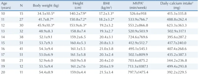
The mean and standard deviation of body wei-ght, heiwei-ght, BMI and daily calcium intake were classiied according to age and are shown in Table 1. Daily calcium intake and physical activity le-vel were positile-vely skewed. After correction, no signiicant differences in these parameters were observed between ages. None of the adolescents reported regular cigarette consumption and only four reported to regularly consume alcoholic beve-rages. In addition, 23 adolescents reported the use of oral contraceptives, but their bone parameters were similar to those not using contraceptives. The mean (± standard deviation) age at menarche was 12.2 ± 1.28 years, corresponding to 79% of the girls studied since 67 have not had their irst menstrual period. Ethnic self-identiication of the participants showed the following distribution: 32.8% (n=108) white, 7.3% (n=24) black, 56.2% (n=185) brown, 1.5% (n=5) yellow, and 2.1% (n=7) Amerindian.
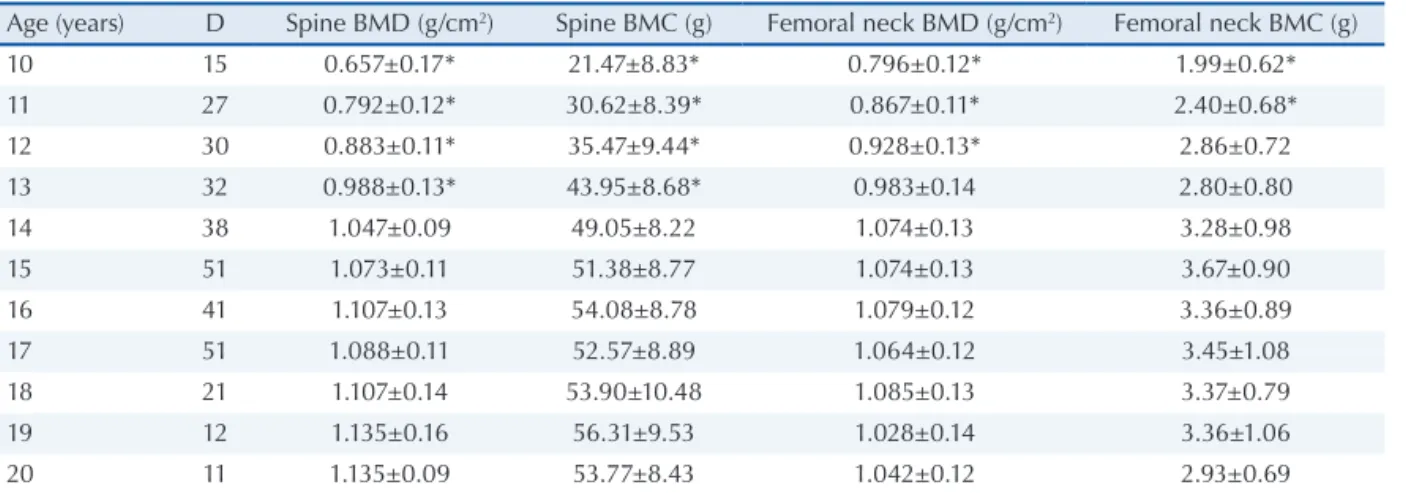
Mean BMD and BMC of the adolescents according to age and pubertal stage are shown in Tables 2 and 3, respectively. Lumbar spine and femoral neck BMD was 58% and 31% higher in postpubertal girls, respectively, when compared to prepubertal adolescents (Table 3).
Table 1. General characteristics of the sample according to age.
Age
(years) N Body weight (kg)
Height (cm)
BMI (kg/m2)
MVPA†
(min/week)
Daily calcium intake†
(mg)
10 15 34.5±10.5* 140.2±7.9* 17.2±3.3* 526.6±918 415.3±335.8
11 27 41.7±8.7* 150.8±7.2* 18.2±3.2* 533.9±766.7 488.8±262.4
12 30 45.9±10.3* 153.9±6.3* 19.2±3.2 553.2±866.8 623.3±363.3
13 32 48.9±8.3 158.8±7.4 19.3±2.7 520.9±503.9 502.9±317.1
14 38 52.1±9.1 159.2±6.5 20.4±3.1 724.6±769.6 395.6±287.2
15 51 53.7±9.3 160.4±5.3 20.8±3.3 412.9±512.7 417.7±240.0
16 41 54.3±9.4 161.1±5.5 21.0±3.8 495.1±545.1 407.4±268.6
17 51 53.0±6.9 161.5±5.8 20.3±2.4 502.1±809.9 435.2±387.3
18 21 52.9±6.0 160.9±5.8 20.4±2.0 703.6±875.2 344.2±236.8
19 12 53.5±9.4 161.2±7.6 20.6±3.9 713.3±1087.1 499.4±293.8
20 11 54.4±8.9 159.0±4.4 21.5±3.4 797.7±1475.4 392.2±229.5
Values are reported as the mean±standard deviation.
BMI: body mass index; MVPA: time (in min) spent per week in moderate/vigorous physical activities. * Significant difference compared to the other ages (p≤0.05).
DISCUSSION
In the present study, body weight and height in-creased signiicantly between 10 and 13 years of age. The mean weight and height of the sample are similar to those reported in a study of students from ive Brazilian regions15. However, comparison
with regional studies showed slightly higher values up to 14 years of age when compared to adolescents from the northeastern region16 and similar values
compared to students from the southern region17,18.
These data suggest that, although body weight and height are within the Brazilian reference range, BMD and BMC can vary between adolescents from different Brazilian regions.
The daily calcium intake observed in the pre-sent study ranged from 26% to 47% of the recom-mended for adolescents (1300 mg/day)19. On the
other hand, the time spent by the participants per-forming MVPA was higher (twice as high for some age groups) than the minimum time recommended for adolescents (300 min of MVPA per week)20.
The relationship between physical activity, calcium intake and BMD gain is still not well understood. However, BMD can increase due to an increase in
blood estrogen levels mediated by physical activity. Estrogen reduces the activity of osteoclasts, the cells responsible for bone-resorption, which leads to an increase of bone mass, and more calcium and phosphorus is then absorbed from blood to bone21.
As a consequence, inadequate calcium intake by adolescents can reduce the amount of circulating calcium in blood and thus compromise BMD gain mediated by MVPA. In this respect, since inade-quate calcium intake has been observed in diffe-rent Brazilian cities10,22, national food reeducation
programs for adolescents are necessary to increase the consumption of foods rich in calcium.
A signiicant increase of femoral neck and lumbar spine BMD was observed in adolescents between the age of 10 and 14 years, with the sta-bilization of femoral neck BMD occurring one year earlier when compared to the lumbar spine BMD curve. In addition, BMD stabilized one year after peak growth velocity and 2 years after menarche (12.2 years). Despite the cross-sectional design of the study, the present results are similar to those reported in longitudinal studies. Peak bone mass gain occurred at 13 years of age in Canadian ado-lescents, approximately one year after peak height
Age (years) D Spine BMD (g/cm2) Spine BMC (g) Femoral neck BMD (g/cm2) Femoral neck BMC (g)
10 15 0.657±0.17* 21.47±8.83* 0.796±0.12* 1.99±0.62*
11 27 0.792±0.12* 30.62±8.39* 0.867±0.11* 2.40±0.68*
12 30 0.883±0.11* 35.47±9.44* 0.928±0.13* 2.86±0.72
13 32 0.988±0.13* 43.95±8.68* 0.983±0.14 2.80±0.80
14 38 1.047±0.09 49.05±8.22 1.074±0.13 3.28±0.98
15 51 1.073±0.11 51.38±8.77 1.074±0.13 3.67±0.90
16 41 1.107±0.13 54.08±8.78 1.079±0.12 3.36±0.89
17 51 1.088±0.11 52.57±8.89 1.064±0.12 3.45±1.08
18 21 1.107±0.14 53.90±10.48 1.085±0.13 3.37±0.79
19 12 1.135±0.16 56.31±9.53 1.028±0.14 3.36±1.06
20 11 1.135±0.09 53.77±8.43 1.042±0.12 2.93±0.69
DMO: Densidade mineral óssea; CMO: Conteúdo mineral ósseo. * Diferença significativa em relação às outras idades (p≤0,05)
Table 3. Bone mineral density and content of adolescent girls according to pubertal stage.
Pubertal stage N Spine BMD† (g/cm2) Spine BMC† (g) Femoral neck BMD† (g/cm2) Femoral neck BMC† (g)
Tanner Ia 32 0.690±0.14 23.2±7.1 0.806±0.12 2.19±0.68
Tanner IIb 25 0.970±0.14 43.5±10.7 1.014±0.14 3.04±0.90
Tanner IIIb 49 0.946±0.15 41.6±11.8 0.978±0.15 2.80±0.78
Tanner IVc 86 1.074±0.13 50.7±8.9 1.063±0.14 3.35±1.08
Tanner Vc 137 1.089±0.12 52.4±8.9 1.059±0.12 3.42±0.87
Values are reported as the mean±standard deviation. BMD: bone mineral density; BMC: bone mineral content. a: prepubertal; b: pubertal; c: postpubertal.
Bone mass in adolescent girls Fonseca et al.
velocity6. In American adolescents, the increase in
total hip BMD reached a plateau at 14 years of age and in lumbar spine BMD at 15 years3. In contrast,
in Swiss adolescents the gain in lumbar spine and femoral neck BMD was only signiicant up to 14 years of age, 2 years after menarche5.
On the basis of the periods of bone mass acqui-sition reported in longitudinal studies, the period of 10 to 14 years observed in the present investigation can correspond to the time of bone mass acquisi-tion for physically active Brazilian adolescent girls but with inadequate calcium intake. However, longitudinal studies involving other populations of adolescents are needed to determine the true gain of bone mass.
The impact of puberty on bone mass acquisi-tion was demonstrated by the signiicant differen-ces in BMD between prepubertal, pubertal and postpubertal girls (Table 3). This fact is directly related to the increased production of sex hor-mones, particularly the already mentioned action of estrogen on osteoclast activity21. In addition,
considering lumbar spine and femoral neck BMD values of 1.200 (g/cm²) and 0.965 (g/cm²)23 during
peak bone mass, postpubertal adolescent girls al-ready reached approximately 90% and 109% of the expected values, respectively. Longitudinal studies also reported that 90% to 100% of peak bone mass is acquired at the end of adolescence5,6. Therefore,
studies evaluating BMD in Brazilian adolescent girls need to control pubertal stages.
Another important factor is that the gain in BMD differs between bone sites. Lumbar spine and femoral neck BMD was increased by 58% and 31%, respectively, in postpubertal adolescents when compared to the prepubertal ones. The same was reported in studies conducted on Lebanese8,
Du-tch24 and Australian25 adolescents, where lumbar
spine BMD has increased more than 60% between pre- and post-pubertal girls. This fact might be related to the effect of sex hormones, which is more pronounced in trabecular bone than in cor-tical bone8. Therefore, in addition to the control
of pubertal stages, at least two bone sites should be used for the analysis of BMD in adolescents, especially in the period which sex hormones have great changes (± 2 years of the age of menarche) since the use of only one bone site could produce equivocal conclusions.
The present study has some limitations. The self-reported race of the participants contributed to the characterization of the sample and cannot be
used for stratiication since the number of subjects in each age group by race would be disproportio-nal. However, in contrast to other countries, the classiication of the Brazilian population according to race using only phenotypic characteristics is di-ficult, mainly because of the interethnic admixed between Europeans, Africans and Amerindians, in which one individual classiied as white, according to phenotypic characteristics, can have African ancestry and another classiied as black can have European one26. There is little information about
the mechanisms by which ethnicity inluences BMD, but it is known that genes related to varia-tions in BMD are race, age and gender speciic27
and that BMD is inluenced by genetic ancestry28.
In a study evaluating BMD in Afro-American women, European genetic ancestry seen in part of the sample was negatively correlated with BMD28.
Therefore, genetic markers for ancestry should be used to identify the relationship between race and BMD in the Brazilian population.
Another limitation was the cross-sectional design of the study, in which BMD and BMC were compared between different subjects and may not represent the true variation in bone mass gain of adolescent girls. Longitudinal follow-up is needed to establish and identify the rate of BMD gain during the growth spurt.
CONCLUSIONS
The BMC and BMD of the Brazilian adolescent girls studied were characteristic of physically active individuals, but calcium intake was considered to be inadequate. Although the body weight and height of the adolescents were within the Brazilian reference range, extrapolation of the BMD and BMC results to other Brazilian regions should be done with caution.
Acknowledgments
This study was supported by Conselho Nacional de Desenvolvimento Científico e Tecnológico (CNPq) (grant MCT/CNPq - 02/2006 – Universal - 475438/2006-0) and Coordenação de Aperfei-çoamento de Pessoal de Nível Superior (CAPES)
REFERENCES
1. Pinto Neto A, Soares A, Urbanetz AA, Souza A, Ferrari A, Amaral B, et al. Consenso Brasileiro de Os-teoporose 2002. Rev Bras Reumatol 2002;42(6):343-54. 2. Ministério da Saúde. SUS gasta quase R$ 81 milhões
com fraturas em idosos em 2009. Brasil, 2010; Available from: http://portal.saude.gov.br/portal/saude/visuali-zar_texto. cfm?idtxt=33674&janela=1. [05/08/2010] 3. Bachrach LK, Hastie T, Wang MC, Narasimhan B, R
M. Bone mineral acquisition in healthy Asian, Hispa-nic, black, and Caucasian youth: a longitudinal study. J Clin Endocrinol Metab 1999;84(12):4702-12. 4. Slemenda CW, Christian JC, Williams CJ, Norton JA,
CC JJ. Genetic determinants of bone mass in adult women: a reevaluation of the twin model, and the potential importance of gene interaction on heritability estimates. J Bone Miner Res 1991;6:561-7.
5. Theintz G, Buchs B, Rizzoli R, Slosman D, Clavien H, Sizonenko PC, et al. Longitudinal monitoring of bone mass accumulation in healthy adolescents: evidence for a marked reduction after 16 years of age at the levels of lumbar spine and femoral neck in female subjects. . J Clin Endocrinol Metab 1992;5(4):1060-5.
6. Bailey DA, McKay HA, Mirwald RL, Crocker PR, Faulkner RA. A six-year longitudinal study of the relationship of physical activity to bone mineral accrual in growing children: the university of Saska-tchewan bone mineral accrual study. J Bone Miner Res 1999;14(10):1672-9.
7. Bachrach LK. Acquisition of optimal bone mass in childhood and adolescence. Trends Endocrinol Metab 2001;12(1):22-8.
8. Arabi A, Nabulsi M, Maalouf J, Choucair M, Kha-lifé H, Vieth R, et al. Bone mineral density by age, gender, pubertal stages, and socioeconomic status in healthy Lebanese children and adolescents. Bone 2004;35(5):1169-79.
9. Fonseca RMC, França NM, Van Praagh E. Rela-tionship Between Indicators of Fitness and Bone Density in Adolescent Brazilian Children. Pediatr Exerc Sci 2008;20(1):40-9.
10. Mesquita WG, Fonseca RMC, França NM. Influ-ência do voleibol na densidade mineral ossea de adolescentes do sexo feminino. Rev Bras Med Esporte 2008;14(6):500-3.
11. Silva CC, Goldberg TBL, Teixeira AS, Dalmas JC. Mi-neralização óssea em adolescentes do sexo masculino: anos críticos para a aquisição da massa óssea. J Pediatr (Rio J) 2004;80(6):461-7.
Ferraz MB, Hilario MO. Bone mineral density of the lumbar spine of Brazilian children and adolescents aged 6 to 14 years. Braz J Med Biol Res 2001;34(3):347-52. 13. Tanner J. Growth at Adolescence. Oxford, UK:
Bla-ckwell Scientiic.; 1962.
14. Guedes D, Lopes C, Guedes J. Reprodutibilidade e vali-dade do Questionário Internacional de Ativivali-dade Física em adolescentes. Rev Bras Med Esporte 2005;11:151-8. 15. Silva DAS, Pelegrini A, Petroski EL, Gaya ACA.
Comparação do crescimento de crianças e adolescentes brasileiros com curvas de referência para crescimento físico: dados do Projeto Esporte Brasil. J Pediatr (Rio J) 2010;86:115-20.
16. Silva R, Silva Júnior A, Oliveira A. Crescimento em crianças e adolescentes: um estudo comparativo. Rev bras cineantropom desempenho hum. 2005;7(1):12-20. 17. Waltrick ACA, Duarte MFS. Estudo das característi-cas antropométricaracterísti-cas de escolares de 7 a 17 anos – uma abordagem longitudinal mista e transversal. Rev Bras Cineantropom Desempenho Hum 2000;2(1):17-30. 18. Glaner M. Crescimento físico em adolescentes do norte
gaúcho e oeste catarinense. Rev Bras Cineantropom Desempenho Hum 2003;5(1):17-26.
19. National Institute Health. Optimal calcium intake. JAMA 1994;272(24):1942-8.
20. Strong WB, Malina RM, Blimkie CJ, Daniels SR, Dish-man RK, Gutin B, et al. Evidence based physical acti-vity for school-age youth. J Pediatr 2005;146(6):732-7. 21. Kemper HC. Skeletal development during childhood
and adolescence and the effects of physical activity. Pediatr Exerc Sci 2000;12:198-216.
22. Lerner BR, Lei DLM, Chaves SP, Freire RD. O Cálcio consumido por adolescentes de escolas públicas de Osasco. Rev Nutr 2000;13(1):57-63.
23. Barros ER, Kasamatsu TS, Ramalho AC, Hauache OM, Vieira JG, M. L-C. Bone mineral density in young women of the city of São Paulo, Brazil: correlation with both collagen type I alpha 1 gene polymorphism and clinical aspects. Braz J Med Biol Res 2002;35(8):885-93. 24. Van Coeverden SC, De Ridder CM, Roos JC, Van’t Hof
MA, Netelenbos JC, HA. D-VdW. Pubertal maturation characteristics and the rate of bone mass development longitudinally toward menarche. J Bone Miner Res 2001;16(4):774-81.
25. Foley S, Quinn S, Jones G. Tracking of bone mass from childhood to adolescence and factors that predict deviation from tracking. Bone 2009;44(5):752-7. 26. Parra F, Amado R, Lambertucci J, Rocha J, Antunes
C, Pena S. Color and genomic ancestry in Brazilians. Proc Natl Acad Sci USA 2003;100(1):177-82.
Bone mass in adolescent girls Fonseca et al.
28. Shaffer JR, Kammerer CM, Reich D, McDonald G, Patterson N, Goodpaster B, et al. Genetic markers for ancestry are correlated with body composition traits in older African Americans. Osteoporos Int. 2007;18(6):733-41.
Address for Correspondence
Nanci Maria de França
Universidade Católica de Brasília QS 07, Lote 1, EPCT, Sala G-119 71966-700 Águas Claras, Taguatinga DF – Brazil