Evaluation of the dental structure loss
produced during maintenance and
replacement of occlusal amalgam
restorations
Abstract: The aim of this in vitro study was to evaluate four different approaches to the decision of changing or not defective amalgam restora-tions in irst primary molar teeth concerning the loss of dental structure. Ditched amalgam restorations (n = 11) were submitted to four different treatments, as follows: Control group - polishing and inishing of the restorations were carried out; Amalgam group - the ditched amalgam restorations were replaced by new amalgam restorations; Composite res-in group - the res-initial amalgam restorations were replaced by composite resin restorations; Flowable resin group - the ditching around the amal-gam restorations was illed with lowable resin. Images of the sectioned teeth were made and the area of the cavities before and after the pro-cedures was determined by image analysis software to assess structural loss. The data were submitted to ANOVA complemented by the Student Newman Keuls test (p < 0.05). The cavities in all the groups presented signiicantly greater areas after the procedures. However, the amalgam group showed more substantial dental loss. The other three groups pre-sented no statistically signiicant difference in dental structure loss after the re-treatments. Thus, replacing ditched amalgam restorations by other similar restorations resulted in a signiicant dental structure loss while maintaining them or replacing them by resin restorations did not result in signiicant loss.
Descriptors: Dentistry, operative; Dental amalgam; Dental restoration, permanent; Dental restoration failure.
Fernanda Sardenberg(a)
Clarissa Calil Bonifácio(b)
Mariana Minatel Braga(c)
José Carlos Pettorossi Imparato(d)
Fausto Medeiros Mendes(d)
(a) MSc Student, Department of Pediatric
Dentistry, School of Dentistry, Federal University of Minas Gerais, Belo Horizonte, MG, Brazil.
(b) MSc Student, Department of Restorative
Dentistry, School of Dentistry, University of São Paulo, São Paulo, SP, Brazil.
(c) PhD Student; (d)Assistant Professors
– Department of Pediatric Dentistry, School of Dentistry, University of São Paulo, São Paulo, SP, Brazil.
Corresponding author:
Clarissa Calil Bonifácio Rua Santa Albina, 154 São Paulo - SP - Brazil Cep: 05518-000 E-mail: [email protected]
Introduction
The replacing of old restorations is still one of the procedures most frequently carried out by den-tists in clinical practice.1,2 These replacements cor-respond to around two-thirds of all operative pro-cedures carried out regularly, and this rate has not been reduced in spite of all the technological ad-vancement in materials.3
One of the major challenges of current opera-tive dentistry is the philosophy of Minimum Inter-vention. According to this philosophy, the repair of defective restorations is placed among other basic procedures, allowing the recovery of existing resto-rations, correcting possible imperfections and con-serving what is conveniently restored, without sacri-icing any remaining healthy dental structures.4
The decision criteria for replacing amalgam res-torations are subjective and dentists often encounter amalgam restorations presenting secondary or re-current caries, open contacts, body fracture, mar-ginal fractures, poor contour/anatomic form, and overhangs.5 Replacing a restoration, however, does not guarantee that the same imperfections will not occur again, nor that new lesions and/or secondary caries lesions will not affect the new restoration.4
Maryniuk, Brunson6 (1989) believe that a mar-ginal defect by itself is not a reason for replacing an amalgam restoration on the occlusal surface. Im-provement in plaque control, diet, and the correct use of luorides are indispensable for the reduction of that type of lesion, since occlusal amalgam res-torations with ditching have similar characteristics to those of pit and issure, and the essential in these cases is to control plaque accumulation.7 In addi-tion, there is research evidence that secondary car-ies occurs more frequently in the gingival third of smooth aspects and proximal areas, demonstrating that a positive correlation between defective mar-gins and secondary caries may not exist.8-10
Currently, there are other options for the effec-tive control of amalgam restoration failure, namely the repair, the re-contouring and the sealing of the restoration.2,11 These alternatives allow the replac-ing of a restoration to be delayed, they minimize the wear of dental structures and involve less clinical time and money.
Considering the options above, it is valid to in-vestigate the loss of dental structure associated to procedures of replacement and maintenance of amalgam restorations in primary molars. Thus, the aim of this in vitro study was to evaluate four dif-ferent approaches to the decision of replacing or not amalgam restorations in primary molars concerning the loss of dental structure.
Material and Methods
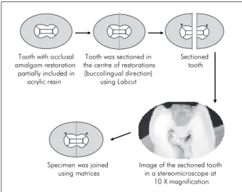
For this study, forty-four human healthy prima-ry irst molars were chosen from the Human Tooth Bank, University of São Paulo (Ethics Committee approval n. 232/04). After being stored in saline so-lution at room temperature, the teeth were cleaned using a low-speed handpiece with pumice/water slurry and rinsed with tap water.
Each tooth was partially embedded in acrylic resin, using rectangular matrices. A class I occlusal cavity was made on each of them with a diamond bur number 3101 (KG Sorensen, São Paulo, SP, Brazil) using a water-cooled high-speed handpiece. Dimensions of the cavities were 2 mm in width (in the buccolingual direction), 4 mm in length (in the mesiodistal direction), and 1 mm in depth (as mea-sured from the enamel-dentine junction). A millime-ter ruler and a K ile were used for standardization.
The restorations were made with encapsulated amalgam (Permite, SDI, Bayswater, Victoria, Aus-tralia). The procedures of insertion, condensation, burnishing and excess removal were carried out according to the manufacturer’s instructions. The sculpture procedure was eliminated to standardize the sample.
After being stored for 24 hours in saline solu-tion, one ditch around all amalgam restorations was made with round bur number 1011 (KG So-rensen, São Paulo, SP, Brasil) using a water-cooled high-speed handpiece. The diameter of the bur de-termined the ditch dimensions. All the procedures were carried out by the same operator.
restora-tions (in the buccolingual direction) using a 0.3 mm-thick diamond saw mounted on a microtome (Lab-cut 1010, Extec Co., Enield, CT, USA).
The specimens were analyzed with a stereomi-croscope at a 10 X magniication and relected light (SZPT Olympus, Tokyo, Japan), and the images of the sectioned teeth were stored in a computer. The areas of the teeth were determined by an image analysis software (Leica Qwin, Leica Microsystems, Heidelberg, Germany) on each side of the sections (in mm²). After that, the specimens were joined us-ing rectangular matrices and the teeth were ran-domly divided into four groups (n = 11).
Control group (CT): burnishing and inishing of the amalgam restorations were performed using a special bur to inish the amalgam restorations, under low-speed.
Amalgam group (AM): removal of the amalgam restorations was performed with a round bur number 1011 (KG Sorensen, São Paulo, Brazil) under water-cooling and high-speed, followed by the making of a new cavity for amalgam, ex-tending up to the ditch and the placing of a new restoration using amalgam (Permite, SDI, São Paulo, Brazil).
Resin group (RS): removal of the amalgam res-torations was performed with a round bur num-ber 1011 (KG Sorensen, São Paulo, Brazil) under water-cooling and high-speed, followed by the placing of new restorations in micro hybrid com-posite resin (Universal Amelogen – Ultradent, UT, USA), also including the ditching; the micro hybrid composite was inserted with the needle provided by the manufacturer into the cavities using an incremental technique, and each incre-ment was light-cured for 30 s.
Flowable composite resin group (FR): inishing and burnishing of the amalgam restorations were carried out with the same bur of CT, followed by the fulilling of the ditching with lowable com-posite resin (Tetric Flow – Vivadent, Schaan, Liechtenstein), inserted with the needle provided by the manufacturer.
After the treatments, the teeth were re-submitted to thermocycling and re-sectioned. New images were made at the same position as previously described
•
•
•
•
and the teeth were measured again by the same ex-aminer using the same software. The loss of dental structure in each one of the studied groups was de-termined by subtracting the area of the teeth before the procedure from the area after the procedures.
The Kolmogorov-Smirnov test was used to evalu-ate if the samples presented normal distribution. As the values were normally distributed, we used the paired Student’s t test to compare the area of each tooth before and after the procedures in each group. The loss of dental structure was evaluated by sub-tracting the tooth areas before and after the proce-dures from each other and, after that, by comparing the obtained differences among the different groups, using ANOVA complemented by the Student New-man Keuls test. The level of signiicance was set at p < 0.05.
Results
In all groups, signiicant differences were ob-served between the areas of the teeth before and after the treatments, indicating loss of dental struc-ture (Figure 1).
The means of the differences between the mea-sures of the teeth areas before and after the treat-ments are expressed in Graph 1. The difference ob-served in AM was signiicantly higher than that of the other groups (Graph 2). The other three groups did not present statistically signiicant differences concerning the loss of dental structure.
Tooth with occlusal amalgam restoration partially included in
acrylic resin
Tooth was sectioned in the centre of restorations (buccolingual direction)
using Labcut
Sectioned tooth
Specimen was joined using matrices
Image of the sectioned tooth in a stereomicroscope at
10 X magnification
* *
* *
Discussion
Currently, the procedures involving the replace-ment of restorations have exceeded the procedures involved in the restoration of new lesions.1 However, the replacement of restorations tends to cause loss of healthy dental structure1 leading to a repeated resto-ration cycle.13
Re-restoring teeth is an important component of operative dentistry, and the perceived presence of secondary caries is a major reason for undertaking it. In the absence of a diagnosis of secondary caries, a morphologic discrepancy at the margin of a restora-tion commonly provides the necessary justiicarestora-tion for
the replacement.13 The ditching in restorations by itself should not be, however, the unique criterion for replac-ing restorations.3 This imperfection may be addressed by repairing or re-contouring the restoration2, thus not requiring the replacement of the restoration.11
Although alternative options to the replacement of restorations are available, there is no consen-sus among dentists about which kind of procedure should be adopted and in which circumstances.2 For those reasons, this study had the objective of evalu-ating different procedures for the repair of amalgam restorations with marginal defects, concerning the loss of sound dental structure.
Loss of dental structure was observed in all the groups. Nonetheless, in those groups in which the restoration was maintained (CT and FR) or the amalgam restoration was replaced by a resin res-toration (RS), the loss of sound tissue was smaller (less than 1 mm²). It can be inferred that the loss of dental structure is a consequence inherent to any treatment. Therefore, dentists must avoid replacing restorations for no reason. This choice must be lim-ited to the cases where secondary caries is present.
The highest loss of healthy dental structure was observed in the group where the amalgam restora-tion with ditching was replaced by another amal-gam restoration. This restoring material requires the preparation of a retentive cavity, following the proper operative principles, leading to greater tooth wear since amalgam does not have adhesion to
den-Graph 1 - Teeth areas before and after the treatments (mean and standard deviation – vertical lines).
Graph 2 - Differences between the means of the teeth ar-eas. The vertical lines indicate the Standard Deviation.
0.00 5.00
18.96 18.62
25.72
22.68
22.78
21.97 22.94 22.20
10.00 15.00 20.00 25.00 30.00 35.00
Control Amalgam Composite Resin Flowable Resin Before After
A
reas
(mm
2)
-1.00 0.00 1.00 2.00 3.00 4.00 5.00 6.00
Control Amalgam Composite Resin Flowable Resin
A
re
as
(mm
2)
0.35 3.04
tal tissues and its retention is obtained by the design of the cavity in which it is inserted.14 Differently, the retention of composite resin is based on adhesion of the adhesive system to the dental structure.15
Dental restorative treatment plays a supporting role in the philosophy of health promotion, whose goals are maximum prevention and minimal inter-vention. The maintenance of a restoration or the sealing of its margins must be considered as treat-ment options since they are approaches that involve minor loss of dental structure and agree with the philosophy of health promotion. The procedures of inishing and burnishing or margin sealing restore dental anatomy, eliminate clinical steps and reduce the consuming of healthy dental structure, increas-ing the useful life of a restoration and minimizincreas-ing the necessity of replacement.3,16-19
The repair of old amalgam restorations in perma-nent teeth has shown clinical success rates similar to those obtained by replacing them after periods of less than 5 years.11,20 On the other hand, after 10 years, repaired amalgam restorations have shown higher in-dexes of imperfection than those of remade ones.20 For primary teeth, however, clinical studies that might corroborate the indings of this in vitro study are required in order to prove the effectiveness of the techniques that cause less loss of dental structure.
Conclusion
In conclusion, all the studied procedures involved loss of healthy dental structure, but the replacing of an old amalgam restoration by a new one made of the same material entailed greater structural loss and should thus be avoided.
References
1. Adegbembo AO, Watson PA. Removal, replacement and place-ment of amalgam restorations by Ontario dentists in 2002. J Can Dent Assoc. 2005;71(8):565.
2. Setcos JC, Khosravi R, Wilson NH, Shen C, Yang M, Mjor IA. Repair or replacement of amalgam restorations: decisions at a USA and a UK dental school. Oper Dent. 2004;29(4):392-7. 3. Anusavice KJ. Criteria for placement and replacement of
den-tal restorations. Fla Dent J. 1988;59(2):30-1.
4. Elderton RJ. Overtreatment with restorative dentistry: when to intervene? Int Dent J. 1993;43(1):17-24.
5. Penning C. Repair and revision 1. Repair or replacement of amalgam. Ned Tijdschr Tandheelkd. 2001;108(2):46-9. 6. Maryniuk GA, Brunson WD. When to replace faulty-margin
amalgam restorations: a pilot study. Gen Dent. 1989;37(6):463-7.
7. Pimenta LA, Navarro MF, Consolaro A. Secondary caries around amalgam restorations. J Prosthet Dent. 1995;74(3):219-22.
8. Kidd EAM, O’Hara JW. The caries status of occlusal amalgam restorations with marginal defects. J Dent Res. 1990;69(6):1275-7.
9. Mjor IA. Repair versus replacement of failed restorations. Int Dent J. 1993;43(5):466-72.
10. Soderholm K, Antonson DE, Fischelsschwerger W. Correlation between marginal discrepancies at the amalgam/tooth inter-face and recurrent caries. In: Anusavice KJ. Quality evaluation of dental restorations / International Symposium on Criteria for Placement and Replacement of Dental Restorations. Lake Buena Vista, Florida, October 19-21, 1987. Chicago: Quintes-sence Book; 1989. p. 97-107.
11. Gordan VV, Riley JL 3rd, Blaser PK, Mjor IA. 2-year clinical
evaluation of alternative treatments to replacement of defective amalgam restorations. Oper Dent. 2006;31(4):418-25. 12. Crim GA, Garcia-Godoy F. Microleakage: the effect of storage
and cycling duration. J Prosthet Dent. 1987;57(5):574-6. 13. Elderton RJ. Clinical studies concerning re-restoration of
teeth. Adv Dent Res. 1990;4:4-9.
14. Goldfogel MH, Smith GE, Bronberg TJ. Amalgam polishing. Oper Dent. 1976;1(4):146-50.
15. Baratieri LN, Ritter AV. Four-year clinical evaluation of pos-terior resin-based composite restorations placed using the total-etch technique. J Esthet Restor Dent. 2001;13(1):50-7. 16. Boyd MA, Richardson AS. Frequency of amalgam replacement
in general dental practice. J Can Dent Assoc. 1985;51(10):763-6.
17. Cardoso M, Baratieri LN, Ritter AV. The effect of finishing and polishing on the decision to replace existing amalgam restorations. Quintessence Int. 1999;30(6):413-8.
18. Cipriano TM, Santos JF. Clinical behavior of repaired amalgam restorations: a two-year study. J Prosthet Dent. 1995;73(1):8-11.
19. Oleinisky JC, Baratieri LN, Ritter AV, Felipe LA, de Freitas SF. Influence of finishing and polishing procedures in the decision to replace old amalgam restorations – An in vitro
study. Quintessence Int. 1996;27(12):833-40.