R E S E A R C H
Open Access
Toxoplasma gondii-skeletal muscle cells
interaction increases lipid droplet biogenesis and
positively modulates the production of IL-12,
IFN-g and PGE
2
Alessandra F Gomes
1, Kelly G Magalhães
2, Renata M Rodrigues
1, Laís de Carvalho
3, Raphael Molinaro
4,
Patrícia T Bozza
4and Helene S Barbosa
1*Abstract
Background:The interest in the mechanisms involved inToxoplasma gondiilipid acquisition has steadily increased during the past few decades, but it remains not completely understood. Here, we investigated the biogenesis and the fate of lipid droplets (LD) of skeletal muscle cells (SkMC) during their interaction withT. gondiiby confocal and electron microscopy. We also evaluated whether infected SkMC modulates the production of prostaglandin E2 (PGE2), cytokines interleukin-12 (IL-12) and interferon-gamma (INF-g), and also the cyclooxygenase-2 (COX-2) gene induction.
Methods:Primary culture of skeletal muscle cells were infected with tachyzoites ofT. gondiiand analysed by confocal microscopy for observation of LD. Ultrastructural cytochemistry was also used for lipid and sarcoplasmatic reticulum (SR) detection. Dosage of cytokines (IL-12 and INF-g) by ELISA technique and enzyme-linked immunoassay (EIA) for PGE2measurement were employed. The COX-2 gene expression analysis was performed by real time reverse transcriptase polymerase chain reaction (qRT-PCR).
Results:We demonstrated thatT. gondiiinfection of SkMC leads to increase in LD number and area in a time course dependent manner. Moreover, the ultrastructural analysis demonstrated that SR and LD are in direct contact with parasitophorous vacuole membrane (PVM), within the vacuolar matrix, around it and interacting directly with the membrane of parasite, indicating that LD are recruited and deliver their content inside the parasitophorous vacuole (PV) inT. gondii-infected SkMC. We also observed a positive modulation of the production of IL-12 and IFN-g, increase of COX-2 mRNA levels in the first hour ofT. gondii-SkMC interaction and an increase of prostaglandin E2(PGE2) synthesis from 6 h up to 48 h of infection.
Conclusions:Taken together, the close association between SR and LD with PV could represent a source of lipids as well as other nutrients for the parasite survival, and together with the increased levels of IL-12, INF-g and inflammatory indicators PGE2and COX-2 might contribute to the establishment and maintenance of chronic phase of theT. gondii infection in muscle cell.
Keywords:Toxoplasma gondii, Lipid droplets, Skeletal muscle cells, Prostaglandin-E2, Cytokines
* Correspondence:[email protected]
1Laboratório de Biologia Estrutural, Instituto Oswaldo Cruz, Fundação
Oswaldo Cruz, Rio de Janeiro, Brazil
Full list of author information is available at the end of the article
Background
T. gondiiis an obligatory intracellular protozoan parasite that resides within a PV, which fails to fuse with host or-ganelles from the endocytic pathway [1,2]. This condi-tion potentially deprives parasites of a large source of nutrients from the host endocytic and exocytic system [3]. It is known that T. gondii alters the metabolism of the host cell during the invasion and replication using host-derived nutrients in their own metabolic pathways [4], and thatT. gondiidoes not synthesize its own chol-esterol but relies mostly on host-derived lipids for their survival [5]. The mechanisms involved inT. gondii lipid acquisition are a matter of interest and are still not com-pletely understood. Some studies show the involvement of organelles such as mitochondria and mainly the endo-plasmic reticulum (ER) of host cell as suppliers of lipids, thus contributing to the increased area of vacuoles mem-brane during the development of the parasite [6]. In addition,T. gondiiinfection leads to increased receptor-mediated cholesterol endocytosis by the low-density lipoprotein (LDL) pathway [1,7].
Recent studies have proposed a dynamic role for LD in the host response to intracellular pathogens. Pathogen-induced increased LD formation has been described in bacterial, viral, fungal and parasitic infections where a role for this organelle in intracellular survival and replication of pathogens has been proposed [8,9]. Of note, a close as-sociation and/or the presence of host-cell LD in pathogen-containing vesicles has been detected in cells infected with
Mycobacterium tuberculosis[10,11],Mycobacterium bovis
BCG [12,13], Mycobacterium leprae[14],Chlamydia[15], as well as with protozoan parasitesPlasmodium falciparum
[16] andTrypanosoma cruzi[17,18], suggesting a role for LD in lipid trafficking during infection.
Structurally, the LD consists of a nucleus of cholesteryl esters and triglycerides surrounded by a single monolayer of phospholipids [19]. The regulated formation of lipid droplets, their protein and lipid content, and their associ-ation with other intracellular organelles have established LD as specialized, inducible cytoplasmic domains that function not only in lipid storage but as organelles with roles in cell signaling and activation, regulation of lipid metabolism, membrane trafficking and control of the syn-thesis and secretion of inflammatory mediators [20,21]. Accordingly, increase of LD numbers produced during in-fection is related to the generation of eicosanoids, where LDs have been shown as sites of compartmentalization of eicosanoid-forming enzymes and domains involved in the mechanisms of enhanced eicosanoid production during inflammatory and infectious conditions [12,17,22] such as prostaglandin E2 (PGE2), a product of cyclooxygenase-2 (COX-2) gene induction [23]. However, the LD formation inT. gondiiand the transference of the host cell lipids to the parasite across the parasitophorous vacuole membrane
(PVM) as well as the participation of ER for the mainten-ance of the intravacuolar parasites were not fully ad-dressed and remain uncertain.
The LDs are also described as sites of storage and synthesis of cytokines. During the past few years SkMC was identified and characterized as a cytokine-producing cell, capable of producing muscle derived cytokines, the myokines, which may participate during infection by intracellular-muscle pathogens such asT. gondii[24]. It is known that some cytokines such as interleukin-12 (IL-12), and particularly interferon gamma (IFN-g) are directly involved in cytogenesis [3], the survival rate of
T. gondii in SkMC [25] and also the integrity of muscle tissue injury [26]. So we studied the formation of LD muscle cells induced by infection withT. gondii and in-vestigated if this infection may modulate the production of IL-12 and IFN-g in this cell type. Besides, some re-searchers have discussed the importance of the host cell type as a determinant for tachyzoite to bradyzoite conversion [27,28]. It has been demonstrated that primary skeletal muscle cells trigger spontaneous T. gondii tachyzoite-to-bradyzoite conversion at higher rates than fibroblasts present in these cultures [29,30]. In the past, little attention had been given to the use of SkMC as potential host cells during the study of the toxoplasmosis, despite its well-known participation during the chronic phase of the disease [31], and its role in the route of parasite transmission via consump-tion of raw or undercooked meat containing Toxoplasma [32]. In the few last years, our group has been working with primary cultures of SkMC as an experimental model for the study of toxoplasmosis in vitro, which opens new perspectives in this field [2,27,29,30,33-36]. Since,T. gondii diverts a large variety of lipid precur-sors from host cytoplasm and efficiently manufactures them into complex lipids to its own benefit [4,37,38], we hypothesized a role for LD biogenesis duringT. gondii
infection.
In this study, we have investigated the role of LD bio-genesis and their interaction with PV, the modulation of IL-12 and IFN-g secretion as well as COX-2 gene ex-pression and PGE2synthesis, duringT. gondii-SkMC in-fection in order to better understand the survival mechanisms of Toxoplasma in muscle cells.
Methods
Primary culture of skeletal muscle cells
Skeletal muscle cell primary cultures were obtained from disaggregated thigh muscles of 18-day-old Swiss-Webster albino mouse embryos. The tissues were minced and incu-bated for 7 min with 0.05% trypsin and 0.01% versene di-luted in phosphate-buffered saline pH 7.2 (PBS). After 5–7 dissociation cycles, the enzymatic digestion was inter-rupted by adding 10% fetal bovine serum at 4°C, the sus-pension was centrifuged at 650 g for 7 min, resuspended in Dulbecco’s modified Eagle medium (DMEM) supple-mented with 10% horse serum, 5% fetal bovine serum, 2% chick embryo extract, 1mM L-glutamine, 1,000 U/ml penicillin, 50 μg/ml streptomycin and then incubated for
30 min at 37°C in a 5% CO2atmosphere. After incubation, the culture flask was gently shaken to release the non-attached cells and the supernatant enriched with myo-blasts was seeded in 0.02% gelatin-treated 24-well culture plates (for fluorescent assays) or in 35 mm-culture plates (for electron microscopy studies), respectively. The cul-tures were maintained at 37°C up to 3–5 days to obtain the muscle fibers and the fresh medium was added every two days. The main characteristics of the cells during myogenesis are: The myoblasts, which are the mononucle-ated precursor cells of muscle fibers, culture divides two to three times and then begins to aggregate and fuse into postmitotic multinucleated muscle fibers. Moreover, fibro-blast phenotype in culture is as follows: the spread exhib-ited outwards, stellate morphology of fibroblast - like cells and freshly isolated cells demonstrates that the fibroblasts were larger than the myoblasts [36].
Parasites
Tachyzoites of T. gondii, RH strain, were maintained in Swiss mice by serial intraperitoneal inoculation of 105 parasites after 48–72 h inoculation. The parasites were harvested in PBS and centrifuged (200 g for 7–10 min) at room temperature in order to discard blood cells and cellular debris. The supernatant was collected and then centrifuged again at 1000 g for 10 min. The final pellet was resuspended in DMEM and used in the parasite-host cell interaction assays.
Lipid droplet staining and counting
Muscle cells infected or not withT. gondii(parasite: host cell approximate ratio of 5:1) after 6, 24 and 48 h were fixed in 3.7% formaldehyde in HBSS (pH 7.4) and stained with osmium tetroxide, or BODIPY. For the osmium staining, the slides were rinsed in 0.1 M cacodylate buffer, incubated with 1.5% osmium tetroxide (OsO4) for 30 min, rinsed in H2O, immersed in 1.0% thiocarbohydrazide for 5 min, rinsed in 0.1 M cacodylate buffer, reincubated in 1.5% OsO4 for 3 min, rinsed in distilled water, and then dried for further analysis. The morphology of fixed cells was observed, and lipid bodies were enumerated by light
microscopy with ×100 objective lens in 50 consecutive cells in each slide. The quantitative analysis was based on 3 independent experiments performed in duplicate with at least 200 cells in each coverslip. The person responsible for counting was blinded to the codes for each slide. Slides were alternatively stained with BODIPY, evidencing the accumulation of neutral lipids in lipid droplets. For the BODIPY staining, the slides were fixed in 3.7% formal-dehyde for 10 min, washed and incubated for 15 min with the vital stain BODIPY-493/503 (4,4-difluoro-1,3,5,7, 8-pentamethyl-4-bora-3a,4a-diaza-s-indacene) or Nile red diluted in PBS, in the proportion of 1:200 and 1:1000 (v/v), respectively. The cultures stained by BODIPY were washed in PBS, incubated for 10 min with 1 μM of
To-PRO_3 iodide-642/661 in PBS to enable the visualization of the nuclei of cells, followed by 4 μg/mL
phalloidin-TRITC (binds to the actin cytoskeleton) for 1 h at 37°C for better visualization of SkMC. The coverslips were mounted over the sections with 2.5% 1.4-diazabicyclo-(2.2.2)-octane (DABCO). The samples were examined using a Zeiss photomicroscope equipped with epifluor-escence and a confocal laser scanning microscope Fluo-view 3.2 Olympus, with objective lens of 63× and of 100× (Farmanguinhos/Fiocruz).
The measurement of the area of lipid droplets was done using the BODIPY fluorescent images, obtained with an objective lens of 63× (at least four fields per slide). The images were scanned and analyzed with 2D image software (LSM Image Browser 5). The spots were determined by automatic detection, and the total area of fluorescent lipid bodies were quantified and compared to the lipid bodies of uninfected cells.
Transmission electron microscopy
SkMC were allowed to interact for 4 to 48 h at 37°C with tachyzoites of T. gondii(parasite: host cell approxi-mate ratio of 5:1). After washing in PBS, the uninfected and T. gondii-infected SkMC were immediately fixed for 30 min at 4°C in 2.5% glutaraldehyde solution (GA) in 0.1 M Na cacodylate buffer containing 3.5% sucrose and 2.5 mM Ca+2, pH 7.2. The cells were then washed in the same buffer and post-fixed for 30 min at 4°C in 1% OsO4in cacodylate buffer. After fixation, the cells were scraped gently from the plastic dish and centrifuged for 5 min at 10,000 g, dehydrated in acetone and embedded in PolyBed 812 resin. Thin sections were contrasted in uranyl acetate and lead citrate and examined in a Zeiss EM10C transmission electron microscope at the Elec-tron Microscopy Platform of Instituto Oswaldo Cruz.
Ultrastructural cytochemistry for lipid detection
addition 3.5% sucrose, pH 7.2. The cultures were washed in the same buffer for 10 min and immediately incu-bated for another 10 min in 0.1 M of imidazole buffer (CH3H4N2), pH 7.5 [39]. After washing, the cultures were post-fixed for 30 min at room temperature in 2% OsO4 diluted in imidazole buffer, pH 7.5, in the dark. After incubation, the cells were washed twice for 10 min in imidazole buffer and then processed for transmission electron microscopy. The ultrathin sections were con-trasted with lead citrate for 1 min and examined in a Zeiss EM10C transmission electron microscope.
Ultrastructural cytochemistry for sarcoplasmatic reticulum (SR) detection
After 4 and 24 h of interaction withT. gondii, the infected SkMC cultures were washed in PBS and fixed for 30 min at room temperature in 2.5% GA in 0.1 M cacodylate (pH 7.2). The cells were washed twice for 15 min in the same buffer and washed again twice for 10 min in 1% potassium iodide (KI) diluted in distilled water. The cultures were then incubated for 48 h (in the dark) at room temperature in 1% OsO4 and 1% KI, washed for 10 min in KI solution diluted in distilled water [40] and finally processed as rou-tine for transmission electron microscopy. The ultrathin unstained sections were examined in a Zeiss EM10C transmission electron microscope.
Cytokine measurement
Cell-free supernatants from muscle cell culture infected by
T. gondiiper 6, 24 and 48 h and controls were harvested and used for IL-12 and INF-g cytokine level measurements by ELISA, following the manufacturer’s recommendations for each kit (Duo Set Kit from R&D systems). The limit of detection of the assay is 40 pg/ml for IL-12 and 30 pg/ml for IFN-g.
PGE2measurement
PGE2 levels were measured directly in the supernatant from uninfected muscle cell cultures andT. gondiiinfected groups after 6, 24 and 48 h of interaction. The PGE2was assayed in the cell-free supernatant by enzyme-linked im-munoassay (EIA), according to the manufacturer’s instruc-tions (Cayman Chemical). The limit of detection of the assay is 15 pg/ml.
Reverse transcriptase polymerase chain reaction (RT-PCR) analysis
Total RNA was extracted from SkMC culture samples harvested at two different time points from experimental
T. gondiiinfection assay (after 3 h and 24 h). For this pur-pose, 106cells were harvested and washed three times in PBS and centrifuged at 10,000 g. The supernatant was completely removed and the pellet obtained was used for RNA extraction with the RNeasy kit (Qiagen California,
CA, USA), according to the manufactor’s recommen-dations. Total mRNA was measured and cDNA was synthesized using oligo(dT) and Superscript III First-Strand System (Invitrogen,cat#18080-051). Real-time PCR was performed on StepOnePlus using Taqman Gene ex-pression assay: COX-2 (Mm01307334_g1) and HPRT1 (Mm01545399_m1) obtained from Applied Biosystems. Efficiency curve showed between 88–92%. All qRT-PCR experiments were performed in duplicate, including no-template controls. The relative expression of COX-2 was determined using the 2(−ddCt)
method.
Statistical analysis
Data were reported as the mean ± S.E. and were analyzed statistically by means of analysis of variance followed by Student’sttest with the level of significance set atp≤0.05.
Results
T. gondiiinfection triggers lipid droplet biogenesis in muscle cells
In order to detect and quantify the incorporation of neu-tral lipids and LD biogenesis in uninfected andT. gondii
time-dependent increase of these structures (about 2–3 times) after 6, 24 and 48 h of interaction withT. gondii(Figure 4). At all times of interaction, in addition to increased LD numbers in observed parasitized SkMC, an increase of about 5.2 times in the LD area was also seen as compared to normal cells (p < 0.001), showing the average size of 22.14μm in uninfected SkMC and about 115.25μm in
in-fected cells.
Sarcoplasmic reticulum and LD interact inT. gondii infection
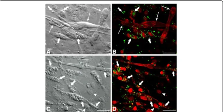
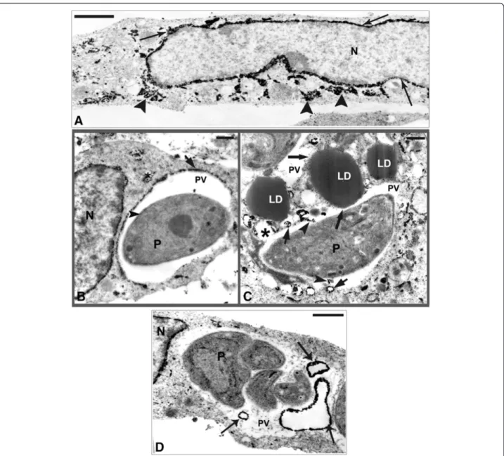
To better understand the results observed by fluores-cence analysis we also investigated the involvement of LD by electron microscopy. In this ultrathin section the ultrastructural analysis of uninfected SkMC shows no LD (Figure 5A), while the infected muscle cell after 6 h of interaction displays the VP containing one parasite and the presence of many light dense cytoplasmic struc-tures with a frequent peripheral rim of electron-dense material consistent with LD in the cell cytoplasm, around the VP and in close contact with a typical PVM (Figure 5B). Analysis after 6 h of interaction shows the conoid of the parasite (P) was in close contact with LD (Figure 6A) and also LD were in intimate contact with the PVM and the membrane of the parasite in Figure 6B and also its integration within the matrix of the vacuole (Figure 6C). After 24 h of infection several darkened LD, as revealed by the imidazole technique, could be seen in close contact with PV and some LD simultaneously associ-ated to two different PV (Figure 6D), like a bridge con-necting them. The ultrastructural cytochemistry of SkMC using the potassium iodide (KI) technique revealed the presence of tubular structures with the electron dense label distributed over the whole cytoplasm and also in the nuclear envelope (Figure 7A). After 4 h of parasite-SkMC interaction it was noted that the reaction product was lo-calized in structures resembling profiles of sarcoplasmic reticulum (SR), which surrounded the PV, dispersed in the host cell cytoplasm and located around the nucleus, as well as in the inner membrane complex of the parasite (Figure 7B). Precipitation of the reaction product for KI could also be observed inside the PV and in association with the PVM and also LD, as revealed by the imidazole technique (Figure 7C). SkMC after 24 h of parasite-host cell interaction showed cells containing two or more para-sites with reaction product for KI surrounding the nucleus of SkMC and in vesicles inside the vacuole containingT. gondii(Figure 7D).
T. gondiiinfection induces cytokine production in skeletal muscle cells
In this study we addressed whether SkMC could produce the cytokines IL-12 and INF-g during myogenesis and T. gondii infection. SkMC showed a gradual decrease in the
synthesis of IL-12, five days after cultivation. However, inT. gondiiinfected-cell cultures, after 6, 24 and 48 h of inter-action a significant increase in the production of this cyto-kine occurred as compared with control. Higher levels of IL-12 were observed in earlier periods of infection analyzed, which were reduced after 48 h of infection (Figure 8A). In-creased INF-g levels were verified in the supernatant ofT. gondii-infected SkMC in all periods analyzed (Figure 8B).
T. gondiiinfection induces eicosanoid generation in skeletal muscle cells
Lipid droplets are stores of the eicosanoid precursor arachidonic acid in different leukocyte subsets, including
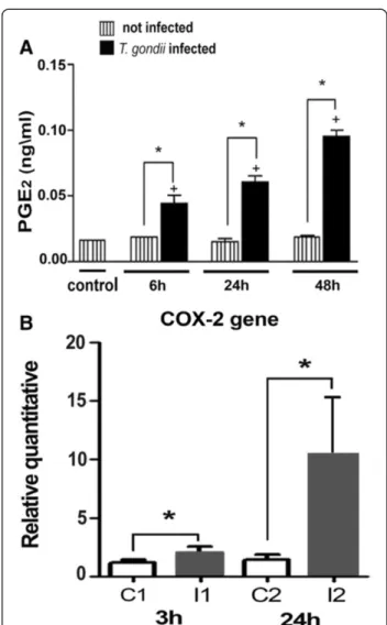
eosinophils, neutrophils, and monocytes, and contain eicosanoid-forming enzymes [12,22]. In this way, we inves-tigated whether T. gondii-infected SkMC would lead to enhanced PGE2production. PGE2quantified in the super-natant from non-infected and infected muscle cells cul-tured withT. gondiiafter 6, 24 and 48 h of interaction and measured by enzyme-linked immunoassay, showed a sig-nificant time-dependent increase in PGE2generation from 6 h up to 48 h, that parallel and positively correlated with LD formation inT. gondii-infected muscle cells but not in uninfected cells (Figure 9A). T. gondiiinfection also trig-gered a time-dependent increase of COX-2 expression (Figure 9B).
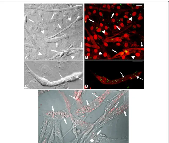
Figure 2Confocal Laser Scanning Microscopy showing LD revealed by BODIPY in uninfected andT. gondii-infected SkMC for different periods of interaction. (A)Control - interferential microscopy revealing the profile of uninfected culture with the presence of myoblasts (double arrows), multinucleated myotubes (arrows) and fibroblasts (arrowhead).(B)Merge showing double marking for actin cytoskeleton and nuclei SkMC (arrows) in red and LD in green. Note discrete distribution of LD across culture.(C)Interferential microscopy showing parasites in
T. gondii-infected SkMC (thick arrows) after 6 h of the interaction.(D)Merge showing actin cytoskeleton and nuclei in red and LD in green in
Discussion
It is known that T. gondii mobilizes lipids resources from the host cells during invasion and its intracellular cycle [4], and although the parasite does not synthesize its own cholesterol it has evolved strategies to divert host cell lipid metabolism to favor its survival [1,7]. Our results suggest that T. gondii-infected SkMC increases
the synthesis of LD as well as their content, providing lipids to the parasite and thus contributing to the growth and maturation of the parasitophorous vacuole, as de-scribed in another cellular model by Caffaro et al.[38]. Initially using the fluorescent dye Nile Red, it was pos-sible to observe the increased formation of LD within the first hour ofT. gondii-SkMC interaction and also the presence of LDs next to the parasite, and at 24 h around the PV.
Our quantitative data showed that T. gondii infection triggers biogenesis of LD within muscle cells. The analysis by light and fluorescence microscopy ofT. gondii-infected SkMC stained with osmium tetroxide or BODIPY clearly showed an increase in the number of LD after 6, 24 and 48 h of the interaction. We suggest that Toxoplasma may be interfering with the lipid metabolism of the host cell stimulating its synthesis. Previous studies have shown se-questration of some phospholipids by Toxoplasma infec-tion whilst in the host cell in order to construct more complex lipids [4,41]. In our experiments it was observed that inT. gondii-infected SkMC, significant increase in the formation of the LD occurs at all times of interaction: 6, 24 and 48 h. It is known that muscle cells may accumulate phospholipids, triacylglycerol and cholesterol in LD, which could have roles in the regulation of cell cycle, migration and myogenesis by activation of proteolytic systems such as the calpain system [42,43]. Thus, we do not discard the
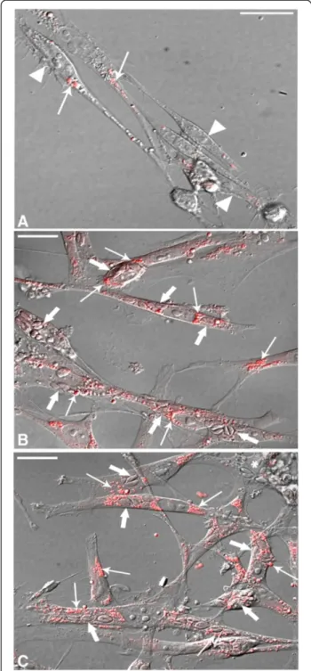
Figure 3Confocal Laser Scanning Microscopy analysis showing LD revealed by BODIPY inT. gondii-infected SkMC after 24 and 48 h of interaction. (A)Image by interferential microscopy showingT. gondii-infected SkMC after 24 h. Note the presence of infected myotubes (thick arrows) and uninfected (thin arrows) in the same culture.(B)Uninfected myotubes (thin arrows) practically does not present LD while cells infected withT. gondii(thick arrows) have numerous LD in green.(C)Interferential microscopy showingT. gondii-infected SkMC after 48 h. Note the presence of infected cells (thick arrows).(D)Double stainingT. gondii-infected SkMC in red and LD in green (arrows). Note the major concentration of LD mainly in infected SkMC. Fibroblasts in culture present few LD infected or not (arrowhead). All bars = 20μm.
Figure 4Biogenesis of LD in SkMC analyzed by osmium stain. Listed columns represent the profile of LD in SkMC control with preservation synthesis during the development of culture. The black columns representT. gondii-infected SkMC. The time-dependent increase of lipid droplets was observed after 6, 24 and 48 h of
hypothesis that the recruitment of LD by the parasite dur-ing its replication, could lead to an increase of LD synthe-sis by the host cell to maintain homeostasynthe-sis of their vital activities. The homeostasis is maintained by a balance be-tween the cholesterol internalized via the LDL receptor and synthesis involving the enzyme: 3-hydroxy-3-methyl-glutaryl-coenzyme A reductase (HMG-CoA) [44]. In cells infected byT. gondii, there is an increase in the synthesis of receptors for internalization of LDL [1], and the activity of HMG-CoA is four times higher [45].
Our ultrastructural cytochemistry analysis demon-strated, for the first time, the direct contact of LD with the vacuolar membrane, its matrix as well as with the parasite membrane during its segregation inside the PV. Studies show the involvement of ER from the host cell in the bio-genesis of LD [21]. This supplement of lipids could con-tribute to the increase of the vacuole membrane area during the intracellular development of T. gondii [6,38]. LD structure and composition, as determined in different cell types and conditions, consists of cholesteryl esters and triglycerides surrounded by a single monolayer of phos-pholipids and contains a variable array of proteins [19,21]. We believe that this recruitment of LD byT. gondii may
be involved in a survival strategy to surmount the defi-ciency of cholesterol and other lipids by the parasite.
The mechanism by which host-cell-derived lipids are transferred across the PVM to the parasite is uncertain [4]. In addition to LD recruitment, our data clearly dem-onstrated: (i) the discharge of the SR to the interior of the PV after 4 h ofT. gondii-SkMC infection; (ii) the pres-ence of vesicles with different diameters and morphology
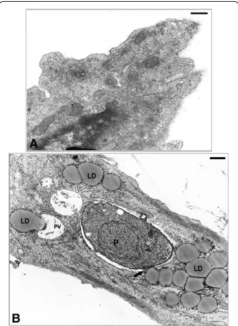
Figure 5Ultrastructure of SkMC in cultures. (A)Uninfected cells without LD.(B)Note the large amount of lipid droplets during the first 6 h ofT. gondii-SkMC interaction. All bars = 10μm.
containing the reaction product for KI localized inside the vacuole after 24 h of parasite-host cell infection. These results are similar to that described by Puzianowska-Kuznicka and Kuznicki [46] who observed by immunoe-lectron microscopy the transfer of SR components into the PV, indicating that the fusion occurs between the two compartments and, (iii) the accentuated decrease of the SR demarcation around the PV, after 24 h of infection. This data suggests that components of the SR can be
incorporated by the intracellular parasites, constituting a source of nutrients and lipid possibly for its development, as proposed previously [4,46].
The ER plays a crucial role in cytoplasmic signaling in a variety of cells. It is particularly relevant to SkMC, where this organelle constitutes the main Ca2+ store for essential functions, such as contraction [47,48]. Our re-sults by electron microscopy showed total reorganization of the SR in T. gondii-infected SkMC. Thus, we believe
that this phenomenon may lead to changes in Ca2+ homeostasis compromising the functionality of SkMC. The importance of juxtapositioning of SR, mitochondria and transverse tubules (T-tubules) in muscle cell for better communication between sites of Ca2+ release which en-sures the contraction of myofibrils are described. There-fore, we suggest thatT. gondiimay be benefiting from the repositioning of the SR sequestered, not only lipids, but also from the Ca2+not used by sarcomeres [49].
The accumulation of the LD within leukocytes in in-flammatory conditions for example: bacterial sepsis, aller-gic lung inflammation, arthritis, and in mycobacterium infections among others has recently been reported [21]. However, mechanisms that regulate LD formation and its functional significance to the cellular biology in T. gondii
infection are not known.
Increased PGE2production in pathological conditions is largely regulated by COX-2 gene induction [23]. By using
qRT-PCR the expression of COX-2 gene was analyzed, showing that after 3 and 24 h of T. gondii-SkMC inter-action, the COX-2 expression was up regulated. Studies have demonstrated that the promoter region of COX-2 has several potential regulatory elements, which can affect gene transcription [50]. In pancreatic beta-cells several transcription factors regulate COX-2 gene expression as for example, the signal transducer and activator of tran-scription 1 (STAT1) that plays a negative role on COX-2 promoter [23]. It was described thatT. gondiican also ma-nipulate the host transcription factors, including inhibiting STAT1 [3]. Thus, we suggest that the presence ofT. gondii Figure 8The graphics show the synthesis of the cytokine
interleukin-12 (IL-12) and interferon-g (IFN-g) inT.
gondii-infected SkMC. (A)SkMC control represented by the columns listed. Note the decreased synthesis of IL-12 during the development of culture. The dark columns represent infection, showing increased synthesis of IL-12 at all times of interaction.(B)Synthesis of the IFN-g inT. gondii-SkMC infected after 6, 24 and 48 h. SkMC control represented by the columns listed with a small increase in the synthesis of this cytokine during myogenesis. The dark columns represent infected cultures demonstrating the maintenance of INF-g synthesis at all times of interaction. t-test: *P≤0,05.
Figure 9PGE2generation and COX-2 gene expression inT. gondii-infected SkMC. (A)Synthesis of PGE2by EIA. The listed
columns represent SkMC control with approximate values in all points. The black columns show infected cultures where there is a time-dependent increase in the production of PGE2.(B)Representation
in SkMC may be inducing the increased COX-2 expres-sion, but whether or not the inhibition of STAT1 also oc-curs in this process still remains uncertain.
Several studies have demonstrated that an increase of LD numbers produced during infections by different pathogens is related to the increased generation of eicos-anoids due to the compartmentalization of the substrate arachidonic acid and the eicosanoid-forming enzymes within newly formed LDs and LDs are major sites of ei-cosanoid production in different inflammatory condi-tions [12,17,18,21,22]. Our results show an increase of PGE2synthesis, from 6 h up to 48 h ofT. gondii-SkMC infection. Of note, theT. gondii-induced increased PGE2 generation occurs in parallel and positively correlates to the increased formation of LDs, suggesting that LDs may have roles in the heightened eicosanoid production during T. gondii infection. However, future studies will be necessary to confirm the involvement of LDs in the PGE2 production triggered by T. gondii. Among other factors, we believe that the success of the infection ofT. gondii in the muscle tissue may be related to the in-creased COX-2 expression, compartmentalization within LD and consequently enhanced production of the eicos-anoid PGE2. In other cell types, studies showed that the enzymatic conversion of free arachidonic acid into pros-taglandin down-modulates the cell-mediated response favoring not only intracellular pathogens, but also the survival of the host [51,52]. Indeed, high concentrations of PGE2potently inhibit the Th1 type response, tumor ne-crosis factor (TNF) and nitric oxide (NO) production, and these changes favor intracellular parasite growth [53,54].
It is worth remembering that SkMC is not a cell of the immune system. The levels of inflammatory markers (PGE2and COX-2) observed in SkMC in our assays are considered quantitatively lower when compared to levels produced by macrophages, despite their increase during Toxoplasma infection [52]. We believe that a moderate SkMC immune response may suppress the replication of parasites favoring the bradyzoite conversion, while a strong immune response may change the cell cycle pro-gression parasite or act as a microbicide activator [55]. These data support our hypothesis that the recruitment of LD byT. gondii, together with the cellular response, may possibly be related to the development of the chronic phase in SkMC.
Accordingly, intracellular pathogens-induced increased LD formation during intracellular infection, including T. cruzi,M. bovisBCG andM. lepraefavouring intracellular pathogen survival through mechanisms which involve LD-derived eicosanoid formation and LD recruitment towards the phagosome [12,14,17]. Similarly, we describe here that the enhanced capacity of muscle cells to generate PGE2in the course of the T. gondii infection correlates to the increased LD formation. And so, the recruitment of
organelles such as LD and SR by the parasite during its host cell interaction, may contribute to the mechanisms that intracellular pathogens have evolved to survive in host cells. Future studies are necessary to characterize the regu-lation and function of the prostaglandins in SkMC and to understand if the presence of SR in PV may be acting as a source of Ca2+that facilitates the preference of muscle tis-sue in the development phase of chronic toxoplasmosis.
In our experiments we observed that SkMC is capable of producing levels of the cytokines IL-12 and IFN-g with a significant increase in their synthesis after 6, 24 and 48 h of interaction with T. gondii. Our results cor-roborate with studies that show the muscle as a cytokine producer by myokines [56]. IL-6 was the first cytokine to be discovered being produced by muscle cells; how-ever, skeletal muscles may produce and express different cytokines families [24,25]. Some authors describe that concerning the synthesis of IFN-g, T. gondii-infected SkMC are able to develop a strong anti-parasitic response, reducing significantly the growth of the parasite [25]. Experiments using mice lacking IFN-g [57] and IL-12 showed absence of appropriate immunity, which rapidly leads to host death [58]. It is most advantageous for the Toxoplasma to keep its host alive until transmission to another host through oral transmission of tissue cysts.
IFN-g is known to induce an inflammatory response and control of parasite load during the early stages of in-fection [59]. During Toxoplasma inin-fection, the host im-mune response is dependent on IFN-g induced by IL-12 production in a variety of cell types. In our study, in the course of myogenesis of SkMC the levels of IFN-g did not change, whereas the concentration of these cytokines in-creased in all times of interaction withT. gondiianalyzed. However, in other cellular models such as macrophages, neutrophils and especially dendritic cells, Langet al. [60] described that T. gondii inhibits production of IL-12. Nickdel et al. [61] showed that during the early stage of oral infection withT. gondiian increase in small-intestine pathology occurred, in addition a reduction in the levels of plasma IL-12 and IFN-g levels was observed. Moreover, Matowicka-Karna et al. [62] studying a group of patients infected with T. gondii, also noted a decrease in IL-12 levels. We believe that the increase in cytokines IFN-g and IL-12 in SkMC-T. gondii infected cells may be related to manipulation of transcription factors of the host by Toxoplasma early on the infection as shown by our results after 3 hours of interaction. Moreover, the increased syn-thesis of IFN-g [25] and IL-12 may be act by reducing the multiplication process of tachyzoites forms (acute phase) favoring, the differentiation to bradyzoites forms, found in tissue cysts (chronic phase).
thus increasing the synthesis of a cytokine that is in-volved in the repair and homeostasis of the host cell and may act as a strategy that favorsT. gondii maintenance. So, Toxoplasma has evolved to exploit own molecules and cellular response of the host, providing a favorable environment for the establishment of chronic infection in SkMC.
Finally, SkMC can be used as an important cellular model for studies on the molecular mechanisms in re-sponse to parasitism byT. gondii, mainly considering its importance as a target cell for encystment and its role in the transmission of the parasite.
Conclusions
In conclusion, our data demonstrated thatT. gondii infec-tion in muscle cells causes a pronounced effect on host-cell lipid metabolism through regulation of LD biogenesis and recruitment of these organelles to PV. The increased LD formation may potentially act as source of prostaglan-din production with implications to the host immune re-sponse and could represent a source of lipids and other nutrients for parasite survival. Thus, the increase the LD, followed by expression of COX-2 and PGE2in the SkMC may be contributing to the control of the synthesis of IL-12 and IFN-g during infection by T. gondii. We believe that the increase of these cytokines involved in the repair, and homeostasis of muscle cells after injury, might con-tribute to the establishment and maintenance of the chro-nic phase ofT. gondiiinfection in SkMC.
Competing interests
The authors declare that they have no competing interests.
Authors’contributions
HSB conceived, participated in the design and coordination of the study and had the general supervision and complete overview of the project. AFG co-conceived the study, carried out most of the experimental work, including the processing of samples and the final illustrations for the manuscript, analyzed data and drafted the manuscript, as part of her PhD thesis. RM participated in the electron microscopy assays. LC participated in the design of the study. RMR carried out the molecular assays. KGM and PTB made substantial contributions to data acquisition, analysis and participated in the revision of the manuscript. All authors analyzed the data and read and approved the final version of the manuscript.
Acknowledgments
The authors thank Carlos Alberto Bizarro Rodrigues from Farmanguinhos/ Fiocruz for the production of interferential microscopy images for the aid with confocal microscopy. We are grateful to Sandra Maria de Oliveira Souza and Genesio Lopes de Faria for technical assistance. This work was supported by grants from Conselho Nacional de Desenvolvimento Científico e Tecnológico (CNPq), Fundação Carlos Chagas Filho de Amparo à Pesquisa do Estado do Rio de Janeiro (FAPERJ), Universidade do Estado do Rio de Janeiro (UERJ), Fundação Oswaldo Cruz (Programa Estratégico de Apoio à Pesquisa em Saúde - PAPES VI), Pronex - Programa de Apoio a Núcleos de Excelência–CNPq/FAPERJ and Instituto Oswaldo Cruz/Fiocruz.
Author details
1
Laboratório de Biologia Estrutural, Instituto Oswaldo Cruz, Fundação Oswaldo Cruz, Rio de Janeiro, Brazil.2Laboratório de Imunologia e
Inflamação, Universidade de Brasília, Brasília, Brazil.3Laboratório Cultura de Células, Depto. Histologia e Embriologia, Instituto de Biologia, Universidade
do Estado do Rio de Janeiro, Rio de Janeiro, Brazil.4Laboratório de
Imunofarmacologia, Instituto Oswaldo Cruz, Fundação Oswaldo Cruz, Rio de Janeiro, Brazil.
Received: 2 December 2013 Accepted: 21 January 2014 Published: 23 January 2014
References
1. Coppens I, Sinai AP, Joiner KA:Toxoplasma gondiiexploits host low-density lipoprotein receptor-mediated endocytosis for cholesterol acquisition.J Cell Biol2000,149:167–180.
2. Andrade EF, Stumbo AC, Monteiro-Leal LH, Carvalho L, Barbosa HS:Do microtubules around theToxoplasma gondii-containing parasitophorous vacuole in skeletal muscle cells form a barrier for the phagolysosomal fusion?J Submicrosc Cytol Pathol2001,33:337–341.
3. Laliberté J, Carruthers VB:Host cell manipulation by the human pathogen
Toxoplasma gondii.Cell Mol Life Sci2008,65:1900–1915.
4. Charron AJ, Sibley LD:Host cells: mobilizable lipid resources for the intracellular parasiteToxoplasma gondii.J Cell Sci2002,115:3049–3059. 5. Nishikawa Y, Ibrahim HM, Kameyama K, Shiga I, Hiasa J, Xuan X:Host
cholesterol synthesis contributes to growth of intracellularToxoplasma gondiiin macrophages.J Vet Med Sci2011,73:633–639.
6. Sinai AP:Biogenesis of and activities at theToxoplasma gondii
parasitophorous vacuole membrane.Subcell Biochem2008,47:155–164. 7. Portugal LR, Fernandes LR, Pietra Pedroso VS, Santiago HC, Gazzinelli RT,
Alvarez-Leite JI:Influence of low-density lipoprotein (LDL) receptor on lipid composition inflammation and parasitism duringToxoplasma gondii
infection.Microbes Infect2008,10:276–284.
8. D’Avila H, Maya-Monteiro CM, Bozza PT:Lipid bodies in innate immune response to bacterial and parasite infections.Int Immunopharmacol2008, 8:1308–1315.
9. van der Meer-Janssen YP, van Galen J, Batenburg JJ, Helms JB:Lipids in host-pathogen interactions: pathogens exploit the complexity of the host cell lipidome.Prog Lipid Res2010,49:1–26.
10. Russell DG, Cardona PJ, Kim MJ, Allain S, Altare F:Foamy macrophages and the progression of the human tuberculosis granuloma.Nat Immunol
2009,10:943–948.
11. Cáceres N, Tapia G, Ojanguren I, Altare F, Gil O, Pinto S, Vilaplana C, Cardona PJ:Evolution of foamy macrophages in the pulmonary granulomas of experimental tuberculosis models.Tuberculosis2009,89:175–182. 12. D’Avila H, Melo RC, Parreira GG, Werneck-Barroso E, Castro-Faria-Neto HC,
Bozza PT:Mycobacterium bovis bacillus Calmette-Guérin induces TLR2-mediated formation of lipid bodies: intracellular domains for eicosanoid synthesis in vivo.J Immunol2006,176:3087–3097. 13. Almeida PE, Silva AR, Maya-Monteiro CM, Töröcsik D, D’Avila H, Dezsö B,
Magalhães KG, Castro-Faria-Neto HC, Nagy L, Bozza PT:Mycobacterium bovis
bacillus Calmette-Guérin infection induces TLR2-dependent peroxisome proliferator-activated receptor gamma expression and activation: functions in inflammation, lipid metabolism, and pathogenesis.
J Immunol2009,183:1337–1345.
14. Mattos KA, Lara FA, Oliveira VG, Rodrigues LS, D’Avila H, Melo RC, Manso PP, Sarno EN, Bozza PT, Pessolani MC:Modulation of lipid droplets by
Mycobacterium lepraein Schwann cells: a putative mechanism for host lipid acquisition and bacterial survival in phagosomes.Cell Microbiol2011, 13:259–273.
15. Cocchiaro JL, Kumar Y, Fischer ER, Hackstadt T, Valdivia RH:Cytoplasmic lipid droplets are translocated into the lumen of theChlamydia trachomatisparasitophorous vacuole.Proc Natl Acad Sci U S A2008, 105:9379–9384.
16. Jackson KE, Klonis N, Ferguson DJ, Adisa A, Dogovski C, Tilley L:Food vacuole-associated lipid bodies and heterogeneous lipid environments in the malaria parasitePlasmodium falciparum.Mol Microbiol2004, 54:109–122.
17. Melo RC, D’Avila H, Fabrino DL, Almeida PE, Bozza PT:Macrophage lipid body induction by Chagas diseasein vivo: putative intracellular domains for eicosanoid formation during infection.Tissue Cell2003,35:59–67. 18. D’Avila H, Freire-de-Lima CG, Roque NR, Teixeira L, Barja-Fidalgo C, Silva AR,
19. Tauchi-Sato K, Ozeki S, Houjou T, Taguchi R, Fujimoto T:The surface of lipid droplets is a phospholipid monolayer with a unique fatty acid composition.J Biol Chem2002,277:44507–44512.
20. Martin S, Parton RG:Lipid droplets: a unified view of a dynamic organelle.
Nat Rev Mol Cell Biol2006,7:373–378.
21. Bozza PT, Magalhães KG, Weller PF:Leukocyte lipid bodies - Biogenesis and functions in inflammation.Biochim Biophys Acta2009,1791:540–551. 22. Pacheco P, Bozza FA, Gomes RN, Bozza M, Weller PF, Castro-Faria-Neto HC, Bozza PT:Lipopolysaccharide-induced leukocyte lipid body formation
in vivo: innate immunity elicited intracellular loci involved in eicosanoid metabolism.J Immunol2002,169:6498–6506.
23. Zhang X, Zhang J, Yang X, Han X:Several transcription factors regulate COX-2 gene expression in pancreatic beta-cells.Mol Biol Rep2007, 34:199–206.
24. Nielsen S, Pedersen K:Skeletal muscle as an immunogenic organ.
Curr Opin Pharmacol2008,8:346–351.
25. Takács AC, Swierzy IJ, Lüder CG:Interferon-γrestrictsToxoplasma gondii
development in murine skeletal muscle cells via nitric oxide production and immunity-related GTPases.PLoS One2012,7:e45440.
26. Cheng M, Nguyen MH, Fantuzzi G, Koh TJ:Endogenous interferon-gamma is required for efficient skeletal muscle regeneration.Am J Physiol Cell Physiol2008,294:1183–1191.
27. Ferreira-da-Silva MF, Barbosa HS, Gross U, Lüder CG:Stress-related and spontaneous stage differentiation ofToxoplasma gondii.Mol Biosyst2008, 4:824–834.
28. Cañedo-Solares I, Calzada-Ruiz M, Ortiz-Alegría LB, Ortiz-Muñiz AR, Correa D: Endothelial cell invasion byToxoplasma gondii: differences between cell types and parasite strains.Parasitol Res2013,112:3029–3033.
29. Ferreira-da-Silva MF, Rodrigues RM, Andrade EF, Carvalho L, Groß U, Lüder CG, Barbosa HS:Spontaneous stage differentiation of mouse-virulent
Toxoplasma gondiiRH parasites in skeletal muscle cells: an ultrastructural evaluation.Mem Inst Oswaldo Cruz2009,140:196–200.
30. Ferreira-da-Silva MF, Takács AC, Barbosa HS, Gross U, Lüder CG:Primary skeletal muscle cells trigger spontaneousToxoplasma gondii
tachyzoite-to-bradyzoite conversion at higher rates than fibroblasts.
Int J Med Microbiol2009,299:281–288.
31. Remington JS, Cavanaugh EN:Isolation of the encysted form of
Toxoplasma gondiifrom human skeletal muscle and brain.N Engl J Med
1965,273:1308–1310.
32. Tenter AM, Heckeroth AR, Weiss LM:Toxoplasma gondii: from animals to humans.Int J Parasitol2000,30:1217–1258.
33. Barbosa HS, Ferreira-Silva MF, Guimarães EV, Carvalho L, Rodrigues RM: Absence of vacuolar membrane involvingToxoplasma gondiiduring its intranuclear localization.J Parasitol2005,91:182–184.
34. Guimarães EV, Carvalho L, Barbosa HS:Primary culture of skeletal muscle cells as a model for studies ofToxoplasma gondiicystogenesis.
J Parasitol2008,94:72–83.
35. Guimarães EV, Carvalho L, Barbosa HS:Interaction and cystogenesis of
Toxoplasma gondiiwithin skeletal muscle cellsin vitro.Mem Inst Oswaldo Cruz2009,104:170–174.
36. Gomes AF, Guimarães EV, Carvalho L, Correa JR, Mendonça-Lima L, Barbosa HS:Toxoplasma gondiidown modulates cadherin expression in skeletal muscle cells inhibiting myogenesis.BMC Microbiol2011,11:110–111. 37. Coppens I:Contribution of host lipids to Toxoplasma pathogenesis.
Cell Microbiol2006,8:1–9.
38. Caffaro CE, Boothroyd JC:Evidence for host cells as the major contributor of lipids in the intravacuolar network of Toxoplasma-infected cells.
Eukaryot Cell2011,10:1095–1099.
39. Argemüller S, Fahimi HD:Imidazole-buffered osmium tetroxide: an excellent stain for visualization of lipids in transmission electron microscopy.Histochem J1982,14:823–835.
40. Locke M, Huie P:The mystery unstained Golgi complex cisternae.
J Histochem Cytochem1983,31:1019–1032.
41. Gupta N, Zahn MM, Coppens I, Joiner KA, Voelker DR:Selective disruption of phosphatidylcholine metabolism of the intracellular parasite
Toxoplasma gondiiarrests its growth.J Biol Chem2005,280:16345–16353. 42. Liu X, Schnellmann RG:Calpain mediates progressive plasma membrane
permeability and proteolysis of cytoskeleton-associated paxillin, talin and vinculin during renal cell death.J Pharmacol Exp Ther2003, 304:63–70.
43. Dedieu S, Poussard S, Mazeres G, Grise F, Dargelos E, Cottin P, Brustis JJ: Myoblast migration is regulated by calpain through its involvement in cell attachment and cytoskeletal organization.Exp Cell Res2004, 292:187–200.
44. Goldstein JL, Brown MS:Regulation of the mevalonate pathway.
Nature1990,343:425–430.
45. Blader IJ, Manger ID, Boothroyd JC:Microarray analysis reveals previously unknown changes inToxoplasma gondii-infected human cells.
J Biol Chem2001,276:24223–24231.
46. Goldszmid SR, Coppens I, Lev A, Caspar P, Mellman I, Sher A:Host ER-parasitophorous vacuole interaction provides a route of entry for antigen cross-presentation inToxoplasma gondii-infected dendritic cells.
J Exp Med2009,206:399–410.
47. Franzini-Armstrong C, Protasi F:Ryanodine receptors of striated muscles: a complex channel capable of multiple interactions.Physiol Rev1997, 77:699–729.
48. Puzianowska-Kuznicka M, Kuznicki J:The SR and ageing II: calcium homeostasis.Ageing Res Rev2009,8:160–172.
49. Rossi D, Barone V, Giacomello E, Cusimano V, Sorrentino V:The
sarcoplasmic reticulum: an organized patchwork of specialized domains.
Traffic2008,9:1044–1049.
50. Sivaramakrishnan V, Niranjali Devaraj S:Morin regulates the expression of INF-kappaB-p65 COX-2 and matrix metalloproteinases in diethylnitrosamine induced rat hepatocellular carcinoma.Chem Biol Interact2009,180:353–359. 51. Rangel Moreno J, Estrada Garcia I, De La Luz Garcia Hernandez M, Aguilar
Leon D, Marquez R, Hernandez Pando R:The role of prostaglandin E2 in the immunopathogenesis of experimental pulmonary tuberculosis.
Immunology2002,106:257–266.
52. Peng BW, Lin JY, Zhang T:Toxoplasma gondiiinduces prostaglandin E2 synthesis in macrophages via signal pathways for calcium-dependent arachidonic acid production and PKC-dependent induction of cyclooxygenase-2.Parasitol Res2008,102:1043–1050.
53. Renz H, Gong JH, Schmidt A, Nain M, Gemsa D:Release of tumor necrosis factor from macrophages: enhancement and suppression are
dose–dependently regulated by prostaglandin E2 and cyclic nucleotides. J Immunol1988,141:2388–2393.
54. Betz M, Fox BS:Prostaglandin E2 inhibits production of Th1 lymphokines but not of Th2 lymphokines.J Immunol1991,146:108–113.
55. Skariah S, McIntyre MK, Mordue DG:Toxoplasma gondii: determinants of tachyzoite to bradyzoite conversion.Parasitol Res2010,107:253–260. 56. Pedersen BK, Akerström TC, Nielsen AR, Fischer CP:Role of myokines in
exercise and metabolism.J Appl Physiol2007,103:1093–1098. 57. Scharton-Kersten TM, Wynn TA, Denkers EY, Bala S, Grunvald E, Hieny S,
Gazzinelli RT, Sher A:In the absence of endogenous IFN-gamma, mice develop unimpaired IL-12 responses toToxoplasma gondiiwhile failing to control acute infection.J Immunol1996,157:4045–4054.
58. Gazzinelli RT, Wysocka M, Hayashi S, Denkers EY, Hieny S, Caspar P, Trinchieri G, Sher A:Parasite-induced IL-12 stimulates early IFN-gamma synthesis and resistance during acute infection withToxoplasma gondii.J Immunol
1994,153:2533–2543.
59. Silva NM, Vieira JC, Carneiro CM, Tafuri WL:Toxoplasma gondii: the role of IFN-gamma, TNFRp55 and iNOS in inflammatory changes during infection.Exp Parasitol2009,123:65–72.
60. Lang C, Gross U, Lüder CG:Subversion of innate and adaptive immune responses byToxoplasma gondii.Parasitol Res2007,100:191–203. 61. Nickdel MB, Roberts F, Brombacher F, Alexander J, Roberts CW:
Counter-protective role for interleukin-5 during acuteToxoplasma gondii
infection.Infect Immun2001,69:1044–1052.
62. Matowicka-Karna J, Dymicka-Piekarska V, Kemona H:DoesToxoplasma gondiiinfection affect the levels of IgE and cytokines (IL-5, IL-6, IL-10, IL-12, and TNF-alpha)?Clin Dev Immunol2009,2009:374696.
doi:10.1186/1756-3305-7-47