Microstructure Evolution and Creep Properties of 2.25Cr-1Mo Ferrite-Pearlite and
Ferrite-bainite Steels After Exposure to Elevated Temperatures
Wagner Ferreira Limaa*, Glaucio Rigueiraa, Heloisa Cunha Furtadoa, Maurício Barreto Lisboaa, Luiz Henrique de Almeidab
Received: August 15, 2016; Revised: November 22, 2016; Accepted: January 02, 2017.
2.25Cr-1Mo steels are widely used in thermoelectric power plants. Under operational temperatures, their properties degrade due to microstructural changes related to carbide coalescence and stoichiometric transformations. The extent of such microstructural changes is controlled by stress, temperature and time. Therefore, these factors can be used to evaluate damage and as life assessment tools for the individual component. In the past, ferrite-pearlite was the predominate microstructure in commercial Cr-Mo steel products, owing to the well-known methodologies for remaining life assessment based degradation. Currently, the ferrite-bainite microstructure obtained through a more economical route is most commonly used for this steel grade. However, there is no consensus in the literature about microstructural changes that can be used as a degradation pattern for ferrite-bainite steels. This paper compares the aged microstructures and creep properties of ferrite-pearlite and ferrite-bainite 2.25Cr-1Mo steels. Aging was conducted at 500, 575 and 600ºC until 2,000 h, and creep tests were performed at 575ºC under a stress of 100 MPa. Microstructural changes were characterized by optical microscopy scanning electron microscopy. Metallographic observations of the ferrite-bainite steel show a more stable behavior at the ageing temperatures and time considered. However, creep tests revealed that the ferrite-pearlite microstructure possesses a better rupture time performance. Carbide size distribution and stoichiometric evolution of the carbides provided by transmission electron microscopy support the creep behavior. These results show that the current techniques for evaluating microstructural degradation of 2.25Cr-1Mo steels must be reconsidered.
Keywords: Microstructural degradation, Aging, Creep, Cr-Mo steels, Carbides
* e-mail: wagnerl80@gmail.com
1. Introduction
Cr-Mo steels are widely used in applications under creep conditions, notably in the petrochemical and power generation industries as pressure vessels, piping, boilers and structural parts. These steels present good resistance to creep and corrosion, high toughness and good weldability, and they have an excellent cost/beneit ratio. At the same time, these materials have two important properties for such applications: a low thermal expansion coeicient and a high thermal conductivity. These factors make Cr-Mo steels very attractive to operate at high temperatures and under low stresses.
The mechanical properties of 2.25Cr-1Mo steel depend on the microstructure, which, in turn, is inluenced by the cooling rate from the austenitizing temperature1. Commercial grade 2.25Cr-1Mo steel can be processed into diferent microstructures as ferrite-pearlite, ferrite-bainite, fully bainite and tempered martensite, with the irst two being typical morphologies for tube fabrication2. A ferrite-bainite microstructure results from
a thermo-mechanical processing involving normalization and tempering steps, while the ferrite-pearlite microstructure results from slow cooling rates after austenitization.
The studies that estimate the remaining life of a component operating under pressure and at high temperatures are based on the data of the literature for 2.25Cr-1Mo steel concerning microstructural evolution and creep behavior. Although life assessment of aged components operating in power plants and process industries has been widely used, the development of an efective, cost-competitive integrity assessment technology has attracted much attention3.
Literature on Cr-Mo steels presents well-established microstructural degradation criteria for a ferrite-pearlite microstructure, such as the Toft and Marsden methodology based on the gradual pearlite spheroidizing and carbide coarsening4. However, a similar criterion speciic to the
ferrite-bainite microstructure has not been formally published. The ferrite-bainite microstructure does not exhibit a clear change during aging, and sometimes, the hardness values remain as those observed in a new material, which makes its assessment using traditional techniques more diicult to apply5,6. This
means that the ferrite-bainite microstructure observed by optical microscopy appeared to be more stable, although some degradation was observed. The main feature is the coalescence of the precipitates. This phenomenon captures elements from
the matrix with a consequent reduction of solid solution hardening5. The similarity between both microstructures when degraded was also veriied, making it diicult to identify them without previous knowledge of the phase distribution in the new material6.
The objective of this paper is to study the behavior of 2.25Cr-1Mo steels with ferrite-pearlite and ferrite-bainite microstructures, aged for diferent durations and at diferent temperatures and creep tested at 575ºC under 100 MPa. Samples were analyzed by optical microscopy, scanning electron microscopy (SEM) and transmission electron microscopy (TEM).
2. Methodology
Table 1 shows the chemical composition of the steels according to the ASTM A355 standard. Both steels were supplied as tubes (324 mm diameter and 25 mm wall thickness), and samples were cut in the longitudinal sections. The aging was conducted at 500, 575, and 600ºC for 100, 500, 1000 and 2000 h. After each aging test samples were polished and etched with Nital 2% followed by observation with optical microscopy and SEM. Precipitates were characterized by TEM using extraction carbon replicas and energy dispersive spectroscopy (EDS). To obtain the extraction carbon replicas, polished samples were etched with Nital 20% and then carbon coated; after they were crossed out and submitted to Vilella’s etchant (5 ml of clorhidric acid, 1 g of picric acid and 100 ml ethanol 95%) and then immersed in ethanol. The precipitates were identiied by EDS analysis by comparing the results with the characteristic spectra of each type of precipitate presented elsewhere6-9. The identiication was done with
the ferrite, pearlite and bainite grains, as well as in the grain boundaries. To compare the mechanical properties between each microstructure, creep tests were conducted on the samples as received. The tests, in accordance to ASTM E-139, were performed at 575ºC under 100 MPa.
3. Results and discussion
Figure 1 shows the creep tests curves. The average minimum creep rate was (4.6 ± 0.8).10-4 %h-1 for ferrite-pearlite
steel and (3.4 ± 1.3).10-3 %h-1 for ferrite-bainite steel. This signiicant diference results in a higher rupture time, as is clearly observed in Figure 1. The rupture time obtained for ferrite-bainite steel in the present work is in agreement with Watanabe et al7.
Table 1: Chemical composition (%wt) of the studied steel and the ASTM A 335 T22 speciication.
Steel C Cr Mo Mn S P Si Fe
Ferritic-pearlitic 0.11 1.90 0.93 0.39 < 0.001 0.009 0.21 Bal.
Ferritic-bainitic 0.11 1.91 0.94 0.38 < 0.001 0.013 0.21 Bal.
ASTM A-335 T22 0.05 – 0.15 1.9 – 2.6 0.87 – 1.13 0.3 – 0.6 0.025max 0.025max 0.5max Bal. Figure 1: Creep curves for the 2.25Cr-1Mo steels tested. Averages minimum creep rates: (3.4 ± 1.3) ×10-3 %h-1 for ferritic-bainitic
microstructure, and (4.6 ± 0.8) ×10-4 %h-1 for ferritic-pearlitic
microstructure.
The microstructure evolution during aging was characterized by optical microscopy and Figure 2 shows them for ferrite-pearlite steel in the as received condition and in the more severe 600ºC for 2000 h, aged steel. Figure 3 shows the microstructure evolution for ferrite-bainite steel in the same conditions as above. The microstructural evolution of 2.25Cr-1Mo steel reveals expected ferrite-pearlite progressive cementite spheroidization until complete pearlite dissolution and, also, precipitation coarsening in the grain boundaries, in agreement with the literature4. The ferrite-bainite microstructure, in turn, does not show a remarkable diference between the as received and aged conditions.
Figure 2: Microstructure of ferritic-pearlitic sample: (a) as received, (b) after 2000 h at 600°C.
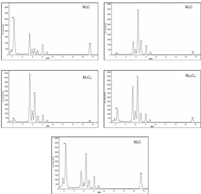
ferrite-pearlite microstructure aged at 600ºC for 1000 hours, which correspond to the ive types of carbides identiied in this steel: M2C, M3C, M7C3, M23C6 and M6C. Figure 9 shows, for both microstructures aged at 600ºC for 1000 h, the carbides’ identiication by colors which enables the observation of their distribution and morphology in the selected regions. The same procedure was adopted for the all aging conditions and for the as received samples and, as result, the precipitation evolution could be studied.
Figure 4 shows the microstructural evolution at 600ºC as a function of aging time, observed by SEM for the ferrite-pearlite microstructure. The white arrows indicate the carbides along the grain boundaries which coalesce along the time. In the same way, Figure 5 shows the microstructural evolution under the same conditions for the ferrite-bainite microstructure, precipitation coarsening is also signaled by the white arrows. Comparing the images of the 100h aging of both microstructures, the coarsening efect in ferrite-bainite is more pronounced, and it is clear that the precipitate distribution in ferrite-pearlite was more efective. That behavior was observed for all ageing samples and it explains the better performance of ferrite-pearlite steels in the creep tests presented at Figure 1.
Figure 4: Ferritic-pearlitic steel scanning electron micrographs at 600°C and 100, 500, 1000 and 2000 h of aging. The arrows indicate the carbides coarsening efect.
Figure 5: Ferritic-bainitic steel scanning electron micrographs at 600°C and 100, 500, 1000 and 2000 h of aging. The arrows indicate the carbides coarsening efect.
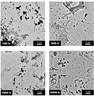
Figures 6 and 7 show TEM carbon replicas images of the precipitation present in, respectively, ferrite-pearlite and ferrite-bainite steels aged at 600ºC for 100, 500, 1000 and 2000 hours. Those images were used to select speciic areas for the phases characterization. According to the literature8-11, carbides present in 2.25Cr-1Mo steels have characteristic EDS spectra, which allow the correct identiication of each type. Figure 8 presents the EDS spectra, obtained from the
Figure 6: Ferritic-pearlitic steel transmission electron micrographs at 600°C and 100, 500, 1000 and 2000 h of aging.
Figure 7: Ferritic-bainitic steel transmission electron micrographs at 600°C and 100, 500, 1000 and 2000 h of aging.
Figure 8: EDS spectra of the carbides identiied in the ferrite-pearlite aged at 600°C for 1000 h sample.
For the ferrite-pearlite microstructure:
M3C + M2C+ M7C3 → M3C + M2C + M7C3 + M23C6 → M3C + M2C + M7C3 + M23C6 + M6C
For the ferrite-bainite microstructure:
M2C + M7C3 + M23C6 → M2C + M7C3 + M23C6 + M6C
As observed, the ferrite-bainite microstructure does not present M3C phase in any moment of its aging, but it initially presents M2C, M7C3 and M23C6 phases. The M23C6 was identiied after 1000h at 550 and 575ºC and after 500h at 600ºC for ferrite-pearlite microstructure. The M6C phase was the last identiied in both microstructures, but the ferrite-bainite presented it before than the ferrite-pearlite, for all conditions of aging. It is important to say that as deleterious phases, like M23C6 and M6C, coalesce, the others decrease their contents, but they
are still present even after the most severe aging conditions. That behavior has already been registered in the literature12:
the authors observed that as the M6C content increased, the M7C3 content decreased, but, in spite of this correlation, the M7C3 carbide remained present in all aged samples.
Figure 9: TEM carbon replicas images of phases identiied in the samples aged at 600°C and 1000 h: (a) and (b) ferritic-pearlitic, (c) and (d) ferritic-bainitic. Carbides are identiied by colors: M2C
(green), M23C6 (black), M7C3 (yellow), M3C (blue), M6C (red).
boundaries, as shown in Figures 4 to 7. The ine precipitation of M2C in the ferrite-pearlite microstructure should also be noted. Finally, it can be emphasized that the M3C precipitate is still present in the ferrite-pearlite steel even when aged at 600ºC for 2.000 h, indicating a slower precipitation evolution for this microstructure.
The creep strength of 2.25Cr-1Mo steels depends on inhibiting the dislocation movement. The controlling mechanisms which contribute to it are solid solution hardening and precipitation hardening13. It is well accepted that as long as a signiicant amount of alloying elements remain in the solid solution and the precipitates are inely dispersed in the matrix and grain boundaries, the creep strength will be high. However, temperature and pressure accelerate the precipitates coalescence at the expense of alloying elements in a solid solution in the matrix. These efects are readily apparent to the ferrite-pearlite steels through the spheroidizing process of pearlite grains observed by optical microscopy. For the ferrite-bainite steel, optical microscopy is not capable of distinguishing the microstructural evolution. In this case, it is more efective to analyze the presence of deleterious phases as M23C6 and M6C using TEM.
4. Conclusions
Although optical microscopy can indicate the microstructural degradation in ferritic-pearlitic 2,25Cr-1Mo steels, this technique is not sensible to identify the loss of mechanical strength at high temperatures for ferritic-bainitic microstructures. However, TEM analysis can provide useful information on the level of material degradation with pearlitic or bainitic microstructure through carbides stoichiometry and morphology.
Creep tests indicate that ferrite-bainite steel is less resistant than ferrite-pearlite steel. The reasons for this diference are because a ferrite-bainite microstructure presents deleterious phases, M23C6 and M6C, earlier and the diference in size and spacing of precipitates.
5. Acknowledgment
The authors would to like to thank Vallourec & Mannesmann Tubes – V&M do Brasil for supplying the 2,25Cr-1Mo steels and the Electric Energy Research Center (Cepel/Eletrobras) for research development.
6. References
1. Kushima H, Watanabe T, Murata M, Kamihira K, Tanaka H, Kimura K. Metallographic Atlas for 2.25Cr-1Mo Steels and Degradation due to Long-term Service at Elevated Temperatures.
OMMI. 2007;4(1):1-13.
2. Wang BQ, Geng GQ, Levy AV. Efect of microstructure on the erosion-corrosion of steels. Wear. 1991;151(2):351-364.
3. Chaudhuri S. Philosophy of integrity assessment of engineering components. Materials Science and Engineering: A. 2008;489(1-2):259-266.
4. Toft LH, Marsden RA. Structural processes in creep: special report no. 70. London: Iron & Steel Institute; 1961. p. 238-244.
5. Bhadeshia HKDH, Honeycombe RWK. Steels: Microstructure and Properties. 3rd ed. Oxford: Butterworths-Heinemann; 2006.
6. Bhadeshia HKDH. Bainite in Steels. Cambridge: Cambridge University Press; 2001.
7. Watanabe T, Yamazaki M, Hongo H, Tabuchi M, Tanabe T. Efect of stress on microstructural change due to aging at 823 K in multi-layer welded joint of 2.25Cr–1Mo steel. International Journal of Pressure Vessels and Piping. 2004;81(3):279-284.
8. Furtado HC, de Almeida LH, Le May I. Precipitation in 9Cr–1Mo steel after creep deformation. Materials Characterization. 2007;58(1):72-77.
9. Xu Y, Zhang X, Tian Y, Chen C, Nan Y, He H, et al. Study on the nucleation and growth of M23C6 carbides in a 10% Cr martensite ferritic steel after long-term aging. Materials Characterization. 2016;111:122-127.
10. Kuimalee S, Chairuangsri T, Pearce JTH, Edmonds DV, Brown AP, Brydson RMD. Quantitative analysis of a complex metal carbide formed during furnace cooling of cast duplex stainless steel using EELS and EDS in the TEM. Micron. 2010;41(5):423-429.
11. Pilling J, Ridley N. Tempering of 2,25 Pct Cr-1 Pct Mo Low Carbon Steels. Metallurgical Transactions A. 1982;13(4):557-563.
12. de Lima CR, Pinto AL, Furtado HC, de Almeida LH, de Souza MFP, Le May I, Quantitative observations of precipitation in 2.25Cr-1Mo steel exposed to diferent creep conditions in a power station. Engineering Failure Analysis. 2009;16(5):1493-1500.


