Pain, masticatory performance and swallowing
threshold in orthodontic patients
Marcos Porto Trein1, Karina Santos Mundstock2, Leonardo Maciel3, Jaqueline Rachor4, Gustavo Hauber Gameiro5
How to cite this article: Trein MP, Mundstock KS, Maciel L, Rachor J, Gameiro GH. Pain, masticatory performance and swallowing threshold in orth-odontic patients. Dental Press J Orthod. 2013 Nov-Dec;18(6):117-23.
Submitted: December 26, 2011 - Revised and accepted: February 19, 2012
» The authors report no commercial, proprietary or inancial interest in the prod-ucts or companies described in this article.
Contact address: Gustavo Hauber Gameiro Rua Sarmento Leite 500, 2° andar – Centro CEP: 90.050-170 – Porto Alegre/RS – Brazil E-mail: [email protected]
1 Specialist in Orthodontics, Federal University of Rio Grande do Sul (UFRGS). 2 PhD in Orthodontics, State University of São Paulo (UNESP). Associate professor
of Orthodontics,UFRGS.
3 Undergraduate student of Dentistry,UFRGS. 4 Undergraduate student of Dentistry,UFRGS.
5 PhD in Orthodontics, University of Campinas (UNICAMP). Associate professor
of Physiology, UFRGS.
Objective:The aim of this study was to assess pain, masticatory performance and swallowing threshold of patients undergoing orthodontic treatment. Methods: Ten patients of both genders (mean age of 17.25 ± 5.21 years), with complete permanent dentition, who underwent orthodontic treatment with ixed appliances were evaluated. The masticatory performance and the swallowing threshold were assessed by patient’s individual capacity of fragmenting an artiicial test food (Optocal) which was chewed and had the resulting particles processed by a standardized siev-ing method, presentsiev-ing the median particle size (MPS) of crushed units. The intensity of pain / discomfort dursiev-ing chewing was evaluated by means of a visual analog scale. All tests were performed at the following times: T0 – before activating the orthodontic appliance; T1 – 24 hours ater activation, and T2 – 30 days ater activation. Results: The results showed a signiicant increase in pain at T1 (T0 – 0.60 ± 0.70 mm; T1 – 66.2 ± 34.5 mm), returning to baseline values at T2 (3.20 ± 3.82 mm). Masticatory performance was also reduced in T1 (MPS 10.15 ± 1.1 mm2) in comparison
to T0 (MPS 7.01 ± 2.9 mm2) and T
2 (MPS 6.76 ± 1.3 mm
2). However, particle size was not afected in the swallowing
threshold test (T0 – 5.47 ± 2.37 mm2; T
1 – 6.19 ± 2.05 mm 2; T
2 – 5.94 ± 2.36 mm
2). Conclusion: The orthodontic
appliances did not interfere in the size of the particles that would be swallowed, even in the presence of pain.
Keywords:Mastication. Malocclusion. Orthodontics.
Objetivo:o objetivo desse estudo foi avaliar a dor, a performance mastigatória e o limiar de deglutição em pacientes submetidos ao tratamento ortodôntico. Métodos: foram avaliados dez pacientes de ambos os sexos (idade média 17,25 ± 5,21 anos), com dentição permanente completa, submetidos ao tratamento ortodôntico com aparelhos ixos. A performance mastigatória e o limiar de deglutição foram avaliados pela capacidade individual de fragmentação de um alimento teste artiicial (Optocal), o qual foi mastigado e as partículas resultantes processadas por um método padroni-zado com peneiras, apresentando o tamanho mediano das partículas (TMP) das unidades trituradas. A intensidade da dor/desconforto foi avaliada com o uso de uma escala analógica visual. Todos os testes foram realizados nos seguintes momentos: T0: antes da ativação do aparelho ortodôntico; T1: 24 após a ativação, e T2: 30 dias após a ativação. Resulta-dos: os resultados demonstraram um aumento signiicativo na dor em T1 (T0: 0,60 ± 0,70mm; T1: 66,2 ± 34,5mm), retornando aos valores basais em T2 (3,20 ± 3,82mm). A performance mastigatória também foi reduzida em T1 (TMP: 10,15 ± 1,1mm2) comparada a T
0 (TMP: 7,01 ± 2,9mm 2) e T
2 (TMP = 6,76 ± 1,3 mm
2). Entretanto, o
ta-manho das partículas não foi afetado no teste do limiar de deglutição (T0: 5,47 ± 2,37mm2; T
1: 6,19 ± 2,05mm 2;
T2: 5,94 ± 2,36mm2). Conclusão: os aparelhos ortodônticos não interferiram no tamanho das partículas que seriam
deglutidas, mesmo na presença da dor.
INTRODUCTION
Orthodontic movement pain is caused by the release of diferent mediators ater the application of forces on the periodontal ligament (PDL). These mediators, in-cluding substance P, histamine, serotonin, glutamate, prostaglandins, leukotrienes, and cytokines may activate nociceptors within the PDL resulting in orthodontic
pain,1 which usually lasts for 2 or 3 days and gradually
reduces by the 5th or 6th day.2 Studies report that 95%
of orthodontic patients experience some pain during treatment and several methods have been used to reduce these symptoms, including low level laser, transcutane-ous electric stimulation, vibratory PDL stimulation and
use of anti-inlammatory drugs.1,3,4 Several factors
asso-ciated with orthodontic pain are still ignored by many clinicians, such as the duration, intensity and functional limitations possibly induced by this symptom.
It has been shown that almost all orthodontic pa-tients report moderate to extreme diiculties in biting and chewing harder foods, and thus tend to choose a less
consistent diet.1 The orthodontic pain is probably the
main responsible for the masticatory limitations associ-ated with ixed appliances. The orthodontic pain occur-ring within 48 hours is so disturbing that approximately 20 per cent of patients report being awakened at night, some of them take medication, and almost all patients
report eating diiculties as a result of pain.5,6 However,
an objective analysis of mastication and deglutition in patients during orthodontic therapy was not performed by previous studies. These analyses are necessary con-sidering that patients normally overestimate their masti-catory ability when they are evaluated only by subjective methods. For example, many individuals with a com-promised dentition and dentures judge their masticatory function as ‘good’ while an objective test resulted in
val-ues much lower than healthy subjects.7 Therefore, the
aims of this study were to evaluate masticatory perfor-mance, swallowing threshold and pain ater orthodontic ixed appliance activation.
MATERIAL AND METHODS
Sample
Ten patients of both genders, ive males and ive fe-males (mean age of 17.25 ± 5.21 years) participated in this study. The following inclusion criteria were con-sidered: Approximately equal number of occlusal units with malocclusions requiring orthodontic treatment,
the presence of complete permanent dentition (except third molars), uneventful medical history and good oral health. Bonding of at least 10 teeth in the maxillary arch and 0.014 (NiTi), 0.014 or 0.016-in (stainless steel) archwires ligated with elastomeric rings were used dur-ing the experimental period. No extractions were per-formed during this period. Four individuals had Class I malocclusions, four had Class III and two had Class II. The exclusion criteria were: previous orthodontic treat-ment and symptoms of temporomandibular joint dys-function. An informed written consent was obtained from all participants or parents prior to their enrolment in the study. The local Institutional Review Board ap-proved the protocol. The study was carried out at the Department of Orthodontics of the Federal Universi-ty of Rio Grande do Sul (UFRGS), in Brazil. Sample size was determined on the basis of clinically relevant masticatory performance data obtained from the
litera-ture,8,9,10 with a power of 80%, α = 0.05, and 10
individ-uals were deemed adequate for this longitudinal study in which each patient served as their own control.
Timing of evaluations
The subjects were analyzed at three time points: T0,
at their irst consultation, before ixed appliances were
installed; T1, 24 hours ater installation and engagement
of the irst archwire; T2, 30 days ater the irst activation
and before reactivation of the appliance. The data col-lected included masticatory performance, swallowing threshold and self-reported pain.
Masticatory performance evaluation
Masticatory performance was evaluated by means of the individual capacity of fragmentation of an
artii-cial test-food (Optocal).11 Subjects were given 17 cubes
The contents of each sieve were then weighed on an analytic scale with a 0.001 g precision. Since the speciic mass of the test-food is known, weight was converted into volume using the Rosim-Rammler equation on Statistical Package for the Social Sciences version 18.0 sotware. The distribution of the particles by weight was described by the cumulative function of median particle size (X50), which represents a virtual sieve mesh where 50% of the particles would pass through. The higher the X50, the worse the masticatory performance.
Swallowing threshold
The individuals were handed another set of 17 Op-tocal cubes and instructed to chew them until they felt the urge to swallow. A trained examiner counted the number of chewing cycles and registered the total time of the cycles, which was measured with a digital stop-watch. The swallowing threshold particles were submit-ted to the same fragment size analysis as it was done for the masticatory performance test, described above.
Pain quantification
Ater the individuals chewed the Optocal cubes they were handed a visual analogic scale (VAS) for registra-tion of the pain experienced on every experimental period. The subjects were instructed to make a mark on the 10 cm line corresponding to the pain experi-enced during chewing. The let limit of the scale was described as “without discomfort” and the right limit as “worst discomfort possible”.
Error of method
The X50 data of 10 subjects with the same age were analyzed with the Dahlberg formula and paired t test ater 2 analyses within a 7 day interval. There was no statistical diference between the evaluations (p > 0.05) and the reproducibility error was less than 10% for the X50 (0.5 mm).
Statistical analyses
Shapiro-Wilks test was used to verify data normal-ity. The variables were analyzed by ANOVA for repeat-ed measures, and by Tukey’s test when they were nor-mally distributed. The Kruskal-Wallis and Dunn’s test were applied when data were not normally distributed. The SPSS sotware was used and the signiicance level was set at p < 0.05.
RESULTS
The results are presented in Figure 1.
Pain and Masticatory Performance
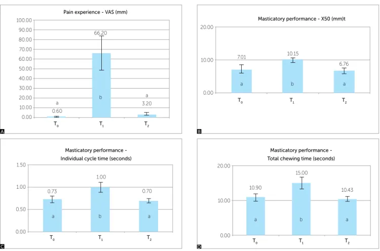
Figure 1 presents the values of pain level, median particle size chewed for 15 cycles, total chewing time and duration of each cycle.
Pain was signiicantly higher at T1 when compared
to T0 and T2 (Kruskal-Wallis + Dunn, p < 0.05).
A sig-niicant reduction in masticatory performance also
occurred in T1 in comparison to T0 and T2
(Kruskal-Wallis and Dunn, p < 0.05). However, masticatory performance levels did not show statistical signiicant
diference between T0 and T2. Total chewing time and
time of each cycle were higher in T1 than in the other
experimental periods (Anova + Tukey, p < 0.05).
Swallowing threshold
Figure 2 demonstrates the X50 of the swallowing threshold evaluation, total chewing time, time for each cycle, and number of cycles until deglutition. The me-dian particle size did not show statistical diference be-tween the timepoints. Total chewing time and time for
each cycle were increased in T1 when compared to T0
but without statistical signiicance. There was a
statisti-cally signiicant reduction when T2 was compared to T1
(Anova + Tukey, p < 0.05). Time taken for each cycle was similar in all 3 timepoints and although there was
an apparent raise in T1 it did not reach statistical
signii-cance (Kruskal-Wallis, p = 0.092).
DISCUSSION
This study evaluated pain, masticatory perfor-mance and swallowing threshold in patients under-going orthodontic treatment with fixed appliances. Although the literature presents some studies on the
possible functional impacts of braces,12,13 no
quantita-tive tool was used for objecquantita-tive evaluation of mastica-tion in these studies.
Objective evaluation of masticatory function is es-sential in clinical trials, since patients tend to overes-timate their chewing ability when evaluated only by subjective methods (e.g. questionnaires). Many pa-tients with compromised dentition or dentures think they have a good chewing ability, even when objec-tive tests show values much lower than in subjects
10.00 20.00 30.00 40.00 50.00 60.00 70.00
0.00
T0 T1 T2
23.30 39.40
26.70
Swallowing threshold -
Number of cycles
a a a
ab a b
Pain experience - VAS (mm)
T0 T1 T2
a 0.60
a 3.20 b
66.20 100.00
60.00 70.00 80.00 90.00
50.00 40.00 30.00 20.00 10.00 0.00
A
T0 T1 T2
0.00 10.00 20.00
10.90
a b a
15.00
10.43
Masticatory performance -
Total chewing time (seconds)
T0 T1 T2
0.00 0.50 1.00 1.50
0.73
a b a
1.00
0.70
Masticatory performance -
Individual cycle time (seconds)
a b a
Masticatory performance - X50 (mm)t
T0 T1 T2
10.15
6.76 20.00
10.00
0.00
a b a
7.01
Figure 1 - Masticatory Performance Results. A) Pain experience expressed by VAS; B) X50 of the particles; C) Total chewing time for the 15 cycles; D) Individual cycle time. T0- Before orthodontic appliance activation; T1- 24 hours after activation; T2- 30 days after activation. Diferent letters = statistical signiicance (Kruskal-Wallis / Dunn or ANOVA / Tukey, p <0.05).
Figure 2 - Swallowing Threshold Results. A) X50 of the particles; B) Total chewing time until urge of swallowing; C) Individual cycle time; D) Number of cycles until swallowing. T0- Before orthodontic appliance activation; T1- 24 hours after activation; T2- 30 days after activation. Diferent letters = statistical
signiicance (Kruskal-Wallis / Dunn or ANOVA / Tukey, p <0.05).
T0 T1 T2
10.00
0.00
5.47 6.19 5.94
Swallowing threshold - X50 (mm)
a a a
T0 T1 T2
10.00 20.00 30.00 40.00 50.00
0.00
19.13
36.72
16.60
Swallowing threshold - Total chewing time (seconds)
a a a
ab a b
T0 T1 T2
0.00
0.73
1.03
0.71
0.50 1.50
1.00
Swallowing threshold -
Individual cycle time (seconds)
a a a
B
B A
C
C
D
The present study demonstrated that orthodontic pa-tients have low masticatory performance when measured one day ater installation and activation of orthodon-tic appliances. This period represents the peak time of orthodontic pain, which tends to decrease signiicantly 3
days ater insertion of archwires.2,3,15,16 Erdinç and
Din-çer17 reported the onset of pain as occurring two hours
ater activation of the device, with a peak within the irst 24 hours, in which the recorded average VAS was 48 mm for the group that received a nickel-titanium alloy archwire of 0.016-in section and 49 mm for the group that received a 014-in NiTi alloy archwire. Tortamano
et al18 demonstrated an average registration of 7.25 and
8.25 on a visual scale on the irst day ater the installa-tion of 0.014-in individualized stainless steel archwires. However, the authors did not use a VAS, but a graduated scale of 1 to 10, in which the subjects had to choose a speciic score, which may have resulted in higher average
scores. Firestone, Scheurer and Bürgin19 found an
aver-age of 27.5 mm VAS scores for pain caused ater the irst archwire was installed. It lasted for 7 days with a
maxi-mum record of 49.1 mm. In Ong, Ho and Miles study,16
the peak of reported pain was reached within 24 hours.
The work of Fernandes, Ogaard and Skoglund,20 carried
out with subjects from 9 to 16 years old, registered pain experience hourly for the irst 11 hours and then on a daily basis for 7 days. The authors found an average of 36 and 37.2 mm VAS scores 24 hours ater installation of the irst archwire (0.014-in NiTi or 0.014-in Sentalloy). These studies were the basis of our choice of evaluating pain and masticatory performance ater 24 hours.
Most studies indicate that there are no gender, age or initial crowding-related diferences for pain ater
orth-odontic appliance activation.2,15,17,21 Firestone, Scheurer
and Bürgin19 did not ind gender-related diferences for
perceived pain. Additionally, Ong, Ho and Miles16 did
not ind any relationship between pain and age, gender
or initial crowding. The indings of Scott et al22
demon-strated no correlation between pain and gender or age, either. Therefore, our data was group regardless of gen-der, age, malocclusion or initial crowding diferences. Our results are in agreement with studies that assessed pain associated with braces, and the pain levels reported
in the present study (66.2 ± 34.5 mm in T1) are very
similar to those found by Polat, Karaman and Durmus3
(59.4 ± 31.2 mm), which also recorded the experience of pain by VAS in patients during mastication.
Pain is often underestimated by orthodontists23
and few studies have assessed the functional impacts
of fixed orthodontic appliances.12,17,24 Pain is often
considered the worst aspect of orthodontic treatment, and is also one of the main reasons why patients drop
out of treatment.1,25,26 Some patients report, for
ex-ample, that the incidence and severity of orthodontic
pain is greater than pain caused by tooth extraction.2
Researchers attribute pain to the hyperalgesia of the periodontal ligament (PDL) caused by induced tooth movement, which is defined as a painful sensation greater than what is expected to a noxious stimulus
and felt over a larger area.27 Orthodontic tooth
pres-sure induces the release of chemical mediators such as histamine, bradykinin, serotonin and prostaglandins, which are capable of activating or sensitizing
noci-ceptors in the PDL.1,4 In addition to hyperalgesia,
orthodontic pain can also be spontaneous or related to non-painful stimuli such as mechanical stimulation of the periodontium during mastication. The pain reported by stimuli that are usually non-painful is
called allodynia,27 and this was clearly observed in our
study, since the pain levels were registered right after the masticatory performance and swallowing thresh-old tests and, after 24 hours, there was significant pain, which is not expected in normal masticatory function. These results are in accordance with those
reported by Erdinç and Dinçer,17 in which
approxi-mately 50% of their patients had problems with their daily activities on days 1 and 2 after orthodontic ap-pliance activation and that the discomfort decreased significantly by the third day. In this same study, however, only subjective evaluations were used. In our study, the results of objective evaluation of mas-ticatory function after 24 hours demonstrated that pa-tients presented difficulties in grinding the test food during the masticatory performance test. This can be seen not only by the significant increase in median size of the crushed particles (X50), but also by the increase in the total chewing time and in the time of each cycle during the test.
to that observed prior to the installation of the
appli-ances (T0). These results suggest that the chewing
dii-culty presented within 24 hours may have been partially balanced by an upward trend in the number of chewing cycles and time. Other ways to compensate the chewing diiculty during the peak of orthodontic pain may also have been employed, such as changes in the dynamics of jaw movements and bite force, but these variables were not evaluated in this study. Another limitation of our study is that only one test food was evaluated. This test food was chosen because it is less consistent in relation
to Optosil® or Cuttersil®,11 and patients in pain usually
avoid harder foods. If we had chosen a harder test-food maybe the results would have pointed towards a more signiicant diference.
The consequences of ineicient chewing for general health have not been fully elucidated. The particle size ingested, which is determined by the performance of the chewing process, may inluence gastric emptying. Some studies suggest that higher masticatory eiciency
accel-erates gastric emptying,28,29 although this issue remains
controversial. Sierpinska et al10 found more severe
chron-ic inlammatory changes and infection by Helchron-icobacter pylori in the gastric mucosa in patients with dyspepsia and impaired mastication. If indeed there is a relation of cause and efect between masticatory eiciency and gas-tric pathologies, orthodontic therapy should not be con-sidered as a potential cause of damage to the patient, since the present results demonstrate that although there is a reduction in masticatory performance within 24 hours, there was no diference in the size of 50X on the verge of swallowing. These indings indicate that orthodontic pa-tients may compensate the functional limitation induced by pain with a more careful mastication until
degluti-tion. To Fontijn-Tekamp et al,30 individuals with poor
masticatory performance tend to swallow larger particles, this observation being similar to that found by English,
Buschang and Throckmorton.13 In the irst article cited,30
adults with good oral health and varied occlusal
condi-tions were evaluated, while the second13 evaluated
indi-viduals with Class I, II and III malocclusions and nor-mal occlusion, but with no braces installed. Both studies reported that individuals with poor masticatory perfor-mance do not compensate this deiciency by increasing the number of cycles until swallowing. However, these results cannot be directly compared to ours, due to the fact that these studies did not evaluate individuals with limitations caused by pain, as in our case, in which the subjects reported signiicant pain during the evaluation period of 24 hours ater orthodontic activation. In the pa-pers mentioned above, the performance was determined only by the dental status of individuals, whereas in our study the experience of pain signiicantly afected the masticatory performance of patients.
CONCLUSIONS
Patients reported a signiicant increase in pain dur-ing chewdur-ing 24 hours ater activation of orthodontic ap-pliances, but ater 30 days there was no diference com-pared to baseline values.
By setting a limit of 15 chewing cycles (masticatory performance test), the median size of crushed particles was higher within 24 hours in comparison to initial and inal values, which indicates a temporary deterioration in masticatory performance, since this decrease was ob-served only at the peak of orthodontic pain.
1. Krishnan V. Orthodontic pain: from causes to management-a review. Eur J Orthod. 2007;29(2):170-9.
2. Jones M, Chan C. The pain and discomfort experienced during orthodontic treatment: a randomized controlled clinical trial of two initial aligning arch wires. Am J Orthod Dentofacial Orthop. 1992;102(4):373-81.
3. Polat O, Karaman AI, Durmus E. Efects of preoperative ibuprofen and naproxen sodium on orthodontic pain. Angle Orthod. 2005;75(5):791-6. 4. Gameiro GH, Pereira-Neto JS, Magnani MB, Nouer DF. The inluence
of drugs and systemic factors on orthodontic tooth movement. J Clin Orthod. 2007;41(2):73-8.
5. Kvam E, Gjerdet N R, Bondevik O. Traumatic ulcers and pain during orthodontic treatment. Community Dent Oral Epidemiol. 1987;15(2):104-7.
6. Scheurer PA, Firestone AR, Bürgin WB. Perception of pain as a result of orthodontic treatment with ixed appliances. Eur J Orthod. 1996;18(4):349-57.
7. van der Bilt A. Assessment of mastication with implications for oral rehabilitation: a review. J Oral Rehabil. 2011;38(10):754-80.
8. Fontijn-Tekamp FA, Slagter AP, van der Bilt A, Van’T Hof MA, Witter DJ, Kalk W, Jansen JA. Biting and chewing in overdentures, full dentures, and natural dentitions. J Dent Res. 2000;79(7):1519-24.
9. van den Braber W, van der Bilt A, van der Glas H, Rosenberg T, Koole R. The inluence of mandibular advancement surgery on oral function in retrognathic patients: a 5-year follow-up study. J Oral Maxillofac Surg. 2006;64(8):1237-40.
10. Sierpinska T, Golebiewska M, Dlugosz J, Kemona A, Laszewicz W. Connection between masticatory eiciency and pathomorphologic changes in gastric mucosa. Quintessence Int. 2007;38(1):31-7. 11. Pocztaruk RL, Frasca LC, Rivaldo EG, Fernandes E de L, Gavião MB.
Protocol for production of a chewable material for masticatory function tests (Optocal - Brazilian version). Braz Oral Res. 2008;22(4):305-10. 12. Bergius M, Kiliaridis S, Berggren U. Pain in orthodontics. A review and
discussion of the literature. J Orofac Orthop. 2000;61(2):125-37. 13. English JD, Buschang PH, Throckmorton GS. Does malocclusion afect
masticatory performance? Angle Orthod. 2002;72(1):21-7.
14. Carlsson GE. Masticatory eiciency: the efect of age, the loss of teeth and prosthetic rehabilitation. Int Dent J. 1984;34(2):93-7.
15. Salmassian R, Oesterle LJ, Shellhart WC, Newman SM. Comparison of the eicacy of ibuprofen and acetaminophen in controlling pain after orthodontic tooth movement. Am J Orthod Dentofacial Orthop. 2009;135(4):516-21.
REFERENCES
16. Ong E, Ho C, Miles P. Alignment eiciency and discomfort of three orthodontic archwire sequences: a randomized clinical trial. J Orthod. 2011;38(1):32-9.
17. Erdinç AM, Dinçer B. Perception of pain during orthodontic treatment with ixed appliances. Eur J Orthod. 2004;26(1):79-85.
18. Tortamano A, Lenzi DC, Haddad AC, Bottino MC, Dominguez GC, Vigorito JW. Low-level laser therapy for pain caused by placement of the irst orthodontic archwire: a randomized clinical trial. Am J Orthod Dentofacial Orthop. 2009;136(5):662-7.
19. Firestone AR, Scheurer PA, Bürgin WB. Patients anticipation of pain and pain-related side efects, and their perception of pain as a result of orthodontic treatment with ixed appliances. Eur J Orthod. 1999;21(4):387-96.
20. Fernandes LM, Ogaard B, Skoglund L. Pain and discomfort experienced after placement of a conventional or a superelastic NiTi aligning archwire. A randomized clinical trial. J Orofac Orthop. 1998;59(6):331-9. 21. Ngan P, Kess B, Wilson S. Perception of discomfort by patients
undergoing orthodontic treatment. Am J Orthod Dentofacial Orthop. 1989;96(1):47-53.
22. Scott P, Sherrif M, Dibiase AT, Cobourne MT. Perception of discomfort during initial orthodontic tooth alignment using a self-ligating or conventional bracket system: a randomized clinical trial. Eur J Orthod. 2008;30(3):227-32.
23. Krukemeyer AM, Arruda AO, Inglehart MR. Pain and orthodontic treatment. Angle Orthod. 2009;79(6):1175-81.
24. Brown DF, Moerenhout RG. The pain experience and psychological adjustment to orthodontic treatment of preadolescents, adolescents, and adults. Am J Orthod Dentofacial Orthop. 1991;100(4):349-56.
25. Oliver RG, Knapman YM. Attitudes to orthodontic treatment. Br J Orthod. 1985;12(4):179-88.
26. Kluemper GT, Hiser DG, Rayens MK, Jay MJ. Eicacy of a wax containing benzocaine in the relief of oral mucosal pain caused by orthodontic appliances. Am J Orthod Dentofacial Orthop. 2002;122(4):359-65. 27. Murray GM. Referred pain, allodynia and hyperalgesia. J Am Dent Assoc.
2009;140(9):1122-4.
28. Holt S, Reid J, Taylor TV, Tothill P, Heading RC. Gastric emptying of solids in man. Gut. 1982;23(4):292-6.
29. Pera P, Bucca C, Borro R, Bernocco C, De LA, Carossa S. Inluence of mastication on gastric emptying. J Dent Res. 2002;81(3):179-81. 30. Fontijn-Tekamp FA, van der Bilt A, Abbink JH, Bosman F. Swallowing