Flexural strength of mini-implants developed for
Herbst appliance skeletal anchorage.
A study in Minipigs br1 cadavers
Klaus Barretto Lopes1, Gladys Cristina Dominguez2, Caio Biasi3, Jesualdo Luiz Rossi4
How to cite this article: Lopes KB, Dominguez GC, Biasi C, Rossi JL. Flexural strength of mini-implants developed for Herbst appliance skeletal an-chorage. A study in Minipigs br1 cadavers. Dental Press J Orthod. 2013 Nov-Dec;18(6):124-9.
Submitted: January 27, 2012 - Revised and accepted: April 17, 2012
Contact address: Klaus Barretto Lopes Rua Visconde de Pirajá, 550/1407 – Ipanema Rio de Janeiro/RJ — Brazil — CEP: 22.410-002 E-mail: klausbarretto@uol.com.br
1 Postdoc in Orthodontics, State University of Rio de Janeiro (UERJ). 2 Full professor, Department of Orthodontics, College of Dentistry, University of
São Paulo (USP).
3 PhD Resident, Veterinary Medicine, University of São Paulo (USP). 4 Postdoc in Materials Science and Engineering, University of Surrey.
» The author Klaus Barretto Lopes asserts to be the developer and patent holder of the mini-implant prototypes used in this study.
Objective:The present study was designed to verify if mini-implant prototypes (MIP) developed for Herbst ap-pliance anchorage are capable of withstanding orthopedic forces, and to determine whether the flexural strength of these MIP varies depending on the site of insertion (maxilla and mandible). Methods: Thirteen MIP were inserted in three minipig cadavers (six in the maxilla and seven in the mandible). The specimens were prepared and submitted to mechanical testing. The mean and standard deviation were calculated for each region. A two-way Student’s t test was used to compare the strength between the sites. A one-way Student’s t test was performed to test the hypothesis. Orthopedic forces above 1.0 kgf were considered. Results: The MIP supported flexural strength higher than 1.0 kgf (13.8 ± 2.3 Kg, in the posterior region of the maxilla and 20.5 ± 5.2 Kg in the anterior region of the mandible) with a significantly lower flexural strength in the anterior region of the mandible (P < 0.05). Conclusion: The MIP are capable of withstanding orthopedic forces, and are more resistant in the anterior region of the mandible than in the posterior region of the maxilla in Minipigs br1 cadavers.
Keywords:Functional appliances. Dental implants. Orthodontic appliances. Orthodontic anchorage procedures. Angle Class II malocclusion.
Objetivo:o presente estudo foi delineado para verificar se protótipos de mini-implantes (PMI) desenvolvidos para a ancoragem esquelética do aparelho de Herbst são capazes de suportar forças ortopédicas e, também, determinar a variação da força de flexão desses PMI de acordo com o local de inserção (maxila ou mandíbula). Métodos: após o cálculo do tamanho da amostra, 13 PMI foram colocados em três cadáveres de Minipigs br1 (seis na maxila e sete na mandíbula). Os corpos de prova foram preparados e submetidos a um teste mecânico. Cálculos da média e o do desvio-padrão foram realizados para cada região. O teste t de Student para duas amostras não pareadas foi utilizado
para comparar a resistência dos PMI entre as regiões de inserção. O teste t de Student para uma amostra foi realizado
para o teste de hipótese. Foram consideradas forças ortopédicas aquelas acima de 1,0kgf. Resultados: os PMI foram capazes de suportar forças de flexão maiores que 1,0kgf (13,8 ± 2,3Kg na região posterior da maxila, e 20,5 ± 5,2Kg na região anterior da mandíbula), apresentando significativa menor força de flexão na região anterior da mandíbula (p < 0,05). Conclusão: os PMI são capazes de suportar forças ortopédicas, sendo mais resistentes quando utilizados na região anterior da mandíbula do que na posterior da maxila, em cadáveres de Minipigs br1.
INTRODUCTION
Implants and mini-implants have been used as orth-odontic anchorages for diferent purposes in diferent locations.1,2,3 Some researchers have suggested the use of
mini-implants as orthopedic anchors in animals4,5 and
in the treatment of Class III malocclusions with retru-sive maxillae in humans.6 However, there is little
infor-mation about the use of mini-implants as orthopedic anchorage in the treatment of Class II malocclusions.
The Herbst appliance has oten been used in the treatment of Class II malocclusions, because of its ef-iciency7 and also because of positive efects in
orth-odontic and orthopedic correction.8 Nevertheless, some
investigators have stated that the correction of Class II malocclusion is a result of anchorage loss, and could be responsible for negative efects, such as protrusion and gingival recession,9 on lower incisors.
Many attempts have been made to reduce the nega-tive efects caused by the Herbst appliance on lower incisors, such as increasing the number of teeth in the mandibular anchorage, using sot-tissue anchorage, splints, and cast splints anchorage.10,11 However, these
attempts were unsuccessful.
With the intention of solving these problems, a mini-implant prototype was developed for Herbst appliance anchorage, and its flexural resistance was measured in an in vitro study.12. However, a question
arose with respect to the resistance strength of this mini-implant prototype when inserted into the bone. The present study was designed to evaluate if the mini-implant prototype developed for Herbst appli-ance anchorage is capable of withstanding orthopedic forces in Minipigs br1, and to compare the prototype resistance between the sites of insertion.
MATERIAL AND METHODS
Thirteen mini-implants (Neodent, Curitiba, Bra-zil), 2 mm in diameter and 10 mm in length, with at-tachment to Herbst appliance telescopic tubes, were inserted in three Minipigs br113,14 (15-month old) ater
they had been euthanized.
A calculation of the sample size15 was carried out by
means of two pilot studies, one for the posterior region of the maxilla and another for the anterior region of the mandible of one minipig.
Based on the performance of the specimens on the graph, the sample size was calculated with the values
ob-tained with a dislocation of 1.2 mm, because ater this point the strength values increased abruptly, indicating the resistance of the metal block.
Aterwards, the following statistical formula was used:
n= (Z x Cv) 2
Er 2
Where n = number of specimens, Z = Standard de-viation from normal distribution, Cv = Coeicient of variation, and Er = Relative error.
Ater sample size calculation, the number of speci-mens needed for the inal study was 6 in the maxilla and 7 in the mandible.
In order to test sample normality, the Kolmogorov-Smirnov test was carried out. To test the hypothesis, a one-way Student’s t test was performed with Minitab 15 (State College, PA, USA). Orthopedic forces above 1 Kgf were considered.16,17
Thereater, the hypotheses of withstanding orthope-dic forces were deined (H0: μ = 1.0 and H1: μ > 1.0).
The criterion to reject the null hypothesis was: tcal > tα, n - 1,
where α =0.05 or calculated: t > t from t table.
To compare the lexural strength between the pos-terior region of the maxilla and the anpos-terior region of the mandible, a two-way Student’s t test was calculated using Minitab 15.
Experiment
The animals were euthanized before the experiment and frozen at a temperature of - 20oC.
Two minipigs received four mini-implants (two in each maxilla and two in each mandible), and one mini-pig received ive mini-implants (two in the maxilla and three in the mandible).
Six specimens with the new mini-implant were ob-tained from the posterior region of the maxilla, and seven specimens were obtained from the anterior region of the mandible, which were the possible places where the mini-implants could be used to anchor the Herbst appliance.
Ater that, the straight telescopic tube was placed in the head of the mini-implant with a suitable screw (Fig 1).
the posterior region of the maxilla, between the roots of the upper irst molar and the anterior region of the man-dible, between the roots of the second premolar and the roots of the third premolar.
To insert the mini-implant, a torque key with a torque meter calibrated at a measure not greater than 30 Kgf.cm was used. When the insertion was complet-ed, new radiographs were taken to check the inal posi-tioning of the mini-implants.
To prepare the specimens, the maxilla and the man-dible were sectioned into small pieces, using an elec-tric cutting machine. Metal blocks were used to protect the specimens during the experiment. Aterwards, an acrylic resin was used to ix the bone fragments with the mini-implants (Fig 2).
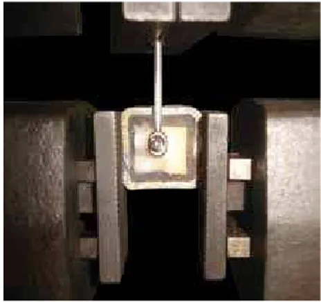
The specimens were placed in an Instron 4400R test machine (Instron, Norwood, MO, USA), with the metal block in the lower part and the telescopic tube in the upper part (Fig 3).
The specimens were submitted to a single cantilever lexure test. Traction was applied at 0.5 mm per minute until 1.5 mm of dislocation was obtained. This value was based on a pilot study carried out by Brettin et al.18
The values were recorded, and a graph of strength x dis-location was constructed.
RESULTS
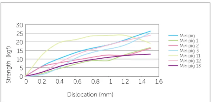
Single cantilever lexure tests were successfully car-ried out on 13 specimens. The graphs illustrate the per-formance of the specimens during the tests in the maxilla and the mandible (Figs 4 and 5). The mini-implant pro-totypes showed a lexural strength of 13.86 ± 2.30 Kgf for the posterior region of the maxilla, and 20.5 ± 5.20 Kgf for the anterior region of the mandible.
The normality of the two samples was conirmed with the Kolmogorov-Smirnov test.
For the hypothesis test, the calculated t was 13.71 (P˂ 0.001) for the posterior region of the maxilla. The critical t obtained from the table was 2.015. The crite-rion used to reject the null hypothesis revealed that:
tcal > tα, n-1, where α = 0.05 → 13.71 > 2.015
Therefore, the value calculated for t for the poste-rior region of the maxilla is outside the region of H0 acceptance. The null hypothesis was rejected, and the hypothesis that mini-implants cannot withstand ortho-pedic forces could not be conirmed for the posterior region of the maxilla.
Using the same criterion for the anterior region of the mandible, the calculated t was 9.94 (P ˂ 0.001). The critical t obtained from the table was 1.943.
The criterion used to reject the null hypothesis re-vealed that:
tcal > tα, n-1, where α = 0.05 → 9.94 > 1.943
Similarly to the maxilla, the value calculated for t is outside the region of H0 acceptance. The null hypoth-esis was rejected, and the hypothhypoth-esis that mini-implants cannot withstand orthopedic forces could not be con-irmed for the anterior region of the mandible.
A statistically signiicant diference was found in the lexural strength between the posterior region of the maxilla and the anterior region of the mandible (P = 0.015): the anterior region of the mandible was sig-niicantly more resistant.
DISCUSSION
One of the purposes of this study was to quantify the flexural resistance of the mini-implant proto-types when inserted in the posterior region of the
Figure 1 - Straight telescopic tube placed in the head of the mini-implant with a suitable screw.
Figure 2 - Bone fragments with mini-implants in-serted which were included in the metal block.
It could be argued that the 2.0-mm diameter mini-implant is too large for the inter-radicu-lar space. However, Poggio et al.20 showed that, in
humans, this size is compatible with the inter-radic-ular distance between the upper first molars and sec-ond premolars and between the lower cuspids and the first premolars (3 mm when the distance from the alveolar crest is 8 mm). This is the location sug-gested for the insertion of the mini-implant proto-types for the Herbst appliance anchorage.
Two pilot studies were performed before the ex-periment to test the researchers’ abilities and to calcu-late the sample size.15 The number of specimens was
calculated in order to obtain scientific validation with the fewest possible specimens and animals.
In the present study, the mini-implant pro-totypes were inserted after the animals had been euthanized. Huja et al21 explained that no healing
period or adaptive response could occur, and resis-tance strength was an indication of primary stabil-ity. Therefore, the values found in the present study are similar to the experiments of immediate loading after placement. Some studies have evaluated bone contact according to the healing period, recom-mending immediate loading after placement.1,22,23
Storage conditions of the specimens used in the study have been associated with diferences in pull-out strengths during mechanical traction testing. Simo-nian et al24 found lower pull-out strengths in frozen
specimens. However, Roe et al25 reported no
signii-cant diference when the test was performed no later than one week ater storage at - 20o C. Therefore, to
avoid any inluence of storage conditions on the re-sults, the specimens used in this study were prepared maxilla and the anterior region of the mandible.
Although studies involving extrapolations from ani-mals to humans should be viewed with caution, the authors’ intention was to assess the strength that the mini-implant prototypes could withstand in specific regions in a Minipig br1. The mini-implant prototype showed a flexural strength of 13.86 ± 23.0 Kgf for the posterior region of the maxilla, and 20.55 ± 5.20 Kgf for the anterior region of the mandible. Another pur-pose of this study was to test the hypothesis that mini-implant prototypes are not capable of withstanding orthopedic forces. The hypothesis was rejected.
Miyawaki et al3 reported that the success rate of
mini-implants with a 1.0-mm diameter was signifi-cantly lower than that of other mini-implants with 1.5-mm or 2.3-mm diameters. This author suggest-ed the use of mini-implants with a 1.5-mm diameter for patients with average-to-low mandibular plane angle, and a 2.3-mm diameter for patients with a high mandibular plane angle (i.e., with a thin corti-cal bone). Miyawaki et al3 did not find a significant
association between the success rate and the mini-implant length. However, Brettin et al18 concluded
that bicortical mini-implants provide superior an-chorage resistance, reduced cortical bone stress, and superior stability when compared with monocorti-cal mini-implants. Also, according to Barros et al19
increases in mini-implant diameters significantly in-fluenced the increases in placement torque and frac-ture torque on quantities that progressively reduced the fracture risk. Therefore, in order to increase the resistance of the mini-implant prototype, the diam-eter was increased to 2.0 mm and the length to 10 mm, so as to achieve bicortical anchorage.
Figure 4 - Graph of strength x dislocation of six specimens tested in the posterior region of the maxilla.
Figure 5 - Graph of strength x dislocation of the seven specimens tested in the anterior region of the mandible.
30
0.2 0.4 0.6 0.8 1 1.2 1.4 1.6
Dislocation (mm)
Specimen 1 Specimen 2 Specimen 3 Specimen 4 Specimen 5 Specimen 6
25
Str
ength (k
gf
)
Str
ength (k
gf
)
20 15 10 5 0 0
30
0.2 0.4 0.6 0.8 1 1.2 1.4 1.6
Dislocation (mm)
Minipig 1 Minipig 2 Minipig 3 Minipig 11 Minipig 12 Minipig 13
25 20 15 10 5 0 0
because of the headgear which functions with 500 g per side.16,17 Other studies showed the use of
or-thopedic forces (from 500 to 800 g) on implants4,5,6
and a few focused on the use of orthopedic forces in mini-implants, such as the study by Büchter et al26
who found that mini-implants resisted up to 900 g without mobility.
The flexural strength differed significantly be-tween the posterior region of the maxilla and the anterior region of the mandible. However, this re-sult may have been influenced by the fact that the mini-implants fixed in the mandible had bicortical anchorage while those fixed in the maxilla apparently did not have this anchorage. Probably, the distance between the cortical bones in the maxilla in the pos-terior region was greater than 6 mm or the cortical-lingual was not accessible due to the palate being too low. Because the mini-implant was 10 mm long, the active part was only 6 mm. This suggests that in fu-ture studies the mini-implants should be constructed according to the distance between the cortical bone, as Brettin et al18 did with human cadavers.
Other forces that could affect the resistance of the mini-implant prototypes could be those originating from muscles and soft tissues of the face, due to man-dibular advancement caused by functional appliances. However, earlier studies27,28 showed that the forces
delivered to the teeth by functional appliances were of low intensity (80 and 160 gf).
The question that arises is whether to use masti-cation forces as a parameter rather than the orthope-dic forces. According to Pancherz and Anehus-Pan-cherz29, there is no contact in the posterior teeth after
the installation of the Herbst appliance. The contact occurs only in the anterior teeth. This is responsible for the decrease in mastication efficiency as well as in temporal and masseter muscle activity in the first three months of treatment. After that, the authors observed an increase in the mastication forces during six months of treatment.
The forces transmitted to the teeth when using the Herbst appliance are probably the best param-eter. However, it was not possible to locate studies with the data that is necessary to carry out a statisti-cal analysis, and the force transmitted to the mini-implants in the skeletal anchorage of the Herbst ap-pliance remains unknown.
on the same day or on the day ater insertion of mini-implants, frozen at - 20o C and tested between one and
seven days ater mini-implant insertion.
A single cantilever flexure test was performed in-stead of a shear force test and traction test, because there was a distance of 4 mm between the block base and the point of force application. In a shear force test, the point of force application should be parallel to the block base. A traction test would not reproduce the perpendicular strength received by the mini-implant when used as an anchorage for the Herbst appliance.
Brettin et al18 also performed a cantilever
flex-ure tests, but in human cadavers. They found lower values than those of the present study (20.55 kgf for bicortical mandibular mini-implants and 13.86 kgf for monocortical maxillary mini-implants in Minipigs, against 11.0 kgf for bicortical mandibular mini-implants and 9.0 kgf for bicortical maxillary mini-implants, 5.0 kgf for monocortical maxillary mini-implants and 7.0 kgf for monocortical man-dibular mini-implants in human cadavers). This dif-ference may be related to the larger diameter of the mini-implants used in the Minipigs (2.0 mm in the Minipigs against 1.6 mm in the human cadavers) or increased cortical bone thickness in the Minipigs.
The cantilever test is a static test. Probably, if dy-namic forces had been applied to the mini-implants, different results would have been obtained. Future in vivo studies using the Herbst appliance anchored in
bone will answer this question.
According to Huja et al21 some possible problems
were related to cantilever tests, namely: standardiza-tion, reproducibility, bone flexure and collision of the mini-implants with adjacent root of the tooth. These problems could negatively influence accuracy of re-sults regarding the flexural strength of the mini-im-plants. In this research, these possible problems were avoided, because the flexural strength of the mini-implants was obtained following the experiment per-formed by Brettin et al18 in which the mini-implants
were tested with dislocation of 1.5 mm. Those au-thors performed a pilot study in cadavers in which they concluded that the mini-implants could present mobility with dislocation above 1.5 mm.
Thus, in vivo and clinical studies are necessary to
assess the possibility of using the Herbst appliance skeletal anchorage in humans.
CONCLUSION
The mini-implant prototypes developed for Herbst appliance skeletal anchorage are capable of withstanding orthopedic forces in Minipigs br1 cadavers. The pro-totypes are more resistant in the anterior region of the mandible than in the posterior region of the maxilla.
1. Deguchi T, Takano-Yamamoto T, Kanomi R, Hartsield JK, Roberts WE, Garetto LP. The use of small titanium screws for orthodontic anchorage. J Dent Res. 2003;82(5):377-81.
2. Kanomi R. Mini-implant for orthodontic anchorage. J Clin Orthod. 1997;(31):763-7.
3. Miyawaki S, Koyama I, Inoue M, Mishima K, Sugahara T, Takano-Yamamoto T. Factors associated with the stability of titanium screws placed in the posterior region for orthodontic anchorage. Am J Orthod Dentofacial Orthop. 2003;124(4):373-8.
4. De Pauw GA, Dermaut L, De Bruyn H, Johansson C. Stability of implants as anchorage for orthopedic traction. Angle Orthod. 1999;69(5):401-7. 5. Smalley WM, Shapiro PA, Hohl TH, Kokich VG, Bränemark PI.
Osseointegrated titanium implants for maxillofacial protraction in monkeys. Am J Orthod Dentofacial Orthop. 1988;94(4):285-95.
6. Enacar A, Giray B, Pehlivanoglu, M, Iplikcioglu H. Facemask therapy with rigid anchorage in a patient with maxillary hypoplasia and severe oligodontia. Am J Orthod Dentofacial Orthop. 2003;123(5):571-7.
7. Bremen JV, Pancherz H. Eiciency of Class II division 1 and Class II division 2 treatment in relation to diferent treatment approaches. Semin Orthod. 2003;(9):87-92.
8. Franchi L, Baccetti T, McNamara J. Treatment and posttreatment efects of acrylic splint Herbst appliance therapy. Am J Orthod Dentofacial Orthop. 1999;115(4):429-38.
9. Yared KF, Senobio EG, Pacheco W. Periodontal status of mandibular central incisors after orthodontic proclination in adults. Am J Orthod Dentofacial Orthop. 2006;130(1):6.e1-8.
10. El-Fateh T, Ruf S. Herbst treatment with mandibular cast splints-revisited. Am J Orthod Dentofacial Orthop. 2011;81(5):820-7.
11. Weschler D, Pancherz H. Eiciency of three mandibular anchorage forms in Herbst treatment: a cephalometric investigation. Angle Orthod. 2005;75(1):23-7.
12. Barretto-Lopes K, Dominguez G, Tortamano A, Rossi JL, Vigorito JW. Avaliação in vitro da resistência à lexão de um protótipo de mini-implante desenvolvido para ancoragem do aparelho de Herbst. Dental Press J Orthod. 2010;15(4):38e1-6. Disponível em: http://www.scielo.br/pdf/dpjo/ v15n4/06.pdf.
13. Mariano M. Minisuíno (minipig) na pesquisa biomédica experimental. O Minipig br1. Acta Cir Bras. 2003;18(5):387-91.
14. Oltramari PVP, Navarro RL, Henriques JFC, Capelozza ALA, Granjeiro JM. Dental and skeletal characterization of the BR-1 minipig. Vet J. 2007;173(2):399-407.
15. Aguilar-Nascimento JE. Fundamental steps in experimental design for animal studies. Acta Cir Bras. 2005;20(1):2-8.
REFERENCES
16. Tanne K, Matsubara S. Association between the direction of orthopedic headgear force and sutural responses in the nasomaxillary complex. Angle Orthod. 1996;66(2):125-30.
17. Almeida-Pedrin RR, Henriques JFC, Almeida RR, Almeida MR, McNamara JA. Efects of the pendulum appliance, cervical headgear, and 2 premolar extractions followed by ixed appliances in patients with Class II malocclusion. Am J Orthod Dentofacial Orthop. 2009;136(6):833-42. 18. Brettin BT, Grosland NM, Qian F, Southard KA, Stuntz TD, Morgan TA, et al.
Bicortical vs monocortical orthodontic skeletal anchorage. Am J Orthod Dentofacial Orthop. 2008;134(5):625-35.
19. Barros SE, Janson G, Chiqueto K, Garib DG, Janson M. Efect of mini-implant diameter on fracture risk and self-drilling eicacy. Am J Orthod Dentofacial Orthop. 2011;140(4):e181-92.
20. Poggio PM, Incorvati C, Velo S, Carano A. “Safe zones”: A guide for miniscrew positioning in the maxillary and mandibular arch. Angle Orthod. 2006;76(2):191-7.
21. Huja SS, Litsky AS, Beck FM, Johnson KA, Larsen PE. Pull-out strength of monocortical screws placed in the maxillae and mandibles of dogs. Am J Orthod Dentofacial Orthop. 2005;127(3):307-13.
22. Huja SS, Rao J, Struckhof JA, Beck FM, Litsky AS. Biomechanical and histomorphometric analyses of monocortical screws at placement and 6 weeks postinsertion. J Oral Implantol. 2006:32(3):110-6.
23. Salmória KK, Tanaka OM, Guariza-Filho O, Camargo ES, Souza LT, Maruo H. Insertional torque and axial pull-out strength of mini-implants in mandibles of dogs. Am J Orthod Dentofacial Orthop. 2008;133(6):790.e15-22. 24. Simonian PT, Conrad EU, Chapman JR, Harrington RM, Chansky HA. Efect
of sterilization and storage treatments on screw pullout strength in human allgraft bone. Clin Orthop Relat Res. 1994:302:290-6.
25. Roe SC, Pijanowski GJ, Johnson AL. Biomechanical properties of canine cortical bone allografts: efects of preparation and storage. Am J Vet Res. 1988:49:6:873-7.
26. Büchter A, Wiechmann D, Gaertner C, Hendrik M, Vogeler M, Wiesmann HP, et al. Load-related bone modelling at the interface of orthodontic micro-implants. Clin Oral Implants Res. 2006;17(6):714-22. 27. Noro T, Tanne K, Sakuda M. Orthodontic forces exerted by activators
with varying construction bite heights. Am J Orthod Dentofacial Orthop. 1994;105(2):169-79.
28. Katsavrias EG, Halazonetis DJ. Intermaxillary forces during activator treatment. Am J Orthod Dentofacial Orthop. 1999;115(2):133-7. 29. Pancherz H, Anehus-Pancherz M. The efect of continuous bite jumping