BJRS
RADIATION SCIENCES
07-03B (2019) 01-04ISSN: 2319-0612 Accepted: 2019-07-01
Sum-peak method with two NaI(Tl) crystals:
68
(Ge + Ga) standardization
Oliveira
aE.M., Iwahara
aA., Poledna
aR., Silva
aC.J., da Cruz
aP.A.L.,
Gomes
aR.S., Delgado
aJ.U., Lopes
bR.T.
a IRD/LNMRI, Rio de Janeiro, RJ, Brasil; b UFRJ/COPPE/PEN, Rio de Janeiro, RJ, Brasil e-mail: estela@ird.gov.br
ABSTRACT
A 68(Ge + Ga) commercial solution has been standardized in LNMRI in Brazil, by sum-peak method, in which a
3”x3” NaI(Tl) gamma-ray detector is positioned at the top of a well-type 5”x5” NaI(Tl) gamma-ray detector, resulting in a set up approximately 4π geometry. In this work the known germanium volatility was tested using three dried sources and three liquid sources in the sum-peak method measurements and the activity results showed a standard deviation of 0.41%. The activities were compared with another primary method: 4πβ−γ live-timed anti-coincidence counting. The two methods gave activity concentration values with differences from the certified value of +0.8 % (anticoincidence method) and -3.4% (sum-peak method).
1. INTRODUCTION
68Ge in equilibrium with its daughter, 68Ga, is a potential surrogate of 18F not only in the
check-ing of the radionuclide calibrators, but also another practices in nuclear medicine [1] because his half-life (270.95 ± 0.16) d [2] is much longer than the 18F half-life (1.8288 ± 0.0003) h [3].
The 68(Ge + Ga) standardization generally is done using liquid scintillation methods [1] to avoid
losses of 68Ge by volatility, if dry sources are done [4]. The purpose of this work was to standardize
a 68(Ge + Ga) solution by sum-peak method, which uses solid sources. Because of the setup system
that requires dry sources, a study for the loss of 68(Ge + Ga) by volatility was done with dry and
liquid sources.
The 68Ge disintegrates 100% by electron capture to 68Ga, producing x-rays and Auger electrons
with energies smaller than 10 keV. 68Ga is a positron emitter and his half-life is (1.1285 ± 0.0010) h
[2]. Despite the presence many gamma rays in the decay scheme of 68Ga, there is just one that
fol-lows up the β+ decay and it has a low intensity, causing a little interference in measurements.
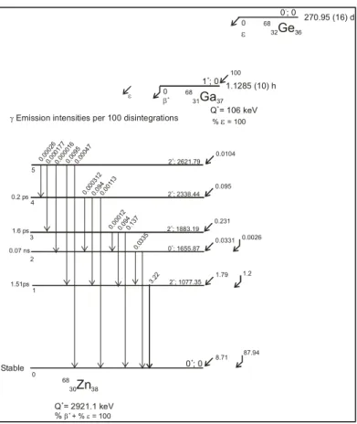
In the Figure 1 it is showed a decay scheme of 68Ge and 68Ga [2], where it is possible to verify
Figure 1. Decay scheme for 68(Ge + Ga).
Source: <http://www.nucleide.org/DDEP_WG/Nuclides/Ga-68_tables.pdf>
2. METHOD
2.1. Sum-peak Method
The sum-peak method was proposed by Brinkman [5, 6] for a well type NaI (Tl) scintillation detector. This method is very simple, using just a single detector, that may be a NaI (Tl) scintillation crystal or an high resolution HP(Ge) detector. To be measured by sum-peak method, the nuclide should be present two or more electromagnetic radiation simultaneously. The radiations may be two gamma rays in coincidence, as 60Co and β+ emitters, or one X-ray followed by one gamma ray, as 65Zn and 51Cr [7].
Equation (1) expresses the fundamental relation of the sum-peak method.
68 32Ge36 270.95 (16) d 0 ; 0 -0 ε Q = 106 keV+ % = 100ε 68 31Ga37 68 30Zn38 1.1285 (10) h Stable
γ Emission intensities per 100 disintegrations 1 ; 0+ Q = 2921.1 keV % + β+ + % = 100ε 0 ; 0+ 2 ; 2621.79+ 2 ; 2338.44+ 2 ; 1883.19+ 0 ; 1655.87+ 2 ; 1077.35+ 87.94 1.2 0.0026 0.0104 100 0.095 0.231 0.0331 1.79 8.71 5 4 3 2 1 0 0.000 26 0.000 177 0.000 016 0.009 5 0.000 47 0.000 312 0.000 12 0.033 5 3.22 0.094 0.094 0.001 13 0.137 0 β+ ε 0.2 ps 1.6 ps 0.07 ns 1.51ps
(1) where, N0 is the source activity, NT is the number of total rate interactions on the spectrum, A1and
A2 are photopeak counting rates and A12 is the sum-peak counting rate, which results from the
sim-ultaneous interaction of the two radiations on detector.
For this type of emitters (pure β+ emitters or β+-γ emitters), the method is possible only if the
source is positioned inside the well, because the geometry must be approximately 4π [5]. This re-striction is due to the angular correlation between the annihilation rays to be extremely strong. Be-cause of this, Brinkman [5] proposed the following equation (2) for the source positioned inside de well when the counting geometry is less than 4π:
(2)
where, W is a coincidence factor, which is equal to 1 if the geometry is very close to 4π.
The W value can be experimentally determined by measuring a 22Na source inside the well of a
NaI(Tl) detector in the geometry equal or less than 4π. In this case three sum-peaks can be observed in the spectrum with the following equations:
(3) (4) (5) As the activity must be the same in the three equations, the W value may be obtained combining equations (3), (4) and (5), as follow:
(6) (7) W N N A A A T 0 12 2 1. ) ( + = 01 1785 1274 511. ) ( N N A A A T = + 02 2276 1274 1022. ) ( N N A A A T = + 03 1022 2 511 ). ( N W N A A T = + 03 02 01 N N N = = ) /( ) /( 1022 2 511 02 1022 2 511 01 AA NT N AA NT N W = + = + 0 12 2 1. N N A A A T = +
The photon spectrum of the 68(Ge + Ga) source placed inside the well presents two photopeaks
that are used in the sum-peak method: 511 keV (A1 and A2) and the peak from the sum of two 511
keV photons. Then, the fundamental sum-peak method equation is:
(8) with
(9)
where: A511keV is the count rate on the photopeak annihilation radiation (511 keV) and A1022keV is
the count rate on the two annihilation rays in coincidence (sum-peak = 511+511 keV).
2.2. 4πβ−γ Live-timed anticoincidence counting
There is no basic difference between coincidence and coincidence counting. The anti-coincidence method is a complementary method of the anti-coincidence method that initially was con-sidered by Bryant [8] in the particular case of nuclides that present meta-stable levels in the decay scheme. Later on, Baerg [9] introduced the use of live time to the anti-coincidence system that elim-inates the correction of dead time using an extending dead time device. In the present version LNMRI uses two modules of MTR2 in its anti-coincidence system [10, 11]. These modules have been introduced in the LNMRI laboratory in December of 2005. In this work it was used the mini-mum dead time of 50 µs in the beta and gamma channels.
The activity of a radionuclide can be determined using equation (10), which is the classic equation of the coincidence method differing only in that Nc, the coincidence count rate that is
determined for one given gamma window as the difference between the gamma rate and uncorrelated gamma rate, represented in (10).
(10) R N N0 = T + keV keV keV A A A R 1022 511 511 . = w i w w N N N N A γ γ γ β − =
Where, Nβ is the count rate in the beta channel;Nγwis the count rate in the gamma window and w
iN
γ is the uncorrelated gamma count rate.
2.2. Uncertainties
The statistical components were evaluated using the equation (8). In this equation there are not nuclear parameters involved. Then, the main components that affect the A-type are: calculation of photopeak net areas; extrapolation to 0 keV energy; and background counts. From the derivation of Equation (8), it is possible to evaluate the statistical uncertainties, according to:
2 2
2
0) ( ) ( )
(∆N = ∆NT + ∆R (11)
where, NT is the sum of counts in the spectrum (E=0 to 1350 keV) The NT value is withdrawn of the spectrum and it is obtained from the gross counts (Ng=SUM(sample spectrum)) stripped the background counts. The equation (8) must be analyzed in parts for clear understanding. The background spectrum live time (Tlive(BG)) and the sample spectrum live time (Tlive(sample spec-trum)) are different. The count rate of the background spectrum T(BG) is expressed by: ) ( ) ( ) ( BG live BG BG T SUM T = (12) if: NT =Ng −BG (13)
and, N =g SUM(samplespectrum) (14)
then, NT = Ng −T(BG).Tlive(samplespectrum) (15)
Taking into account the extrapolation to 0 keV energy: ) 0 / . ( . ( ) ) ( T extr p E keV T N NT = g − BG live samplespectrum + = (16)
Following the rules for uncertainties: 2 2 ) ( ) ( 2 2 ( ) [ ( . )] ( 0) )
(∆NT = ∆Ng + ∆ TBG Tlivesamplespectrum + ∆ext→ (17)
Nγ , T(BG).Tlive (sample spectrum), and (ext 0) are counts and from the Poisson distribution the relations
below can be used:
g g N N = ∆ )2 ( (18) ) ( ) ( 2 ) ( ) ( ). ] . (
[∆ TBG Tlivesamplespectrum =TBG Tlivesamplespectrum (19)
0 ) 0 (∆ext→ 2 =ext→ (20) Then, ) 0 ( . )
(∆NT 2 =Ng +T(BG)Tlive(samplespectrum)+ ext→ (21) The uncertainty in R is:
12 2 1 N N N R = (22) 2 12 12 2 2 2 2 1 1 2 ∆ + ∆ + ∆ = ∆ N N N N N N R R (23)
R is obtained when N0 is determined, having ∆R/R and R:
( )
2 2.R2 R R R ∆ = ∆ (24)The total square uncertainty is the sum of the equations (14) and (17).
R N N N N N N = T + = T + 12 2 1 0 (25)
(
)
( )
2 2 0 2 2 0 2 0) ( R N N N N N N R T T ∆ ∂ ∂ + ∆ ∂ ∂ = ∆ (26)(
)
2 2( )
2 2 2 0 1 N 1 R N = ∆ T + ∆ ∆ (27)Besides the A-type uncertainties derived from N0, others uncertainties components also affect
the accuracy of the activity of 68(Ge + Ga) solution (sample weight, dilution factor and decay
cor-rection) considered in this work. According to the masses used to prepare 68(Ge + Ga) sources for
sum-peak method, the sample weighting component was 0.10 %, and for the anti-coincidence method it was 0.05 %. The uncertainty dilution factor component may be neglected.
The decay correction component (half-life) is evaluated by the Equation (7).
2 / 1 2 / 1 1/2 . . 2 ln T t ST T µ ∆ = (28)
where: ∆t is the time interval between counting date and reference date; µT1/2 is the uncertainty in
the half-life value [2]; and T1/2 is the half-life [2].
3. EXPERIMENTAL PROCEDURE
3.1.Sum-peak Method
The 68(Ge + Ga) original solution was in the form of a solution in 0.1 mol/l HCl, carrier free.
Three sources were prepared from a 68Ge/68Ga diluted solution (dilution factor of 20.622298) by
dropping deposition of known masses onto a cavity in the center of an acrylic disk fixed in a 0.05 mm thick polystyrene film, as show in the Figure 2. The masses were determined in a Mettler Tole-do MT5 micro analytical balance (readability: 1 µg; weighing capacity: 5100 mg) using the pyc-nometer differential weighing technique. The sources were dried in an infrared lamp and immedi-ately after drying, the sources were covered by the same polystyrene film. In the same way more three sources were prepared, but they were not dried. After weighing, the sources were covered by the same film. The six sources were measured with the purpose to investigate if there are losses of
68Ge by volatility (known behavior of 68Ge described in [8]). In order to achieve the decay chain
Figure 2. Scheme for 68(Ge + Ga) source preparation in sum-peak method measurements.
Source: Oliveira et al, 2012 [12].
The sources were placed inside the well type 5”x5” NaI (Tl) gamma ray detector (bottom). In the top of this well type detector there was a 3”x3” NaI (Tl) gamma-ray detector resulting in a set up approximately 4π counting geometry. Figure 3 illustrates the experimental arrangement used for the sum-peak method.
Figure 3. Experimental arrangement used in sum-peak measurements including shield, detectors
and electronic modules.
Source: Oliveira et al, 2012 [12]. NaI(Tl) Detector
3”x3”
Well type NaI(Tl) Detector 5”x5” Pre-amplifier Pre-amplifier Amplifier Amplifier Dual Sum inverter MCA Shield: Pb-10.0 cm
Adhesive polystyrene film 0.05 mm thick Acrylic disk
After dried
Both sides covered with the same film Deposition (2-3 mm)
1.0 mm 25 mm
Measurements were done using the coincidence 511 keV, resulting in a 1022 keV (sum-peak). The measuring time of spectra varied from 2900 to 6000 seconds, which was enough to achieve a statistical uncertainty around 0.03 - 0.04 %. The peak area evaluation and its uncertainties associat-ed were carriassociat-ed out using data acquisition software (Maestro) [13], incorporatassociat-ed within a commer-cial multichannel analyzer. These peak area evaluation in each analyzed spectrum also included corrections for dead-time and background subtraction. In order to minimize the undesirable but pre-sent instrumental pile-up effect that distorts the γ-ray peaks the sources were prepared with low count rate intensity.
The correction factor for decay during counting related to the initial count time is done obeying the equation (29) below:
C t c D e t F λ −λ − = 1 (29)
where, λis the decay constant (λ= ln(2)/T1/2) and tc is the period of time.
The photon spectrum is shown in the Figure 4. The spectrum exhibits two photopeaks which were used for the activity determination, from the equation (1).
Figure 4. 68(Ge + Ga) gamma energy spectrum measured with a 5”x5” NaI (Tl) well type detector
covered by a 3”x3” NaI (Tl) detector resulting in 4π geometry for sum-peak measurements.
Energy (keV) 100 600 1100 1600 0 10000 20000 30000 40000 50000 60000 70000 80000 90000 C ou nt s pe r c ha nn el 511 keV 1022 keV
Due to the low resolution of NaI (Tl) detector, counts from the 1077 keV gamma ray from the decay of 68Ga not appear in the spectrum but interferes in the counts of 1022 keV sum peak and
must be accounted for as follow:
2 2 0 2 1 2 1 0 P N P N ) sum ( N =
ε
γ +ε
γ (30)Where, N(sum) includes the contribution from 1077 keV represented by N0ε2Pγ2 [5].
This interference is small, but the following procedure to correct was adopted. First, it was es-tablished that the lines for 1077 and 1115 keV from 65Zn decay have the same photopeak efficiency.
A 65Zn reference source was used (in the same geometry measurements conditions that 68Ge
sources) to obtain the value of photopeak efficiency. After knowing the interference counts, it was obtained the counts values for 1022 keV alone, which corresponds an increase of 0.26 % in the final value of activity concentration.
To prove that the coincidence factor (W) is 1 in this set up, it was used a 22Na reference solution
due to the fact that this nuclide emits both annihilation radiation and one γ-rays (1274 keV). So, it was possible to determine the value of W from equation (7). For these measurements, five sources from a 22Na solution were prepared in the same way that 68Ge. The W value found is 1.0058 ±
0.0005.
3.2. Live-timed anti-coincidence counting 4πβ−γ
To the anticoincidence measurements nine sources were prepared from the same diluted solu-tion used in the sum-peak method. A set of vials were prepared by adding know masses of the dilut-ed solution to 15 ml commercial scintillator cocktail. It was usdilut-ed three commercial scintillators: Ultima Gold, HI Safe III and Instagel Plus (three vials of each cocktail).
The measurements in the anticoincidence system have been made using liquid scintillation counting in the beta channel and a NaI (Tl) scintillation detector in the gamma channel. In both beta and gamma channels were used the MTR2 modules [14, 10]. The MTR2 modules allow operating with dead time values from 25 to 200 µs. In the measurements of 68Ge it was used a minimum dead
discrimination and extrapolated for 1.0. In order to avoid the contribution of electron capture events from 68Ga and 68Ge, we cut the extrapolation curve in the region of low energy (adjusted to 20
keV), in our experimental condition its mean 1.5 V. The NaI (Tl) was gated on 511 keV region us-ing a sus-ingle channel analyzer in this condition the mainly γ-ray counts events were due positon-annihilation decay. But a contribution from Compton scattering of 1077 keVγ-rays correspond to electron capture events and not due positron emission therefore a correction to the intercept was necessary. In order to determine this correction, it was used a 60Co source for this factor in a similar
proceeding proposed by Zimmerman [1].
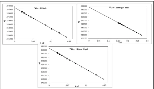
Figure 5 presents one extrapolation curve obtained for each cocktail.
Figure 5. Extrapolation curve counting for each cocktail (HiSafe, Ultima Gold and Instagel Plus)
used in anticoincidence method.
245000 250000 255000 260000 265000 270000 275000 280000 285000 290000 0 0.05 0.1 0.15 N β 1- εβ 68Ge - HiSafe 200000 210000 220000 230000 240000 250000 260000 270000 280000 290000 0 0.05 0.1 0.15 0.2 0.25 0.3 N β 1-εβ 68Ge - Instagel Plus 240000 245000 250000 255000 260000 265000 270000 275000 280000 285000 290000 0 0.05 0.1 0.15 N β 1- εβ 68Ge - Ultima Gold
4. RESULTS AND DISCUSSION
The six sources (dry and liquid sources) prepared for sum-peak method showed activity concentration values with standard deviation of 0.41 %.
The results of each method and the reference value are showed in the Table 1(Reference time: January 1st, 2011-12 h – Official time of Brasília).
Table 1. Result of the standardization of 68(Ge + Ga) solution with the sum-peak method and
comparison with the value obtained by anti-coincidence method. (Reference time: January, 1st,
2011-12 h – official time of Brasília).
MEASUREMENT SYSTEM ACTIVITY/MASS (kBq/g) U (%) k=1 Live- timed anticoincidence counting 4πβ−γ 6.624 0.18 Certified value (traceable to NIST-Gamma
spectrometry) 6.572 1.4
SUM-PEAK METHOD 6.347 0.14
Table 2 shows the main uncertainty components considered in the activity determination by the sum-peak method.
Table 2.Uncertainty components evaluated in the determination of the activity concentration of 68(Ge + Ga) solution using sum-peak method.
Uncertainty Component Type u (%)
Mass determination B 0.10
Live time B 0.01
Decay corrections B 0.01
Statistic counts* A 0.10
Combined uncertainty (k=1) 0.14 *including background and extrapolation to zero keV uncertainties
Table 3 presents the uncertainties components considered in the activity determination by the an-ti-coincidence method.
Table 3.Uncertainty components evaluated in the 68(Ge + Ga) activity determination by anti-coincidence
method.
Uncertainty Component Type u (%)
Statistics counts A 0.02 Fitting procedures A 0.05 Mass weighing B 0.05 Live time B 0.01 Background B 0.04 Decay corrections B < 0.01 Decay branch B 0.13
Correction due to detection
of 1077 keV photons B 0.10
Combined uncertainty (k=1) 0.18
5. CONCLUSION
The small value for the standard deviation (0.41%) for activity values in the sources dried and liquids shows that if the drying is fast and the source is immediately covered, there aren’t signifi-cant losses of 68Ge due to the volatility. The cocktails HiSafe III and Ultima Gold were more
suita-ble for preparing samples for liquid scintillation counting resulting in higher detection efficiencies than those prepared with Instagel plus.
The value of W proves that β+ and β+-γ emitters can be measured in this experimental
arrange-ment.
The difference between results obtained by sum-peak method and 4πβ−γ live-timed anticoinci-dence counting shows that an improvement should be done in the sum-peak method arrangement.
Due to the better resolution of the Ge detector compared to the NaI (Tl) detector, an option to im-prove the measurements of the sum peak method will be to exchange the 5”x5” NaI (Tl) well type detector and the 3”x3” NaI (Tl) detector by well type Ge detector and a planar Ge detector. Thus, the peak 1077 keV of the 68Ge can be separated from the peak of 1022 keV (sum), eliminating the
interference that is probably causing the difference in the results between the sum peak method and the anticoincidence method.
REFERENCES
[1] ZIMMERMAN, B. E.; CESSNA, J. T.; FITZGERALD, R. Standardization of 68Ge/68Ga using
three liquid scintillation counting based methods. Journal of Research of the National Insti-tute of Standards and Technology, vol.113, p 265-280, 2008.
[2] SCHÖNFELD, E. DDEP- Decay Data Evaluation Project Data. Available at:
<http://www.nucleide.org/DDEP_WG/Nuclides/Ge-68_tables.pdf>. Last accessed:January 1st,
2011.
[3] CHISTÉ, V. and BÉ, M.M. DDEP-Decay Data Evaluation Project Data. Available at:
<http://www.nucleide.org/DDEP_WG/Nuclides/F-18_tables.pdf>.Last accessed: January 1st,
2011.
[4] GRIGORESCU, E. L.; NEGUT, C. D.; LUCA, A.; RAZDOLESCU, A. C.; TANASE, M. Standardization of 68(Ge + Ga). Applied Radiation and Isotopes, vol.60, p. 429-431, 2004.
[5] BRINKMAN, G. A.; ATEN JR, A. H. W.; VEENBOER, J. Th. Absolute standardization with a NaI(Tl)crystal—I: calibration by means of a single nuclide. International Journal of Applied Radiation and Isotopes, vol.14, p. 153-157, 1963.
[6] BRINKMAN, G. A.; ATEN JR., A. H. W. Absolute Standardization with a NaI(Tl) Crystal-III – Calibration of β+ - Emitters. International Journal of Applied Radiation and Isotopes.Vol.14,
p. 503-510, 1963.
[7] ALMEIDA, M. C. M.; IWAHARA, A.; POLEDNA, R.; DA SILVA, C. J.; DELGADO J. U. Absolute disintegration rate and 320 keV γ-ray emission probability of 51Cr. Nuclear Instru-ments and Methods in Physics Research A, vol.580, p. 165-168, 2007.
[8] BRYANT, J. Anticoincidence counting method for standardizing radionuclides emitting delayed gamma rays. Applied Radiation and Isotopes, vol.13, p. 273-276, 1962.
[9] BAERG, A. P.; MUNZENMAYER, K.; BOWES, G.C. Live-timed anticoincidence counting with extending dead-time circuitry. Metrologia, vol.12, p.77-80, 1976.
[10] DA SILVA, C. J.; IWAHARA, A.; POLEDNA, R., BERNARDES, E. M. O.; DI PRINZIO, M. A. R. R.; LOPES, R. T. Standardization of Ga-67, Cr-51 and Fe-55 by live-timed anti-coincidence counting with extending dead time. Applied Radiation and Isotopes, vol. 66, p.
231-235, 2008.
[11] DA SILVA, C. J.; IWAHARA, A.; POLEDNA, R.; BERNARDES, E. M. O.; DI PRINZIO, M. A. R. R.; DELGADO, J. U.; LOPES, R. T. Standardization of 241Am, 124Sband 131I by
live-timed anticoincidence counting with extended dead time. Applied Radiation and Isotopes,
vol. 66, p. 886-889, 2008.
[12] OLIVEIRA, E. M.; IWAHARA, A.; POLEDNA, R.; DELGADO, J.U.; DA SILVA, C. J.; DA SILVA, R. L.; LOPES, R. T. Standardization of 65Zn by sum-peak method. Applied Radia-tion and Isotopes, vol. 70, p. 2087 - 2090, 2012.
[14] BOUCHARD, J. A new set of electronic modules (NIM standard) for a coincidence system using the pulse mixing method. Applied Radiation and Isotopes, vol. 56, p. 269-273, 2002.
[13] MAESTRO II, 2002. Software Operator’s Manual. Maestro for Windows. Version 5.33. EG&G. ORTEC.