Article
Printed in Brazil - ©2016 Sociedade Brasileira de Química0103 - 5053 $6.00+0.00*e-mail: [email protected]
#Maristela de F. S. Peres and Karina Nigoghossian contributed equally to this paper.
Bacterial Cellulose Membranes as a Potential Drug Delivery System for
Photodynamic Therapy of Skin Cancer
Maristela F. S. Peres,a,# Karina Nigoghossian,b,# Fernando L. Primo,a,c Sybele Saska,b
Ticiana S. O. Capote,d Raquel M. S. Caminaga,d Younes Messaddeq,b
Sidney J. L. Ribeirob and Antonio C. Tedesco*,a
aCentro de Nanotecnologia, Engenharia Tecidual e Fotoprocessos Voltado a Saúde, Grupo de
Fotobiologia e Fotomedicina, Departamento de Química, Faculdade de Filosofia, Ciências e Letras de Ribeirão Preto (FFCLRP-DQ), Universidade de São Paulo, Campus Ribeirão Preto,
14040-901 Ribeirão Preto-SP, Brazil
bInstituto de Química, Universidade Estadual Paulista “Júlio de Mesquita Filho” (UNESP),
CP 355, 14801-970 Araraquara-SP, Brazil
cDepartamento de Bioprocessos e Biotecnologia, Faculdade de Ciências Farmacêuticas,
Universidade Estadual Paulista “Júlio de Mesquita Filho” (UNESP), 14801-902 Araraquara-SP, Brazil
dDepartamento de Morfologia, Faculdade de Odontologia, Universidade Estadual Paulista
“Júlio de Mesquita Filho” (UNESP), 14801-903 Araraquara-SP, Brazil
The development of drug delivery systems for photodynamic therapy (PDT) is increasingly demanded due to the hydrophobicity presented by most of photosensitizers molecules. Bacterial cellulose (BC), a highly pure cellulose produced by bacteria, possesses the essential features for applications in drug delivery systems, such as large surface area and excellent loading capacity. BC membranes prepared containing a photosensitizer, chloroaluminum phthalocyanine (ClAlPc), were tested aiming applications as a drug delivery system for PDT skin cancer protocols. BC membranes production was optimized regarding thickness and optical transmission. Thinner membranes lead to higher relative incorporation efficiencies. Skin permeation and retention in vitro tests were
performed by using pig’s ears as a skin model. ClAlPc was retained at stratum corneum and epidermis/dermis, showing adequate properties for topical administration of ClAlPc. Photophysical studies showed that singlet oxygen production was not affected for ClAlPc compartmentalized in the BC array. BC-ClAlPc membranes did not present cytotoxic effects in vitro.
Keywords: bacterial cellulose, phthalocyanine, drug delivery system, photodynamic therapy
Introduction
A variety of new approaches and therapeutic protocols has been emerging in the last years as a consequence of the progresses in the field of applied nanobiotechnology to treat a huge spectrum of diseases, through improved targeting or delivery of the therapeutic agent. In the case of application for cancer therapeutics, these innovative technologies tend to be less invasive and more effective when compared with the most conventional antitumor treatment techniques:
surgery, radiotherapy and chemotherapy.1-3 Photodynamic
therapy (PDT) has been focus of intense research during the last decades as an emerging therapeutic modality. Despite the fact of being a novel technique, PDT is a well-established procedure mainly used in the treatment
of neoplastic and non-neoplastic diseases.4 This therapy is
focused mainly in skin cancer treatment, from early stage until melanoma one (there is no indication for the use of this therapy to treat melanoma). PDT is already used as a clinical tool for treating tumors and a considerable number of pathological conditions, such as arthritis, skin disorders
and many other non-oncological diseases.5-8 The technique
synthetic sources, in the presence of a specific drug delivery
system) followed by visible light activation.9 The classical
photosensitizers are chemical compounds, which absorb visible light in a specific wavelength. Upon irradiation, the photosensitizer is promoted from its ground singlet state to its triplet excited state followed by reactions that occurs in a complex mechanistic pathway through classical
photochemical reactions,10 leading to the production of
reactive oxygen species (ROS), of which singlet oxygen is one of the main active species in photodynamic processes.
Most of the photosensitizers used in PDT present difficulty for administration in physiological environment due to their hydrophobicity. Therefore, it is necessary to develop drug delivery systems capable to overcome the
tendency to aggregate in aqueous media.11 The development
of new drug delivery systems based in nanobiotechnology has improved the therapeutic and toxicological properties of existing chemotherapeutic and photochemotherapeutic agents and fostered the implementation of new agents. A wide assortment of biomaterials with suitable biological properties is offered today as a potential tool to be used as a drug delivery system, but considerable attention has been drawn to the use of biocompatible polymers based ones. By combining drugs with different polymers, either synthetic or natural, it is possible to optimize pharmacokinetics and biodistribution of the agents, and, consequently efficacy and
safety of therapy are improved.12 Additionally, the polymer
offers protection against enzymatic, hydrolytic and other
types of chemical degradation.13
Polymeric release systems can be designed in many forms, including matrices or membranes in which
the active ingredient is dispersed or dissolved.14-16
The administration route, carrier formulation, release mechanism and physiochemical properties of drug molecule are determinant factors, which influence on drug release rate and thus must be considered when selecting a
suitable polymer of a release device.17 Moreover, the ideal
polymers for the development of dry delivery systems should be chemically inert to the drug action and present
appropriate physical features.18
Bacterial cellulose (BC) is a polysaccharide of glucose
produced by Gluconacetobacter sp. that is superior to
plant cellulose due to its purity and nano-morphology. BC presents high water-holding capacity, large surface area, and high crystallinity, besides being renewable, biocompatible
and biodegradable.19 The incorporation of organic/inorganic
compounds in its structure is possible due to the network of ribbon-shaped nanosized cellulose fibrils and the high presence of water. A number of studies in the literature report the successful use of BC membranes in biomedical
applications20 and, more specifically, drug delivery
systems21 due to their unique physical and mechanical
properties.22 Such membranes are particularly advantageous
in topical or transdermal drug delivery systems, as they have the ability to absorb exudates and adhere to irregular skin
surfaces, such as the oral mucosa.23-26 Moreover, a previous
study reported the good skin tolerance of BC membranes.27
As the majority of transdermal patches are manufactured by superimposing different materials, a system composed of fewer or even a single layer, such as a BC film, could simplify the preparation procedure and lower production
costs.28 Recently, our group has reported a system based
on BC membranes incorporated with the photosensitizer chloroaluminum phthalocyanine (ClAlPc) and luminescent upconversion nanoparticles, which emits light at the wavelength range of ClAlPc absorption under infrared
irradiation, within the biological transparency window.29
Chitosan, which is chemically related to cellulose and plant
cellulose nanocrystals, has been studied by Schmitt et al.30
and Drogat et al.31 in photodynamic therapy. In view of
these facts, the development of new delivery systems that can efficiently deliver ClAlPc could enable its clinical use for topical PDT.
The aim of the present study was to show the feasibility of bacterial cellulose as a potential drug delivery system for photodynamic therapy. The BC-ClAlPc membranes were evaluated regarding the incorporation efficiency of
photosensitizer and in vitro diffusion studies with Franz
cells. The BC-ClAlPc membranes were tested as a setup to activate photoprocesses useful for treat neoplastic and non-neoplastic diseases susceptible to the photoactivation process. The cytotoxicity potential of BC-ClAlPc was evaluated aiming a safe use in humans.
Experimental
Microorganisms
The strains used were Acetobacter xylinum (ATCC 23760)
and other isolated in the laboratory identified by Centro Pluridisciplinar de Pesquisas Químicas, Biológicas e Agrícolas (CPQBA), Universidade Estadual de Campinas
(UNICAMP) as Gluconacetobacter sp.(GL). BC obtained
from each microorganism were produced in two different stages of growth, in order to vary the thickness of the
membranes: ATCC(2 and 3 days) and GL (1 and 3 days).
Chemicals
microbiological culture media. ClAlPc, dimethyl sulfoxide (DMSO), phosphate buffer saline solution (PBS), sodium 3’-[1-(phenylaminocarbonyl)-3,4-tetrazolium]-bis (4-methoxy-6-nitro) benzene sulfonic acid hydrate (yellow tetrazolium salt XTT), cell culture medium Ham’s F-10 (HAM-F10):Dulbecco’s modified Eagle’s (DMEN), 1:1 and doxorubicin were purchased from Sigma-Aldrich (St. Louis, MO, USA). Fetal bovine serum (FBS) and DMEM medium without phenol red were purchased from Cultilab (Campinas, SP, Brazil). XTT:electron solution 50:1 cell proliferation kit II (XTT) was acquired from Roche Molecular Biochemicals (Basel, Switzerland).
Bacterial cellulose production
The BC membranes were produced by growing the bacteria at 30 °C for 24 h in liquid medium under static
conditions. Culture medium was composed of 50 g L−1
glucose, 4 g L−1 yeast extract, 2 g L−1 potassium phosphate
monobasic anhydrous, 0.73 g L−1 magnesium sulphate
heptahydrate, and 20 g L−1 ethanol.32 After this period,
the flask was vigorously agitated and 10% of the culture media was withdrawn and inoculated into a new liquid production medium at 30 °C over different periods of time (24, 48 and 72 h).
Bacterial cellulose purification
After the incubation time, BC membranes were withdrawn from the culture medium and treated with a
0.1 mol L−1 NaOH solution, at 80 °C, for 30 min to eliminate
all attached cells. Then, the membranes were washed with distilled water to remove components of the culture media and other residues until its whitening and reaching pH 7.0. BC membranes were dried at 30 °C and then stored in a desiccator. Measurements of the thickness of the membranes were performed in a Formtracer profilometer, model SV-CS25 (Mitutoyo, Kawasaki, Japan).
Chloroaluminum phthalocyanine (ClAlPc) in bacterial cellulose membranes
The BC wet membranes (5 × 5 cm) were weighed.
Moderate pressure was applied to the surface of the
membranes to remove water until the mass loss of 50%.
The drained BC membranes were then soaked for 5 h in ClAlPc solutions in ethanol at concentrations of 1, 5 and
10 µmol L−1. The membranes were dried at 28 °C in a
ventilated oven for 6 h.
To facilitate the understanding of the results, it is important to highlight that BC refers to bacterial cellulose
membranes and BC-ClAlPc refers to the bacterial cellulose membranes incorporated with the photosensitizer ClAlPc.
The samples are referred according to three factors: (i) the
strains from which membranes were produced: ATCC(A)
or GL(G); (ii) thestages of growth: one, two or three days
(1d, 2d and 3d, respectively); (iii) when applicable, the
theoretical concentrations of ClAlPc (1, 5 and 10 µmol L−1)
are indicated by the numbers 1, 5 and 10 at the end of samples names. Table 1 presents the sample names according to these variables.
The quantification of ClAlPc was carried out according to a validated methodology based in fluorescence spectroscopic technique (Spex-FluoroLog 3, Horiba Jobin Yvon, Edison, NJ, USA) with excitation at fixed wavelength
(λex 615 nm) and emission at 680 nm (slits were adjusted
to 10 nm).33
The ClAlPc content was analyzed in two steps. Firstly, BC-ClAlPc (1 mg) was immersed in 5 mL of acetonitrile at 60 ºC for 15 min to determine the free ClAlPc content (non-incorporated drug). Subsequently, the membrane was immersed in 5 mL of acetonitrile and homogenized in an IKA Ultra-Turrax T-8 Basic (Staufen, Germany) at 11000 rpm for 5 min to quantify ClAlPc retained into BC fibers. The concentrations were obtained from a calibration standard curve. Total ClAlPc concentration corresponds to the sum of the values found in sequential steps described above. Efficiency of incorporation (EI) of the photosensitizer into BC membranes was calculated by the following equation
(1)
The experiments were performed in duplicate. One-way analysis of variance (ANOVA; OriginPro 8.0, OriginLab,
Table 1. List of sample names and corresponding variables
Sample Strain growth / dayStage of Theoretical concentration of ClAlPc / (µmol L−1)
G1d1 GL 1 1
A2d1 ATCC 2 1
G3d1 GL 3 1
A3d1 ATCC 3 1
G1d5 GL 1 5
A2d5 ATCC 2 5
G1d10 GL 1 10
A2d10 ATCC 2 10
Northampton, UK) was used for statistical analysis at a
significance level p < 0.05.
Scanning electron microscopy images of BC and BC-ClAlPc surfaces were obtained with a LEO equipment model 440 (Leica, Wetzlar, Germany) with an Oxford detector. Fourier transform infrared (FTIR) spectra were obtained with a PerkinElmer Spectrum 2000 Fourier transform infrared spectrophotometer (Waltham, MA, USA) using KBr pellets. Thirty-two scans were acquired
over the range 4000-370 cm−1 with a resolution of 2 cm−1.
In vitro skin diffusion studies
PBS + ethanol 10% (v/v; 7 mL, pH 7.4) were used
as the acceptor medium.34 A calibration standard curve
of ClAlPc in the acceptor medium was constructed as a reference. Aliquots of ClAlPc were added with a microsyringe (Hamilton, Ocala, FL, USA) directly in medium in a 1.0 cm quartz cell under constant stirring. The fluorescence emission spectra were determined in the
range of 0.219-1.314 µg mL−1 (n = 10).
The skin was extracted from the dorsal surface of pig’s ears, obtained directly from a slaughterhouse (Olho d’Água Ind. e Com. de Carnes Ltda., Ipuã, SP, Brazil), to be used as a model of skin. The front ear was
dissected. The skins were applied in the tests in natura.
These tissues were stored in a freezer up to a maximum
of 90 days before their use in all in vitro procedures as
an animal model. In vitro retention studies were carried
out using the skin tissues fixed on Franz diffusion cells
(1.77 cm2 diffusion surface areas) maintained at 37 °C by
a circulating water bath and stirring speed of 300 rpm. Samples were directly applied topically to the exposed area of skin. After 6, 12 and 24 h of diffusion test, the system was dismounted and the skins were carefully removed to perform the tape-stripping analyses. In this second step, the stratum corneum (SC) was extracted from the diffusion surface by using 15 standardized stripping tapes (Scotch 3M, Maplewood, MN, USA). The tape-strips were placed in 5 mL of acetonitrile in glass tubes, stored overnight and then stirred for 1 min before filtration. The remaining skin (epidermis + dermis) was cut in small pieces and added to 5 mL of acetonitrile in a tissue homogenizer, sonicated for 20 min and then centrifuged for 15 min at
5000 rpm.35 ClAlPc amounts present in tape-strips and
remaining skin (epidermis + dermis) were assayed by fluorescence standard curve. Assays were performed in three independent experiments with five replicates on each. The statistical analysis was performed using the software OriginPro 8.0 (OriginLab, Northampton, UK) by the
method one-way ANOVA at a significance level p < 0.05.
Photophysical studies
Photophysical studies were performed to determine singlet oxygen generation. Lifetime was calculated from kinetic analysis of the mono-exponential decay for transient species obtained at 355 nm. The spectrophotometer used for time-resolved measurements was an Edinburgh analytical instruments, model FL9000CD (Livingston, UK). The source of irradiation was a pulsed Nd:YAG laser from Continuum (Surelite I-10, Continuum, Santa Clara, CA, USA) adjusted for the third harmonic (355 nm). The pulse length was 8 ns and the repetition rate was 10 Hz. The pulse energy was 15 mJ, measured by a power meter (FieldMaster, Coherent, Santa Clara, CA, USA). The decay kinetics was measured at a single wavelength using a monochromator M300 and a photomultiplier R928P from Bentham Instruments (Livingston, UK). Singlet oxygen was detected by phosphorescence emission signal at 1270 nm using a germanium photodetector of North Coast Scientific Corporation, model 823 (North Coast Scientific, LLC., Santa Rosa, CA, USA). The software provided by Edinburgh instruments (L900, Livingston, UK) was used to obtain and analyze the exponential decay curves, based on iterative algorithm Marquadt (analysis of nonlinear least squares). The BC membrane produced by ATCC
with theoretical ClAlPc concentration of 5.0 µmol L−1 was
analyzed. ClAlPc in ethanol was adopted as a reference. The experiments were done using a solution of pheophorbide
A in ethanol as a standard for relative calculations.36 The
concentrations of reference solutions were adjusted based on the absorbance at 355 nm (excitation wavelength), fixed in 0.3 to avoid any internal filter effects. The measurements were performed under air-equilibrated conditions.
Cytotoxicity test
The potential cytotoxic effect of the new biomaterial obtained was evaluated by XTT assay that quantifies the cells by measuring their metabolic activity. The methodology is based on the cleavage of the yellow tetrazolium salt XTT by metabolically active cells, forming an orange formazan dye that can be measured by its absorbance at 492 nm. Thus, this conversion occurs only in viable cells due to the
activity of mitochondrial dehydrogenases.37
The BC-ClAlPc membranes were tested using an eluate prepared according to the ISO 10993-12:2007(E),
considering the surface area (6 cm2 mL−1). The membranes
membranes were removed from the medium and this resulting eluate was used to carry out the treatment.
Chinese hamster ovary cells (CHO-K1) were cultured in the medium HAM-F10:DMEN 1:1 supplemented with
10% of FBS at 37 ºC and 5% of CO2. Cells were used after
the third passage. Cytotoxicity test was performed in three independent replicates. Each treatment, including positive controls (PC) and negative controls (NC), was carried out in triplicate using cell culture plates.
Cells (2 × 10⁴) were seeded in 24-well plates in culture
medium (1 mL, HAM-F10:DMEN 1:1) supplemented with
10% of FBS at 37 ºC and 5% of CO2. After 24 h, the cells
were washed with PBS, and then treated with the eluates. The cells were exposed to the eluates for 24 h. NC were cells CHO-K1 without any treatment (untreated controls),
while PC were treated with doxorubicin (3 µg mL−1)
for 24 h.
After the treatment, the cultures were washed with PBS (250 µL) and inserted in the culture medium supplemented with FBS. After 24 h of incubation, the cells were washed with PBS (250 µL). DMEM medium without phenol red was added (1 mL), followed by 60 µL of XTT/electron solution 50:1 that remained in culture at 37 ºC for 3 h. The culture medium was then transferred to a 96-well plate, and the absorbance was measured by a microplate reader (VersaMax, Molecular Devices, Sunnyvale, CA, USA) at 492 and 690 nm. The absorbance is directly proportional to the number of viable cells in each treatment after 24 h of exposure.
For each analyzed sample, the results from three individual experiments (each one made in triplicate) were subjected to one-way ANOVA followed by Tukey’s test. Dunnett’s test was also applied to compare data from treated groups to the negative control. BioEstat statistical package version 5 was used (Universidade Federal do Pará, Belém, PA, Brazil) to perform the tests. Differences were
considered statistically significant when p < 0.05.
Results and Discussion
Bacterial cellulose membranes characterization
The bacterial cellulose membranes were obtained with different characteristics regarding thickness and transparency in accordance with the bacteria strain (ATCC or GL) and the growth time (1, 2 or 3 days). The thickness of pure bacterial cellulose membranes (Figure 1) were characterized by using a profilometer. One of the goals of this work was to produce BC membranes with different characteristics to select the most appropriate for the intended application. Thickness is a structural
property of the membranes that influences loading and releasing efficiency of drug into the polymeric matrix. Note that the membranes produced by ATCC (with stage of growth of 2 and 3 days, A2d and A3d) present greater values of thickness (103.2 ± 6.9 and 156.0 ± 22.4 µm) due to a greater amount of cellulose fibers present. The membranes produced by the strain GL (with stage of growth of 1 and 3 days, G1d and G3d) are thinner (thickness of 39.7 ± 5.3 and 62.2 ± 4.6 µm).
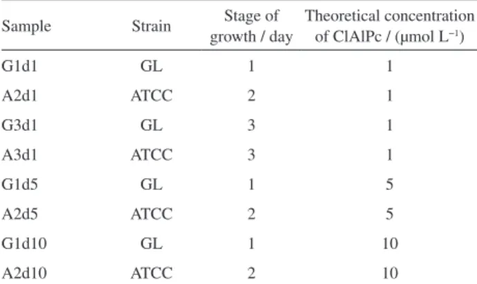
The electronic spectroscopy in the visible region was applied to evaluate the optical transmission of the membranes. Figure 2 shows the spectra of BC membranes, confirming the greater transparency at lesser thickness. We observed similar transparency for BCs produced by different strains. It is expected that a higher amount of fibers favors the incorporation of a greater amount of drug. The amount of fibers is improved with prolonged incubation of
Figure 1. Thickness of pure bacterial cellulose (BC) membranes.
400 500 600 700
40 50 60 70 80 90 100
T
ra
n
s
m
it
ta
n
c
e
/
%
Wavelength / nm
A2d G1d
A3d G3d
Figure 2. Optical transmission spectra of pure bacterial cellulose (BC)
the bacteria. On the other hand, thicker membranes obtained from longer cultivation have lower transparency. The highly transparent media for incorporation of the photosensitizer is favorable for the transmission of the source of light used for excitation.
ClAlPc incorporation in bacterial cellulose membranes
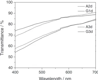
The strategy used here was the incorporation of ClAlPc into the bacterial cellulose membrane, for use in PDT trials for cancer treatment. The pure BC wet membrane (Figure 3a) was soaked in a solution of ClAlPc (Figure 3b) and then dried (Figure 3c). The color of the membranes turned from white to blue after incorporation of ClAlPc. The morphology was investigated by scanning electron microscope (SEM) as shown in Figure 3d. The micrographs of BC-ClAlPc showed the tridimensional fibrillar network characteristic of BC and the absence of aggregates formation, which indicated no precipitation of ClAlPc.
Figure 4 displays the FTIR spectra of ClAlPc, pure BC dried membrane and BC-ClAlPc. The characteristic vibrational frequencies assigned to cellulose were
observed at 3500 to 3200 cm−1 (OH stretching), 2908 cm−1
(CH stretching of CH2 and CH3 groups), 2700 cm−1
(CH2), 1645 cm−1 (water OH bending), 1435 cm−1 (CH2
symmetric bending), 1370 cm−1 (CH bending), 1160 cm−1
(anti-symmetric bridge C–O–C stretching), 1111 and
1056 cm−1 (skeletal vibrations involving C–O stretching).19
The phthalocyanines present the following characteristic
bands: at around 518 and 760 cm−1 (Al−N stretching),
1329 cm−1 (C−N−C stretching), 1121 cm−1 (C=C stretching
of benzene rings) and 489 cm−1 (stretching Cl−Al).38 The
spectrum of BC-ClAlPc is basically formed by the sum of the bands present in the BC and ClAlPc spectra. The
decrease in intensity of the band with a peak at 2908 cm−1
(CH stretching of CH2 and CH3 groups) on BC-ClAlPc
spectrum suggests that the presence of the ClAlPc affected the cellulose groups, due to the interactions between the hydrophobic ClAlPc molecule and CH groups of cellulose, and confirms the strong interaction between the BC and ClAlPc.
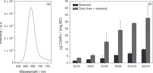
The incorporation efficiency of ClAlPc from the solutions of different concentrations into membranes with different characteristics was studied. Fluorescence spectra (excitation at 615 nm) were measured after the steps of extraction of free ClAlPc in acetonitrile and homogenization of BC membranes. The ClAlPc well known emission at 680 nm, obtained under 615 nm excitation, is shown in the Figure 5a. The presence of the characteristic emission of ClAlPc confirmed the incorporation of this phthalocyanine
derivative into BC fibrils.33,39
Figure 5b shows the ClAlPc amount retained in the fibrils (measured at homogenization step) and total ClAlPc for membranes with different theoretical concentrations
(1, 5 and 10 µmol L−1). These membranes were produced
with the minimal time of growth stage that was enough to obtain membranes with satisfactory mechanical resistance and good transparency (that is 1 day for GL and 2 days for ATCC). The incorporation efficiencies of ClAlPc are listed in Table 2. The statistical analysis (ANOVA)
confirmed a significant difference (p > 0.05) between the
membranes. It was possible to determine an efficient and
Figure 3. Photographs of pure bacterial cellulose (BC) wet membrane
(a); chloroaluminum phthalocyanine (ClAlPc) wet (b); dried BC-ClAlPc membrane (c); and scanning electron microscope (SEM) image of BC-ClAlPc (d).
Figure 4. Fourier transform infrared (FTIR) spectra of chloroaluminum
validate quantification method for analyses of ClAlPc
loaded into cellulose.33 Studies have investigated the
incorporation efficiency of ClAlPc in different bacterial cellulose membranes. A2d showed higher concentrations in steps of extraction and homogenization for all theoretical
concentrations studied (1, 5 and 10 µmol L−1). Figure 5b
shows that, as the theoretical concentration of ClAlPc increases, the amount of ClAlPc retained in the cellulose fibers increases in a less pronounced way in respect to the total amount of incorporated drug (free + retained). Thus, membranes with lower ClAlPc theoretical concentration retain the drug more efficiently.
As expected, A2d1 showed the highest incorporation efficiency (87.4%; Table 2). The membranes grown for shorter time (A2d and G1d) have higher incorporation efficiency than A3d and G3d, and therefore they were selected for the skin permeation and retention tests. The
structural characteristics of the three-dimensional network formed by cellulose nanofibrils, such as fiber density and surface area, influence on the interaction between drug molecule and polymeric matrix. Such properties influence on drug incorporation efficiency and release kinetics according to each biomaterial obtained and may be adjusted by varying the BC production process parameters. The higher amount of drug incorporated by thinner membranes (A2d and G1d) can be related to its larger surface area. In addition to this fact, a greater amount of cellulose fibers is another feature that favors higher concentrations, as
observed for the membrane A2d.40
Skin permeation and retention in vitro studies
For in vitro permeation and skin retention, it was
necessary to develop a standard calibration curve to quantify the ClAlPc present in the environment of the receiver solution (PBS pH 7.4, and ethanol 10% v/v). Serial dilutions were made with known concentrations of ClAlPc and the emission spectra were obtained under the same conditions. The curves were plotted from the emission intensities at 680 nm as a function of ClAlPc concentration. Five curves were obtained with high coefficients of determination
(R2), greater than 0.99. The calibration standard curve
was obtained (y = 310357x + 218237, R2 = 0.99) in the
concentration range from 0.219 to 1.314 µg mL−1.
In studies using Franz cells, the permeation profile is drawn from the quantification of ClAlPc present in the receiver solution. The tests performed for BC membranes G1d1 and A2d1 and control test (without the bacterial
Table 2. Percentage of chloroaluminum phthalocyanine (ClAlPc)
incorporated into the cellulose
Sample Incorporation / %
G1d1 47.5 ± 6.7
A2d1 87.4 ± 3.4
G3d1 44.6 ± 8.6
A3d1 41.7 ± 31.9
G1d5 25.7 ± 17.3
A2d5 41.8 ± 8.0
G1d10 27.1 ± 2.8
A2d10 34.0 ± 3.2
640 660 680 700 720 0
1 × 10⁶ 2 × 10⁶ 3 × 10⁶
4 × 10⁶ 5 × 10⁶
In
te
n
s
it
y
/
a
.u
.
Wavelength / nm
G1d1 A2d1 G1d5 A2d5 G1d10 A2d10 0
5 10 15 20 25 30 35 40 45
Retained
Total (free + retained)
(b) (a)
µ
g ClAlPc / (mg BC)
Figure 5. Chloroaluminum phthalocyanine (ClAlPc) fluorescence emission spectrum under 615 nm excitation (a); and ClAlPc retained and total
cellulose matrix) showed no detectable amount of ClAlPc by fluorometry technique in the receiver solution until the period of 12 h. The absence of ClAlPc in the receiving solution may be considered positive for topical application of the photosensitizer, because it prevents its systemic absorption, which may cause a generalized
photosensitization on the patient.41,42 Stratum corneum
is an efficient barrier from the external environment, controlling the flux of endogenous components outside and inside, acting as a first layer of skin with a highest
lipophilic potential.42 The effective tissue penetration is
constantly associated with direct interaction between drugs and SC. For cutaneous diseases is indispensable a higher SC penetration and adequate bioaccumulation for efficient
therapies and biological response.41
In our work, the profile of cutaneous retention in SC and skin-deep layers (epidermis + dermis = E + D)
was carried out. Samples were applied topically under
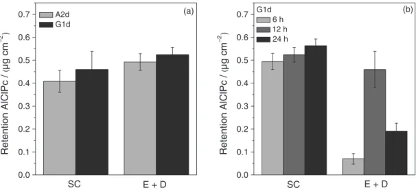
biomimetic conditions in a skin animal model. After hours of administration, the apparatus were dismounted and the skin treated as described in Experimental section. Tape-stripping protocols and tissue homogenization allowed determining the amount of ClAlPc penetrated and retained in skin layers. The retention profiles are shown in Figure 6 and the results are summarized in Table 3. The ClAlPc retained in the skin layers (SC and E + D) was quantified after 12 h of testing. The retention profiles of the samples A2d1 and G1d1 are illustrated in Figure 6a. ClAlPc concentrations were present in the
SC 0.492 ± 0.037 µg cm−2 and 0.524 ± 0.032 µg cm−2 and in
the E + D 0.408 ± 0.047 µg cm−2 and 0.459 ± 0.079 µg cm−2
to the membranes A2d1 and G1d1, respectively. The statistical analysis (ANOVA) confirmed that there is no
significant difference (p > 0.05) between the retention
profiles of the two membranes studied in both skin layers analyzed (SC and E + D). The study of ClAlPc retention in the skin layers as a function of time was conducted for the sample G1d1 for 6, 12 and 24 h (Figure 6b). The tests showed no ClAlPc in the receiver solution until the 24 h test. The retention profiles are shown in Figure 6b. ClAlPc concentrations detected in the SC for the
different test times are: 6 h, 0.495 ± 0.035 µg cm−2; 12 h,
0.524 ± 0.032 µg cm−2; and 24 h, 0.563 ± 0.030 µg cm−2.
Statistical analysis showed that the profile of the 12 h test is not different from the others, i.e., there is no significant difference between 6 and 12 h and between 12 and 24 h; and 6 and 24 h tests are different. In the E + D were quantified:
6 h, 0.070 ± 0.023 µg cm−2; 12 h, 0.459 ± 0.079 µg cm−2;
and 24 h, 0.190 ± 0.036 µg cm−2 (E + D). Statistical analysis
showed a significant difference (p > 0.05) between the
profiles of retention in the epidermis and dermis for the three different test times studied. Large variations in the retention values for the deeper skin layers (epidermis and dermis) were observed between the tests of 6, 12 and 24 h. The use of pig’s ear skin as skin model may have led to variation in experimental results due to the enormous heterogeneity that exists between ears samples from different animals, or even in different regions of the same ear. However, it should be considered that this biological variability reflects the reality and it was possible to find a characteristic retention for the system. The use of human
skin as a model26 would be ideal for skin permeation/
retention studies in vitro. However, this material obtained
from plastic surgery presents limitations to its use, as the low availability and the need to undergo the experiment to
Research Ethics Committees.43-45 Alternatively, the animal
skins, such as primate, pig, rat, guinea pig and snake, are
widely used.46 Three dimensional cultures of human cells47
Figure 6. Penetration profiles of chloroaluminum phthalocyanine (ClAlPc) into the skin layers: epidermis + dermis (E + D) and stratum corneum (SC) for
are also an option, however these materials are deficient in skin-associated epithelial structures (appendages), as
pilosebaceous units, hair follicles and sweat glands.48
Synthetic membranes with defined pore sizes are also employed in assays to Franz cells to reduce inter assay
variation due to biological variability of the skin tissue.49
The pig ear skin was used in this study considering the factors described above. It was also taken into account its high availability with relative ease to obtain and the low cost, once it is a by-product of the food industry. Besides being the animal model that more closely resembles histologically and biochemically to human tissue, after
the primates.46
In PDT, these phenomena related to skin permeation and retention are decisive for treatment of neoplastic cells. Before laser irradiation, it is necessary that an appropriated amount of photosensitizer penetrates the SC to a better interaction with target tissues, to accumulate in malignant cells, to promote an adequate biological response and, thus, to obtain an efficient therapy. An effective delivery system for PDT by topical administration should carry the photosensitizer beyond the stratum corneum to the malignant cells present in
viable layers of the epidermis.41 A global analysis of
this study permits the inference that approximately 0.5
µg cm−2 of ClAlPc remained in the furrows of the stratum
corneum and 0.3 µg cm−2 in the epidermis/dermis. The
drug delivery system containing a derivative of chlorine temoporfin (the synthesis and purification of Foscan® (5,10,15,20-tetra(m-hydroxyphenyl)chlorin) was carried out by Prof PhD Philippe Maillard, coordinator of the Chimie Bioorganique’s Laboratoire, Institute Curie,
Orsay, France) developed by Primo et al.50 promoted a
retention of 0.5 µg cm−2 drug in the stratum corneum
and 0.6 µg cm−2 in the epidermis/dermis. Thus, the
results showed an interesting alternative for delivery of photosensitizers in PDT based on bacterial cellulose, a polymer well known by a wide range of features that favors its use for biomedical applications, as mentioned
above. Particularly in this case, BC-ClAlPc present advantages as a drug delivery system, as the very simple approach of preparation and the absence of cytotoxicity, according to the XTT assay.
Photophysical characterization
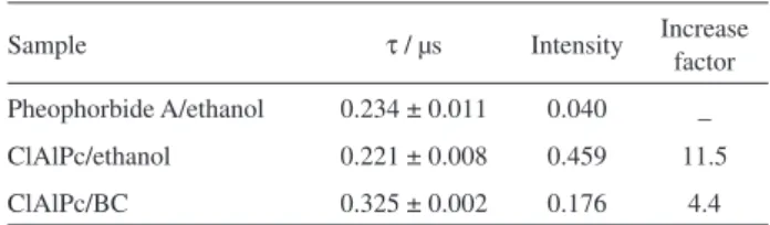
The singlet oxygen analysis was based on the direct detection of its luminescence at 1270 nm to measure the
luminescence-decay kinetics. Lifetimes (τ) and maximum
intensities observed in the decay curves are shown in Table 4. ClAlPc solution in ethanol was also evaluated for the purpose of comparing the effect of the photosensitizer free in solution. Pheophorbide A in ethanol was adopted as a standard solution for relative calculation. The lifetime values found are very close, except for the BC-ClAlPc membrane, which showed a longer lifetime. This difference may be due to the solid form of the sample.
Photophysical studies of BC-ClAlPc were performed for the major reactive species responsible for the destruction of tumor cells, singlet oxygen. The results shown in Table 4 are
consistent with those of Siqueira-Moura et al.,39 which have
found similar lifetime (0.22 µs) for ClAlPc in polymeric nanocapsules. Higher luminescence intensity was obtained for BC-ClAlPc (4.4 times) in respect to the standard solution (pheophorbide A in ethanol). This compound is a derivative bacteriological hydrochloric which produces high yield of the species being studied and is widely used as
a reference in spectroscopic studies.36 Therefore, these data
suggest that BC does not interfere with the production of singlet oxygen, an important condition in the PDT process.
Cytotoxicity assay (XTT cell viability assay)
Evaluation of BC-ClAlPc cytotoxicity was performed by cell viability test using Chinese hamster ovary cells (CHO-K1) because its structures and functions are common to most types of cells, i.e., the objective was to determine the basal cytotoxicity potential of BC-ClAlPc
membrane.51 The cell viability is related to the absorbance
Table 3. Chloroaluminum phthalocyanine (ClAlPc) concentrations
detected in epidermis + dermis (E + D) and stratum corneum (SC) for the samples A2d and G1d after 12 h, and for the G1d tested for 6, 12 and 24 h
Sample time / h SC / (µg cm−2) E + D / (µg cm−2)
A2d 12 0.492 ± 0.037 0.408 ± 0.047
G1d
6 0.495 ± 0.035 0.070 ± 0.023 12 0.524 ± 0.032 0.459 ± 0.079
24 0.563 ± 0.030 0.190 ± 0.036 Mean ± SD (n = 5).
Table 4. Fluorescence lifetimes (τ) and maximum intensities in the decay
curves for the samples
Sample τ / µs Intensity Increase factor
Pheophorbide A/ethanol 0.234 ± 0.011 0.040 _ ClAlPc/ethanol 0.221 ± 0.008 0.459 11.5
ClAlPc/BC 0.325 ± 0.002 0.176 4.4
measure. Negative control was considered 100% cell viability. Figure 7 shows the cell viability (%) expressed as mean and standard error. Cells treated with the eluate from BC-ClAlPc membrane showed 32% of lower cell viability than negative control (without any treatment). Therefore, cell viability of the BC-ClAlPc membrane is not
significantly different from NC (p > 0.05; Dunnett). Thus,
the BC-ClAlPc membrane does not significantly affect cell viability, being non-cytotoxic.
For a safe use in humans, biomaterials should not present cytotoxic effects. The cytotoxicity potential of pure BC was
already evaluated by Saska et al.,52 which demonstrated
absence of in vitro cytotoxicity effects of BC membranes.
In the present study, cytotoxicity assay demonstrated that BC-ClAlPc membrane was non-cytotoxic in CHO-K1 cells. The non-cytotoxic profile is an essential consideration when developing a material for safe use in a biomedical application. Thus, cytotoxicity assay is the first step toward ensuring the biocompatibility of a biomaterial. This fact combined with the skin permeation/retention profiles are indicators of the strong potential of BC-ClAlPc membranes as a topical drug delivery system for PDT.
Conclusions
The study showed the feasibility of using BC as a matrix for incorporation and controlled release of photosensitizers. BC membranes with different properties were obtained by varying the bacterial strains and production times. The interaction between the BC and ClAlPc was confirmed by FTIR spectra. The results showed that the structural properties of membranes (such as thickness, fibers amount, surface area) correlates with the drug incorporation
efficiency. The permeation/retention profiles observed for BC-ClAlPc confirm the possibility of using this system in topical administration in the process of PDT. The photophysical properties of ClAlPc are not affected after its incorporation in BC membranes. Moreover, these
membranes demonstrated no in vitro cytotoxicity effects,
suggesting their potential for safe biological use.
Acknowledgments
This study was supported by the Brazilian agency FAPESP (process No. 2011/15759-7). Rede CON-NANO-CAPES awarded a fellowship to Maristela de F. S. Peres.
References
1. Seigneuric, R.; Markey, L.; Nuyten, D. S. A.; Dubernet, C.; Evelo, C. T. A.; Finot, E.; Garrido, C; Curr. Mol. Med. 2010, 10,640.
2. Wilson, B. C. In P h o t o n - B a s e d N a n o s c i e n c e a n d Nanobiotechnology; Dubowski, J.; Tanev, S., eds.; Springer: Dordrecht, 2006, ch. 7.
3. Barreto, J. A.; O’Malley, W.; Kubeil, M.; Graham, B.; Stephan, H.; Spiccia, L.; Adv. Mater. (Weinheim, Ger.) 2011, 23,H18.
4. Dolmans, D. E.; Fukumura, D.; Jain, R. K.; Nat. Rev. Cancer 2003, 3, 380.
5. Trauner, K. B.; Gandour-Edwards, R.; Bamberg, M.; Shortkroff, S.; Sledge, C.; Hasan, T.; Photochem. Photobiol. 1998, 67,133.
6. Mitra, A.; Stables, G. I.; Photodiagn. Photodyn. Ther. 2006, 3, 116.
7. Jori, G.; Fabris, C.; Soncin, M.; Ferro, S.; Coppellotti, O.; Dei, D.; Fantetti, L.; Chiti, G.; Roncucci, G.; Lasers Surg. Med. 2006, 38, 468.
8. Dai, T.; Huang, Y. Y.; Hamblin, M. R.; Photodiagn. Photodyn. Ther. 2009, 6, 170.
9. Agostinis, P.; Berg, K.; Cengel, K. A.; Foster, T. H.; Girotti, A. W.; Gollnick, S. O.; Hahn, S. M.; Hamblin, M. R.; Juzeniene, A.; Kessel, D.; Korbelik, M.; Moan, J.; Mroz, P.; Nowis, D.; Piette, J.; Wilson, B. C.; Golab, J.; Ca-Cancer J. Clin. 2011, 61, 250.
10. Castano, A. P.; Demidova, T. N.; Hamblin, M. R.; Photodiagn. Photodyn. Ther. 2004, 1, 279.
11. Konan, Y. N.; Gurny, R.; Allémann, E; J. Photochem. Photobiol., B 2002, 66, 89.
12. Ernsting, M. J.; Murakami, M.; Roy, A.; Li, S. D.; J. Controlled Release 2013, 172, 782.
13. Yamagata, T.; Morishita, M.; Kavimandan, N. J.; Nakamura, K.; Fukuoka, Y.; Takayama, K.; Peppas, N. A.; J. Controlled Release 2006, 112, 343.
CN CP BC-ClAlPc
0 20 40 60 80 100
C
e
ll
v
ia
b
ili
ty
/
%
*
Figure 7. Cell viability obtained from XTT test for negative control
14. Lancer, R.; Acc. Chem. Res. 1993, 26, 537.
15. McHugh, A. J.; J. Controlled Release 2005, 109, 211. 16. Sugibayashi, K.; Morimoto, Y.; J. Controlled Release 1994, 29,
177.
17. Jawahar, N.; Meyyanathan, S.; Int. J. Health Allied Sci. 2012, 1, 217.
18. Rios, M.; Pharm. Technol. 2005, 29, 42.
19. Percoraro, E.; Manzani, D.; Messaddeq, Y.; Ribeiro, S. J. L. In Monomers, Polymers and Composites from Renewable Resources; Belgacem M. N.; Gandini, A., eds.; Elsevier: Amsterdam, 2008, ch. 17.
20. Czaja, W. K.; Young, D. J.; Kawecki, M.; Brown, R. M.; Biomacromolecules 2007, 8, 1.
21. Amin, M. C. I. M.; Ahmad, N.; Halib, N.; Ahmad, I.; Carbohydr. Polym. 2012, 88, 465.
22. Torres, F. G.; Commeaux, S.; Troncoso, O. P.; J. Funct. Biomater. 2012, 3, 864.
23. Huang, L.; Chen, X.; Nguyen, T. X.; Tang, H.; Zhang, L.; Yang, G.; J. Mater. Chem. B 2013, 1, 2976.
24. Bodhibukkana, C.; Srichana, T.; Kaewnopparat, S.; Tangthong, N.; Bouking, P.; Martin, G. P.; Suedee, R.; J. Controlled Release 2006, 113,43.
25. Stoica-Guzun, A.; Stroescu, M.; Tache, F.; Zaharescu T.; Grosu, E.; Nucl. Instrum. Methods Phys. Res., Sect. B 2007, 265, 434.
26. Trovatti, E.; Freire, C. S. R.; Pinto, P. C.; Almeida, I. F.; Costa, P.; Silvestre, A. J. D.; Neto, C. P.; Rosado, C.; Int. J. Pharm. (Amsterdam, Neth.) 2012, 435, 83.
27. Almeida, I. F.; Pereira, T.; Silva, N. H.; Gomes, F. P.; Silvestre, A. J.; Freire, C. S.; Lobo, J. M. S.; Costa, P. C.; Eur. J. Pharm. Biopharm. 2014, 86, 332.
28. Padula, C.; Colombo, G.; Nicoli, S.; Catellani, P. L.; Massimo, G.; Santi, P.; J. Controlled Release 2003, 88, 277. 29. Nigoghossian, K.; Peres, M. F. S.; Primo, F. L.; Tedesco, A. C.;
Pecoraro, E.; Messaddeq, Y.; Ribeiro, S. J. L.; Colloids Interface Sci. Commun. 2014, 2, 6.
30. Schmitt, F.; Lagopoulos, L.; Käuper, P.; Rossi, N.; Busso, N.; Barge, J.; Wagnières, G.; Laue, C.; Wandrey, C.; Juillerat-Jeanneret, L.; J. Controlled Release 2010, 144, 242.
31. Drogat, N.; Granet, R.; Le Morvan, C.; Bégaud-Grimaud, G.; Krausz, P.; Sol, V.; Bioorg. Med. Chem. Lett. 2012, 22, 3648. 32. Bionext Produtos Biotecnológicos Ltda. (Brazil). Farah,
L. F. X.; Podlech, P.A.S.; Archanjo, C. R.; Coral., L. A.; US 2009/0017506.
33. Siqueira-Moura, M. P.; Primo, F. L.; Peti, A. P. F.; Tedesco, A. C.; Pharmazie 2010, 65, 9.
34. Sartorelli, P.; Andersen, H. R.; Angerer, J.; Corish, J.; Drexler, H.; Göen, T.; Griffin, P.; Hotchkiss, S. A.; Larese, F.; Montomoli, L.; Perkins, J.; Schmelz, M.; van de Sandt, J.; Williams, F.; Environ. Toxicol. Pharmacol. 2000, 8, 133.
35. Godin, B.; Touitou, E.; Adv. Drug. Delivery Rev. 2007, 59, 1152. 36. Krasnovsky Jr, A. A.; Neverov, K. V.; Egorov, S. Y.; Roeder, B.;
Levald, T.; J. Photochem. Photobiol., B 1990, 5, 245. 37. Silva, W. J.; Seneviratne, J.; Parahitiyawa, N.; Rosa, E. A. R.;
Samaranayake, L. P.; del Bel Cury, A. A.; Braz. Dent. J. 2008, 19, 364-369.
38. Basova, T. V.; Kiselev, V. G.; Plyashkevich, V. A.; Cheblakov, P. B.; Latteyer, F.; Peisert, H.; Chassè, T.; Chem. Phys. 2011, 380, 40.
39. Siqueira-Moura, M. P.; Primo, F. L.; Espreafico, E. M.; Tedesco, A. C.; Mater. Sci. Eng., C 2013, 33, 1744.
40. Rezaei, A.; Nasirpour, A.; Fathi, M.; Compr. Rev. Food Sci. Food Saf. 2015, 14, 269.
41. Rossetti, F. C.; Lopes, L. B.; Carollo, A. R. H.; Thomazini, J. A.; Tedesco, A. C.; Bentley, M. V. L. B.; J. Controlled Release 2011, 155,400.
42. Shah, V. P. In Drug Permeation Enhancement - Theory and Applications; Hsieh, D. S., ed.; Marcel Dekker: New York, 1994, ch. 2.
43. Schmook, F. P.; Meingassner; J. G.; Billich, A.; Int. J. Pharm. (Amsterdam, Neth.) 2001, 215, 51.
44. Rigg, P. C.; Barry, B. W.; J. Invest. Dermatol. 1990, 94,235. 45. Baby, A. R.; Haroutiounian Filho, C. A.; Sarruf, F. D.;
Tavante Júnior, C. R.; Pinto, C. A. S. O.; Zague, V.; Arêas, E. P. G.; Kaneko, T. M.; Velasco. M. V. R.; Rev. Bras. Cienc. Farm. 2008, 44, 233.
46. Godin, B.; Touitou, E.; Adv. Drug. Delivery Rev. 2007, 59, 1152. 47. Primo, F. L.; Reis, M. B. C.; Porcionatto, M. A.; Tedesco, A. C.;
Curr. Med. Chem. 2011, 18, 3376.
48. Netzlaff, F.; Lehr, C. M.; Wertz, P. W.; Schaefer, U. F.; Eur. J. Pharm. Biopharm. 2005, 60, 167.
49. Coulman, S. A.; Barrow, D.; Anstey, A.; Gateley, C.; Morrissey, A.; Wilke, N.; Allender, C.; Brain, K.; Birchall, J. C.; Curr. Drug Delivery 2006, 3,65.
50. Primo, F. L.; Michieleto, L.; Rodrigues, M. A. M.; Macaroff, P. P.; Morais, P. C.; Lacava, Z. G. M.; Bentley, M. V. L. B.; Tedesco, A. C.; J. Magn. Magn. Mater. 2007, 311, 354. 51. Ekwall, B.; Silano, V.; Paganuzzi-Stammati, A.; Zucco, F.
In Short-Term Toxicity Tests for Non-Genotoxic Effects; Bourdeau, P.; Sommers, E.; Richardson, G. M.; Hickman, J. R., eds.; Wiley: Chichester, 1990, 7, 75.
52. Saska, S.; Scarel-Caminaga, R. M.; Teixeira, L. N.; Franchi, L. P.; dos Santos, R. A.; Gaspar, A. M. M.; Oliveira, P. T.; Rosa, A. L.; Takahashi, C. S.; Messaddeq, Y.; Ribeiro, S. J. L.; Marchetto, R.; J. Mater. Sci: Mater.: Med 2012, 23, 225
Submitted: December 29, 2015
Published online: March 17, 2016