Camargo BLC, Domingues TC, Niji VM, Nai GA. Comparison between the concentration of mast cells and risk criteria of malignancy in intestinal adenomas. J Coloproct, 2012;32(1): 26-33.
AbstrACt: Intestinal adenomas are benign neoplasms that present a risk of malignancy associated with three independent characteristics: the polyp size, the histological architecture and the severity of epithelial dysplasia (or atypia). Current evidence suggests that mast cells (CM) contribute to the tumorigenesis of colorectal carcinomas. Objective: Compare the concentration of CM in intestinal adenomas and risk criteria for malignancy in these tumors (size, histological type and degree of cellular atypia). Methods: We conducted a retrospective study with 102 anato-mopathological reports of intestinal adenoma excision. We selected parafin blocks with the central area of the tumor. The CM were stained with toluidine blue. Results: In most cases (89.2%, n=91), the mast cells concentration (MC) was less than 6 CM/10 high power ield (HPF) (p=0.0001). Most adenomas, regardless of their histological type, showed 0 CM/10 HPF (p=0.083). In most adenomas, regardless of their size, MC was 0 CM/10 HPF (p=0.665). Presence or absence of atypia was associated, in most cases, with MC of 0 CM/10 HPF (p=0.524). Conclusion: This study did not show association between the MC and histological type, size or presence of atypical cells in intestinal adenomas.
Keywords: mast cell; adenoma; intestinal cancer; histology; pathology.
rEsUMO: Adenomas intestinais são neoplasias benignas que apresentam risco de malignização relacionado a três características inde-pendentes: o tamanho do pólipo, a arquitetura histológica e a gravidade da displasia (ou atipia) epitelial. Evidências atuais sugerem que os mastócitos contribuem para a tumorigênese do carcinoma colorretal. Objetivo: Analisar comparativamente a concentração de mastócitos em adenomas intestinais e os critérios de risco para malignização nesses tumores (tamanho, tipo histológico e grau de atipia celular). Métodos: Realizou-se um estudo retrospectivo, com seleção de 102 laudos anatomopatológicos de exérese de adenoma intestinal. Foram selecionados os blocos de paraina com a área central da neoplasia para a realização da coloração de azul de toluidina para evidenciar os mastócitos. Resultados: Na maioria dos casos (89,2%, n=91) a concentração de mastócitos (CM) foi menor que 6 mastócitos/10 campos de grande aumento (CGA) (p=0,0001). A maioria dos adenomas, independente do tipo histológico, mostrou 0 mastócito/10 CGA (p=0,083). A maioria dos adenomas, independentemente do tamanho, tinha CM de 0 mastócito/10 CGA (p=0,665). A presença ou a ausência de atipias esteve associada, na maioria dos casos, a CM de 0 mastócito/10 CGA (p=0,524). Conclusão: Este estudo não mostrou associação entre a concentração de mastócitos e tipo histológico, tamanho ou presença de atipias celulares nos adenomas intestinais.
Palavras-chave: mastócito; adenoma; câncer intestinal; histologia; patologia.
Comparison between the concentration of mast cells and risk
criteria of malignancy in intestinal adenomas
Bruna Luz Custódio Camargo1, Thaísa Consorte Domingues1, Vanessa Miguel Niji1, Gisele Alborghetti Nai2
1Academician in Medical Sciences, Universidade do Oeste Paulista (UNOESTE) – Presidente Prudente (SP), Brazil. 2Doctor in Pathology; Professor of the Department of Pathology, Universidade do Oeste Paulista (UNOESTE) –
Presidente Prudente (SP), Brazil.
Study carried out at the Department of Pathology of the Universidade do Oeste Paulista (UNOESTE) – Presidente Prudente (SP), Brazil. Financing source: none.
Conlict of interest: nothing to declare.
Submitted on: 10/17/2011 Approved on: 11/23//2011
INtrODUCtION
In terms of incidence, colorectal cancer is the third most frequent cause of cancer in the world, in both genders, and the second cause in developed countries. Around 9.4% of all cancers are colorectal
The family history of colorectal cancer and the genetic predisposition to developing chronic bowel disease (such as APC) are the most important risk fac -tor for the development of this type of neoplasm1,2.
The early detection of colorectal adenomatous polyps (precursors of colorectal cancer) and local cancers is possible through the fecal occult blood test and endoscopic methods1. The natural history of col-orectal cancer enables ideal conditions for its early detection. However, even countries with abundant re -sources have had dificulties in performing diagnostic assessments using endoscopic exams in patients with the presence of fecal occult blood, not allowing the population screening. The purpose of this strategy is not to diagnose more polyps or more lat lesions, but reduce the incidence and mortality of this type of can-cer in the target population1.
Mast cells (CM) were described iniltrating the interface between developing tumors and healthy tis-sues in 1891 by Westphal, a student of Paul Erlich (apud3,4). CM are common in several human cancers, including carcinoma of Merkel cells, breast, lung, skin and Hodgkin’s lymphoma5. Some studies suggest that CM can promote tumor growth in some types of can-cers, but opposed results in others6,7.
Several studies have evaluated the association be-tween CM and colorectal cancer, showing greater con-centration of CM in tumors when compared to adjacent healthy tissues3, high level of metalloproteinase (MMP) of CM in normal tissue adjacent to tumor8, signiicant correlation between MC and microvascular density9, greater concentration of CM in poorly differentiated tu-mors, but no correlation with invasion depth10, longer survival in patients with lower concentration of CM and lower vascular density9,11 and better prognosis in pa-tients with lower concentration of CM in the tumor12.
The study conducted by Gounaris et al.13 showed a MC in adenomatous polyps (lymphocyte-indepen-dent), which is required for polyp formation, the ini-tial step for colon cancer.
Understanding the mechanisms that lead to neo-plasm aggressiveness is important when trying to insti-tute more effective therapeutic methods that mainly pre-vent its local iniltration and systemic dissemination. In this context, the tumor microenvironment plays an im-portant role, and knowing possible differences in the mi-croenvironment of cancer-precursor lesions is essential.
The literature does not have studies that compare MC to risk criteria for malign transformation into in-testinal adenomas.
Based on that, the purpose of this study was to compare MC in intestinal adenomas and the risk crite-ria for malignancy in these tumors (size, histological type and degree of cellular atypia).
MEtHODs
selection of cases
A retrospective study was conducted, with the selection of 102 anatomopathological reports of in-testinal adenoma excision made by the Laboratory of Pathological Anatomy at the Universidade do Oeste Paulista (UNOESTE), between January 2005 and Au -gust 2009.
The cases of intestinal adenoma incision biopsy were excluded, as they are performed for the preop-erative diagnosis of lesions, but do not allow to assess the lesion size.
Data related to patients’ gender, adenoma size, histological type (tubular, tubulovillous and villous), presence and degree of cellular atypia (absent, mild, moderate and severe) and presence of associated sion in another bowel area and, if present, what le-sion was found, were obtained from the exam reports. Then, the adenomas were sorted according to their size into the following categories: adenomas smaller than 1.0 cm, 1.0–1.9 cm, 2.0–2.9 cm, 3.0–3.9 cm and 4 cm or larger.
The evaluation of histological parameters (histo-logical type and atypia) was made by the same pa-thologist (GAN).
Determination of mast cell concentration
Parafin blocks with the central area of the tu -mor were selected for CM staining with toluidine blue (Merck, Darmstadt, Germany) to evidence the CM, as determined by Michalany14. The CM count of each plate was made in the adenoma stroma, with 10 HPF (objective lens 40×), which corresponds to around 1 mm2, using an optical microscope. Regardless of the adenoma size, all of them presented a proportional quantity of stroma.
statistical analysis
The quantitative variables were primarily tested to verify the adequacy to normal distribu-tion, through the Kolmogorov-Smirnov test and, as they did not present normal distribution, non-parametrical tests were used. The Kruskal-Wallis test was used to compare the histological type and cellular atypia in the adenomas to the patients’ age; the histological type to the adenoma size; the ad-enoma size to cellular atypia; and the MC to the histological type of adenomas and cellular atypia. The Mann-Whitney test was used to compare the adenoma size to the patients’ gender, the adenoma size to the presence or absence of adenoma-related lesions and the MC to the presence or absence of adenoma-related lesions. The Spearman correlation was used to verify the association between the MC and the adenoma size and the patients’ age, and the association between the patients’ age and the ad -enoma size. The χ2 test was used to compare the histological type of adenomas to cellular atypia; and the patients’ gender and the histological type to the presence or absence of associated lesions. The likelihood ratio was used to compare the histologi-cal type to cellular atypia and cellular atypia to the presence or absence of adenoma-related lesions, as in the χ2 test, 20% or more of boxes presented expected frequencies lower than 5. All tests were made considering the signiicance level of 5% us -ing the Statistical Package for the Social Sciences
(SPSS), version 15.0.
Approval from the research Ethics Committee
This study was approved by the Research Eth-ics Committee of the Universidade do Oeste Paulista (CEP/UNOESTE – Process n° 584/10).
rEsULts
The patients’ age varied between 22 and 94 years, mean age was 63.96 years (standard deviation of 13.27 years and median value of 66 years), and 56.9% of them were males.
Regarding the histological type, 39.2% (n=40) were tubular adenomas, 42.1% (n=43) were tubulo -villous adenomas and 18.7% (n=19) were -villous
ad-enomas. The adenoma size ranged from 0.20 to 4.50 cm in the greatest axis (mean of 1.29 cm, standard de-viation of 0.84 cm and median of 1.00 cm), with most cases (81.3%, n=83) measuring less than 2 cm and only two cases measuring 4 cm or more. Regarding cellular atypia, 22.5% (n=22) had no atypia, 37.2% mild atypia, 29.4% (n=30) moderate atypia and 10.9% (n=11) had severe atypia.
A statistically signiicant difference was observed between the histological type of adenomas and the pa-tients’ gender (p=0.019), with tubular and tubulovillous types more frequent in women, and also in relation to age (p=0.025). No statistically signiicant difference was observed between cellular atypia and the patients’ gender (p=0.495) or the patients’ age (p=0.241). Re -garding the adenoma size, a difference was observed in relation to the patients’ gender (p=0.035), but not in relation to the patients’ age (p=0.555).
Most cases did not present other associated le-sions in the bowel (78.4%, n=80). Nineteen cases were associated with adenocarcinoma (18%) and three (2.9%) with hyperplastic polyp.
Most tubular adenomas measured less than 2 cm (92.5%, n=37) and presented mild atypia (45%, n=18) or no atypia (35%, n=14). Tubulovil -lous adenomas measured between 1.0–1.9 cm in 48.8% of the cases (n=21) and presented moderate atypia in 34% (n=15) of the cases. The two adenomas measuring 4 cm or more were tubulovillous and pre -sented mild and moderate atypia. Only one villous adenoma measured between 3.0–3.9 cm, the others measured up to 1.9 cm and most of them presented mild (42.1%, n=8) or moderate atypia (42.1%, n=8) (Figure 1). A statistically signiicant difference was observed between the histological type of adenomas and the size (p=0.0001) and cellular atypia (p=0.007) of the adenomas.
When comparing the size to cellular atypia, most cases of all categories presented mild atypia (p=0.414).
Figure 1. (A) Tubular adenoma of 1 cm (hematoxilin-eosin, 100x enlargement). (B) Absence of cellular atypia (hematoxilin-eosin, 400x enlargement). (C) Concentration of mast cells (CM) above 25 (toluidine blue staining, 400x enlargement). (D) Tubulovillous adenoma of 1.5 cm (hematoxilin-eosin, 100x enlargement). (E) Absence of cellular atypia (hematoxilin-eosin, 400x enlargement). (F) CM between 6 and 15 (toluidine blue staining, 400x enlargement). (G) Villous adenoma of 1 cm (hematoxilin-eosin, 100x enlargement). (H) Absence of cellular atypia (hematoxilin-eosin, 400x enlargement). (I) CM of 0 (toluidine blue staining, 400x enlargement). (J) Tubulovillous adenoma of 4.5 cm (hematoxilin-eosin, 100x enlargement). (K) With mild cellular atypia (hematoxilin-eosin, 400x enlargement). (L) CM between 1 and 5 (toluidine blue staining, 400x staining).
A
C
E
G
I
K
B
D
F
H
J
40%
7% 3% 1%
49%
0 1 to 5 6 to 15 16 to 25 > 25
49%
Figure 2. Distribution of cases according to the concentration of
mast cells in the intestinal adenomas (n=102), p=0.0001.
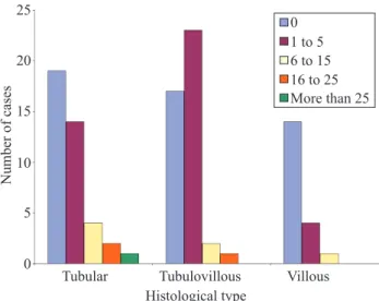
0 1 to 5 6 to 15 16 to 25 More than 25
Tubular 0
5 10 15 20 25
Tubulovillous Histological type
Number of cases
Villous
Figure 3. Distribution of cases according to the concentration
of mast cells and histological type of the intestinal adenomas (n=102), p=0.083.
0 1 to 5 6 to 15 16 to 25 More than 25
0
<1 1–1.9 2–2.9
Size of adenomas (cm)
3–3.9 4
5 10 15 20 25
Number of cases
Figure 4. Distribution of cases according to the concentration of
mast cells and the size (in cm) of the intestinal adenomas (n=102), p=0.665.
0 1 to 5 6 to 15 16 to 25 >25
0
Absent Mild
Cellular atypia
Moderate Severe
2 4 6 8 10 12 14 16 18 20
Number of cases
Figure 5. Distribution of cases according to the concentration
of mast cells and the degree of cellular atypia of the intestinal adenomas (n=102), p=0.524.
Figure 6. Distribution of cases according to the concentration of
mast cells and the intestinal lesion in other areas associated with the adenomas (n=102), p=0.202.
Adenocarcinoma Hyperplastic polyp Without other associated lesions
0 0 5 10 15 20 25 30 35 40 45
1 to 5 6 to 15 16 to 25 >25
Concentration of mast cells
Number of cases
between associated lesions and histological type of adenomas (p=0.019) and size (p=0.016), but no dif-ference was observed in relation to cellular atypia (p=0.51) of adenomas.
The MC ranged from 0 to 28 CM/10 HPF (mean was 2.28, standard deviation of 4.57 and median was 1) (p=0.0001) (Figures 1 and 2). No statistically signiicant difference was observed between the MC and the patients’ age (p=0.790) and gender (p>0.05).
DIsCUssION
Intestinal polyps can be non-neoplastic (in-lammatory, hamartomatous or hyperplastic) and neoplastic (adenomatous polyps or adenomas). Ad-enomas can be of three types, according to their histological architecture: 1) tubular adenomas, composed of tubular glands similar to the mucosa topology, are the most frequent types; 2) villous ad-enomas, with villous projections, corresponding to 1% of the adenomas; 3) tubulovillous adenomas, a combination of the two types described above, cor-responding to 5 to 10% of the adenomas15. In this study, most cases of intestinal adenomas occurred in male patients, with median age of 66 years, as described in the literature. However, tubulovillous adenomas were predominant (42.1%), followed by tubular adenomas (39.2%). The median size was 1 cm and mild atypia predominated in the studied sample (29.4%). Such data show that the adenomas had early diagnosis, reducing the risk of malignan-cy of these tumors.
In this study, the histological type of adenomas was associated with the patients’ gender and age, while the adenoma size was associated with gen-der only, and cellular atypia was not associated with either parameter. In addition, a statistically signii -cant difference was observed between the histologi-cal type and size (p=0.0001) of adenomas and his-tological type and cellular atypia (p=0.007), but no association was observed between the adenoma size and cellular atypia (p=0.414). The absence of asso -ciation observed between adenoma size and cellular atypia may be explained by the fact that most cases had adenomas measuring up to 1.9 cm, with moder-ate and severe cellular atypia as the most frequent types observed in adenomas larger than 2 cm15. On the other hand, most cases had tubular and tubu-lovillous adenomas, which tend to be smaller and with lower degree of cellular atypia15, which pre-dominated in this study.
The risk of malignancy for an adenoma is as-sociated with three independent characteristics: polyp size, histological architecture and epithelial dysplasia (or atypia) severity, described as follows: 1) cancer is rare in tubular adenomas with diam-eter of less than 1 cm; 2) the probability of cancer
is high in villous adenomas with diameter of more than 4 cm, reaching up to 40%; 3) severe dysplasia, when present, is frequently found in villous areas15. This study showed a statistically signiicant differ -ence between the pres-ence of associated lesions and the histological type of adenomas (p=0.019) and adenoma size (p=0.016), partially conirming the data above, as no association was observed between these other lesions and the degree of cellular atypia of the adenomas (p=0.51). This fact may have oc -curred due to the small number of adenocarcinomas (18%) in the sample of associated lesions, as well as the small number of adenomas with severe atypia (10.9%), which are more associated with the prog-ress into malign neoplasm.
The role of stroma-epithelium interaction in the initial events of carcinogenesis was proposed around 30 years ago6. An interaction between tumor cells and their microenvironment is important for their growth and survival. In this context, the involvement of in-lammatory cells in the initiation, promotion and pro -gression of cancer has indicated a new therapeutic op-portunity in the treatment of cancer. The main immune cells involved in tumor-associated inlammation are macrophages, dendritic cells, lymphocytes, neutro-phils, eosinophils and CM16.
CM are metachromatic cells from hematopoietic pluripotent stem cells of the bone marrow17, irst de -scribed by Erlich, in 18783,4. The CM can be found in most tissues, but are found in greater number in the skin, airways and digestive tract4. The study con-ducted by McGinnis et al.18 showed that the use of a histochemical method, with toluidine blue staining, or the immunohistochemical method for CM detection, has similar results.
New roles of the CM have been identiied, show -ing that these cells have a critical role, in innate im-munity, or adaptive, normal or pathological immunity (e.g., acute or chronic bacterial or parasital infections, autoimmune diseases, pregnancy), including the im-mune tolerance16,19-22.
Tumor angiogenesis is essential for growth above 1 mm3, invasion and metastases in solid tumors4,9,16. Several evidences have shown that the CM have an important role in tumor angiogenesis9,16. The CM secrete the vascular endothelial growth fac-tor (VEGF), interleukin (IL)-8 and growth facfac-tors, which enable the formation of new vessels6. The in-iltration of CM is well associated with tumor angio -genesis and metastases in gastric cancer, colorectal cancer, pulmonary adenocarcinoma, renal cell cancer and prostate cancer5.
It has been suggested that, in the context of de-veloping tumors, the tissue remodeling ability of CM is subverted, leading to the rupture of adjacent extra-cellular matrix, thus, increasing the tumoral dissemi-nation, mainly through greater release of MMP-94.
Immunosuppression is another basic inding in the tumor microenvironment. Although the im-munological vigilance occurs in the initial stages of tumorigenesis, the tumor establishment irst in -duces immune tolerance. An absolute suppression of the immune response is generated in the tumoral microenvironment only in late stages of the tumor23. The CM have been identiied as having a critical role in the suppression of immune responses23. The hista-mine released by the CM can cause the tumor cells to proliferate through H1 receptors and suppress the immune response through H2 receptors6. SCF (stem cell factor)-activated CM accentuate the tumoral im-munosuppression through the release of adenosine, which inhibits the production of interferon (IFN)-γ and IL-2 by TCD4+ cells and the increase of T regu-latory (Treg) cells, which release IL-10, a cytokine with immunosuppressant effect23,24.
Mutations in the tumoral suppressor, the colon adenomatous polyposis (APC) gene, are required to initiate hereditary or sporadic colorectal cancers. However, angiogenesis and tissue remodeling are re -quired for the tumoral expansion19.
The bowel, just as other mucosae exposed to the external environment, is an area where inlammation and cancer are closely linked. Bacterial infections,
ex-posure to toxic molecules that damage the epithelial barrier, genetic predisposition and increased immune reactivity can promote chronic colitis, which induces cellular proliferation, stromal remodeling, neoangio-genesis and suppression of the adaptive antitumor im-mune response25.
In the bowel, the CM play an important role in different processes, including cleaning of enteric pathogens, allergy to food, visceral hypersensitivity and intestinal cancer22.
CM in the human bowel are the greatest source of tumoral necrosis factor (TNF)-α of the gastroin-testinal tract26. The TNF is required for the growth of adenoma, the colorectal cancer precursor lesion. Thus, the TNF released by the CM can act in an au -tocrine manner. In the absence of CM, the geneti-cally altered epithelium is not able to develop into a complete tumor13. In this study, most cases (89.2%) presented concentration of mast cells in intestinal ad-enomas of up to 5 CM/10 HPF, with 50% of them not presenting CM in the studied material. Then, no as-sociation of the MC was observed with the patients’ age or gender, nor with the histological type, size and cellular atypia of the adenomas, nor with the pres-ence of associated lesions. The sample evaluated in this study presented predominance of tubulovil-lous and tubular adenomas, of less than 2 cm and with mild cellular atypia, which would tend to have a lower concentration of CM, for presenting lower risk of developing into an adenocarcinoma. Perhaps the small number of villous adenomas, adenomas of more than 3 cm and adenomas with severe cellular atypia inluenced the non association of risk criteria for malignancy of adenomas with MC.
CONCLUsION
rEFErENCEs
1 Brasil. Ministério da Saúde. Secretaria de Atenção à Saúde. Instituto Nacional do Câncer. Estimativa 2010: Incidência de câncer no Brasil. Rio de Janeiro: INCA, 2009 [cited: 2011 maio 15]. Available from: http://www.inca.gov.br
2 Zandoná B, Carvalho LP, Schimedt J, Koppe DC, Koshimizu RT, Mallmann ACM. Prevalência de adenomas colorretais em pacientes com história familiar para câncer colorretal. Rev Bras Coloproct 2011;31(2):147-54.
3 Kashiwase Y, Inamura H, Morioka J, Igarashi Y, Kawai-Kowase K, Kurosawa M. Quantitative analysis of mast cells in benign and malignant colonic lesions: immunohistochemical study on formalin-ixed, parafin-embedded tissues. Allergol Immunopathol 2008;36(5):271-6.
4 Maltby S, Khazaie K, McNagny KM. Mast cells in tumor growth: angiogenesis, tissue remodeling and immune-modulation. Biochim Biophys Acta 2009;1796(1): 19-26. doi:10.1016/j.bbcan.2009.02.001.
5 Gounaris E, Blatner NR, Dennis K, Magnusson F, Gurish MF, Strom TB, et al. T-regulatory cells shift from a protective anti-inlammatory to a cancer-promoting proinlammatory phenotype in polyposis. Cancer Res 2009;69(13):5490-7. 6 Conti P, Castellani ML, Kempuraj D, Salini V, Vecchiet J,
Tetè S, et al. Role of mast cells in tumor growth. Ann Clin Lab Sci 2007;37(4):315-21.
7 Nechushtan H. The complexity of the complicity of mast cells in cancer. Int J Biochem Cell Biol 2010;42(5):551-4. 8 McKerrow JH, Bhargava V, Hansell E, Huling S, Kuwahara T,
Matley M, et al. A functional proteomics screen of proteases in colorectal carcinoma. Mol Med 2000;6(5):450-60. 9 Acikalin MF, Oner U, Topçu I, Yaşar B, Kiper H, Colak E.
Tumour angiogenesis and mast cell density in the prognostic assessment of colorectal carcinomas. Dig Liver Dis 2005;37(3):162-9.
10 Taweevisit M. The association of stromal mast cell response and tumor cell differentiation in colorectal cancer. J Med Assoc Thai 2006;89(Suppl 3):S69-73.
11 Yodavudh S, Tangjitgamol S, Puangsa-Art S. Prognostic signiicance of microvessel density and mast cell density for the survival of Thai patients with primary colorectal cancer. J Med Assoc Thai 2008;91(5):723-32.
12 Gulubova M, Vlaykova T. Prognostic signiicance of mast cell number and microvascular density for the survival of patients with primary colorectal cancer. J Gastroenterol Hepatol 2009;24(7):1265-75.
13 Gounaris E, Erdman SE, Restaino C, Gurish MF, Friend DS, Gounari F, et al. Mast cells are an essential hematopoietic component for polyp development. Proc Natl Acad Sci USA 2007;104(50):19977-82. DOI: 10.1073_pnas.0704620104. 14 Michalany J. Técnica histológica em anatomia patológica. Com
instruções para o cirurgião, enfermeira e citotécnico. 3a ed. São Paulo: Editora Michalany Ltda.; 1998. p. 153-5.
15 Robbins. Basic Pathology. Kumar K, Abbas AK, Fausto N, Mitchell RN (editors). Oral cavity and gastrointestinal tract. Chapter 15. 8th ed. Rio de Janeiro: Elsevier; 2008. p. 672-81.
16 Groot Kormelink T, Abudukelimu A, Redegeld FA. Mast cells as target in cancer therapy. Curr Pharm Des 2009;15(16):1868-78.
17 Nemolato S, Cabras T, Fanari MU, Cau F, Fraschini M, Manconi B, et al. Thymosin beta 4 expression in normal skin, colon mucosa and in tumor iniltrating mast cells. Eur J Histochem 2010;54(1):e3.
18 McGinnis MC, Bradley Jr. EL, Pretlow TP, Ortiz-Reyes R, Bowden CJ, Stellato TA, et al. Correlation of stromal cells by morphometric analysis with metastatic behavior of human colonic carcinoma. Cancer Res 1989;49:5989-93.
19 Kalesnikoff J, Galli SJ. New developments in mast cell biology. Nat Immunol 2008;9(11):1215-23. DOI:10.1038/ni.f.216. 20 Rao KN, Brown MA. Mast cells: multifaceted immune cells
with diverse roles in health and disease. Ann N Y Acad Sci 2008;1143:83-104.
21 Kumar V, Sharma A. Mast cells: Emerging sentinel innate immune cells with diverse role in immunity. Mol Immunol 2010;48(1-3):14-25.
22 Shea-Donohue T, Stiltz J, Zhao A, Notari L. Mast cells. Curr Gastroenterol Rep 2010;12(5):349-57.
23 Huang B, Lei Z, Zhang G-M, Li D, Song C, Li B, et al. SCF-mediated mast cell iniltration and activation exacerbate the inlammation and immunosuppression in tumor microenvironment. Blood 2008;112(4):1269-79. DOI:10.1182/blood-2008-03-147033.
24 Blatner NR, Bonertza A, Beckhovea P, Cheonc EC, Krantzc SB, Strouchc M, et al. In colorectal cancer mast cells contribute to systemic regulatory T-cell dysfunction. Proc Natl Acad Sci USA 2010;107(14):6430-5. DOI: 10.1073/pnas.0913683107. 25 Colombo MP, Piconese S. Polyps wrap mast cells
and Treg within tumorigenic tentacles. Cancer Res 2009;69(14):5619-22.
26 Bischoff SC, Lorentz A, Schwengberg S, Weier G, Raab R, Manns MP. Mast cells are an important cellular source of tumour necrosis factor alfa in human intestinal tissue. Gut 1999;44:643-52.
Correspondence to: Gisele Alborghetti Nai