Freitas CD, Sartor MC, Correa MFM, Froehner Junior I, Martins JF, Kotze PG, Zanoni ECA, D’Assunção MA. Appropriateness of colonoscopy indication for colorectal neoplasm detection in patients under 50 years old with hematochezia. J Coloproct, 2012;32(1): 40-9.
AbstrACt: Objective: Evaluate the appropriateness of colonoscopy indication for neoplastic lesion detection in patients under age 50 with hematochezia. Methods: Retrospective and cross-sectional study in patients who underwent colonoscopy, from 2002 to 2009. Inclusion criteria included patients with hematochezia over 20 years old. Exclusion criteria were: history of inlammatory bowel disease (IBD), polypectomy, family history of colorectal cancer (CRC), fecal occult blood (FOB), anemia, weight loss and personal history of cancer. Neoplastic lesions were stratiied into proximal or distal to splenic lexure. results: 683 patients met the inclusion criteria in 5,000 colonoscopies registered. Median age was 49.46 years old (20 to 94 years old) and 486 patients (71.2%) were females. No proximal colon cancer was detected in the proximal group under 50 years old. Proximal advanced adenomas were diagnosed in one (0.9%) patient in the group of 30 to 40 years old (n=113) versus 7 (3.75%) in the group of 40 to 50 years old (n=187), with p=0.268. Conclusions: Malignant neoplastic lesions and advanced adenomas are uncommon and predominantly distal in the population between 30 and 50 years old, with hematochezia without risk factors for colorectal cancer (CRC). Therefore, lexible sigmoidoscopy appears to be suficient as the initial method for evaluating these patients.
Keywords: colonoscopy; colorectal neoplasms; adults; gastrointestinal hemorrhage.
resuMO: Objetivo: Avaliar a propriedade da indicação da colonoscopia para pesquisa de lesões neoplásicas em pacientes com menos de 50 anos com hematoquezia. Métodos: Estudo retrospectivo e transversal, realizado em pacientes submetidos à colonoscopia, de 2002 a 2009. Foram incluídos pacientes com hematoquezia com idade igual ou superior a 20 anos. Os critérios de exclusão foram: história de do -ença inlamatória intestinal, polipectomia, história familial de câncer colorretal, sangue oculto nas fezes, anemia, emagrecimento e história pessoal de neoplasia. Lesões neoplásicas foram estratiicadas em proximais ou distais ao ângulo esplênico. resultados: Obedeceram aos critérios de inclusão 683 pacientes dentro de 5.000 colonoscopias registradas. A média de idade foi 49,46 anos (20 a 94 anos) e 486 pacien
-Appropriateness of colonoscopy indication for colorectal
neoplasm detection in patients under 50 years old
with hematochezia
Cristiano Denoni Freitas1, Maria Cristina Sartor2, Marco Fábio Maia Correa3, Ilario Froehner Junior4, Juliana Ferreira Martins5, Paulo Gustavo Kotze6, Esdras Camargo Andrade Zanoni7, Marco Aurélio D’Assunção8
1Former resident physician at the Service of Coloproctology, Hospital Universitário Cajuru (SECOHUC) at
Pontifícia Universidade Católica do Paraná (PUCPR) – Curitiba (PR), Brazil; Digestive Tract Surgeon and Coloproctologist at the Imperial Hospital de Caridade and Hospital Governador Celso Ramos – Florianópolis
(SC), Brazil. 2Coloproctologist and Preceptor Physician at SECOHUC - PUCPR and at the Service of Coloproctology, Hospital de Clínicas da Universidade Federal do Paraná (UFPR) – Curitiba (PR), Brazil; Technical Director of Clínica Lucano – Curitiba (PR), Brazil. 3Doctor in Zoology, UFPR; Biologist and professor of statistics at UFPR – Curitiba (PR), Brazil. 4Resident physician at SECOHUC - PUCPR – Curitiba (PR), Brazil. 5Coloproctologist and Preceptor Physician at SECOHUC - PUCPR – Curitiba (PR), Brazil. 6Head of SECOHUC -
PUCPR – Curitiba (PR), Brazil. 7General Surgeon and Digestive Tract Surgeon at the Hospital Cajuru da PUCPR; Colonoscopist at Clínica Lucano – Curitiba (PR), Brazil. 8Endoscopist at the Hospital Sírio-Libânes – São Paulo
(SP) and Clínica Lucano – Curitiba (PR), Brazil.
Study carried out at the Service of Coloproctology, Hospital Universitário Cajuru (SECOHUC) at Pontifícia Universidade Católica do Paraná – Curitiba (PR), Brazil.
The studied patients were from Clínica Lucano (a private Coloproctology clinic in Curitiba). Funding source: none.
Conlict of interest: nothing to declare.
INtrODuCtION
Colonoscopy is considered the most effective method for colorectal cancer (CRC) screening in the population over 50 years old with sporadic risk1. In
addition, it allows to remove adenomas, reducing the incidence of CRC and, consequently, causing a true impact on the natural history of this disease2,3.
The number of colonoscopies performed world -wide has increased progressively. Inadequate
indica-tions and abusive uses of this procedure are some of the reasons for increasing and unachievable demand at the public health centers4. The exam is safe, but it
can involve complications such as: intestinal perfora-tion, hemorrhage and cardiopulmonary alterations in terms of sedation, which may lead to death in case of late diagnosis5.The correct indication, based on well deined clinical criteria, is essential for a proper cost-beneit ratio and minimized complications.
The guidelines of the American Society for Gas-trointestinal Endoscopy (ASGE), issued in 2000, and of the European Panel on the Appropriateness of Gas-trointestinal Endoscopy II (EPAGE II) indicate the
CRC screening, preferably through colonoscopy, to
asymptomatic patients without family history of CRC over age 50, each ten years6,7.
In symptomatic patients, hematochezia is the fre-quent indication for this exam. It is considered an impor-tant sign of anorectal and colonic disease. Physicians and patients should take it as a warning of the risk for CRC, as up to 25% of the patients present such diagnosis8.
Talley and Jones, while studying a speciic popu
-lation, described up to 20% of individuals with that
diagnosis in the period of 12 months. This complaint
seemed to be more common in patients under 50 years
old. Less than half of these patients searched for medi-cal support and, when they did it, it was usually for other gastrointestinal complaints9.
Hematochezia, before or after defecation, is more suggestive of distal colorectal lesions. Bright red bleed -ing on the paper or underwear cloth-ing is more related
to anorectal diseases, such as issures and hemorrhoids. Blood in stool should be taken by physicians as a warn -ing of more proximal lesions to colon, such as
diver-ticular diseases, inlammatory diseases and CRC. The practice remains indeinite in the literature
for patients with hematochezia under 50 years of age and without other risk factors for CRC.
The EPAGE II and the ASGE make a speciic ref -erence to patients under 40 years old with hematochezia.
For this group, when the proctologic exam and lexible rectosigmoidoscopy show evidences that the case is be -nign and distal, there is no indication of colonoscopy7,10.
In France, the Agence Nationale d’Accréditation et d’Evaluation en Santé (ANAES) does not recom-mend colonoscopy to patients under 50 years old who
complain of isolated hematochezia, prescribing lex
-ible rectosigmoidoscopy instead. Colonoscopy is in
-dicated when bleeding is chronic, associated with sev -eral episodes, regardless of the age11.
The irst publications on colonoscopic indings in
patients with hematochezia were retrospective. They
con-cluded that colonoscopy would be adequate in this group of patients due to the high rate of signiicant lesions12-14.
Acosta et al. published the irst study that evalu
-ated the colonoscopic indings in young adults with hematochezia. They found 21% of the indings were signiicant and concluded that colonoscopy would be justiiable in patients under 40 years of age15.
Lewis, Shih and Blecke analyzed, in a retrospec-tive study, 570 patients with hematochezia under 50 years of age. The authors concluded that the patients
above 40 years old with hematochezia required an in
-vestigation with colonoscopy, due to the considerable number of neoplastic lesions. However, in patients un
-der 40 years of age, the number of advanced neoplastic
tes (71,2%) pertenciam ao gênero feminino. Nenhum câncer do cólon proximal foi detectado no grupo com menos de 50 anos. Adenomas avançados proximais foram diagnosticados em 1 (0,9%) paciente no grupo de 30–40anos (n=113) versus 7 (3,75%), no de 40–50 anos (n= 187), com p=0,268. Conclusões: As lesões neoplásicas malignas e os adenomas avançados são pouco frequentes e predominantemente distais na população entre 30–50 anos, com hematoquezia, sem fatores de risco para o câncer colorretal. A retossigmoidoscopia lexível, portanto, parece ser suiciente como método inicial para a avaliação de tais pacientes.
lesions was low. Then, the indication of colonoscopy
to this age group should be on a case-by-case basis16. The habitual use of colonoscopy as an initial in
-vestigation of hematochezia in young adults has been questioned by several authors.
Mulcahy et al., in a retrospective series of studies that analyzed 1,766 patients with hematochezia, also questioned the role of colonoscopy for the same indi-cation. No proximal CRC was detected in this series.
Then, they concluded that lexible rectosigmoidosco
-py would be suficient as the initial method17.
Eckardt et al., in a prospective control-case study, evaluated the prevalence of neoplastic lesions in the group with hematochezia without risk factors for CRC.
Hematochezia presented OR=1.2 for proximal lesions, with lexible rectosigmoidoscopy suficient as the ini -tial method. The patients with positive fecal occult
blood (FOB) test or bleeding with clots would have to be submitted to colonoscopy, as these groups presented
increased risk for proximal neoplastic lesions18.
Carlo et al., in a prospective study, analyzed 417 patients with hematochezia, without risk factors for CRC. The patients were sorted into two groups: over and under 45 years old. No proximal neoplastic lesion was detected in the second group. They concluded that
lexible rectosigmoidoscopy could be used as the initial
method for patients with hematochezia in this group19.
Spinzi et al., in multicenter prospective study,
an-alyzed 622 patients between 30 and 50 years old with
hematochezia. No proximal CRC was detected and less
than 1% of advanced proximal adenoma was observed
in the group of 40–50 years old. The authors suggest that
lexible rectosigmoidoscopy is suficient as the initial
method of investigation in patients with hematochezia
under 40 years old, and that colonoscopy should not be habitual for patients in the group of 40–50 years old20.
In 1991, Church evaluated, in a prospective study,
patients with hematochezia, positive FOB test, bleeding in stool and lower gastrointestinal bleeding. Only one adeno
-ma proxi-mal to splenic lexure was found in the patients
with hematochezia21. Then, he concluded that lexible rectosigmoidoscopy would be suficient in patients with
hematochezia during or after defecation. In 2008, Eric Mardestein and James Church came to the same conclu-sion in a prospective study that analyzed 703 patients22.
Based on these facts, there is no absolute agree
-ment between the guidelines for the initial evaluation
of patients under 50 years of age, with hematochezia. The purpose of this study was to analyze the appro-priateness of colonoscopy indication for young adults with hematochezia, without risk factors for CRC.
ObJeCtIVes
The primary purpose was to analyze the appropriate-ness of colonoscopy for neoplastic lesion detection in pa-tients under 50 years old with hematochezia. The
second-ary objectives included: describe epidemiological data;
analyze the frequency of the indication of colonoscopy for
hematochezia; describe the signiicant indings of colonos -copy exams performed in this population and analyze the
prevalence and anatomical distribution of colorectal neo -plasms in patients under 50 years old with hematochezia.
MetHODs
The research project of this study was approved
by the Research Ethics Committee of the Pontifícia Universidade Católica do Paraná (PUCPR), under number 0004024/10.
That was a retrospective, cross-sectional and obser
-vational study conducted by the Service of Coloproctol
-ogy of the Hospital Universitário Cajuru (SECOHUC). The studied population included patients submit -ted to colonoscopy at Clínica Lucano (a private ser-vice of Coloproctology and Digestive Endoscopy), in
Curitiba, between January 2002 and December 2009. The database information was taken from the
clinical records, colonoscopy reports and
anatomo-pathological exams. Data collection was performed by
three investigators (two resident physicians and one coloproctologist), according to the study instrument.
The inclusion criteria were: patients over 20 years old, complaining of hematochezia. The exclusion
cri-teria were: clinical reports or records with insuficient data; patients under 20 years old; history of inlam
-matory bowel disease (IBD); previous polypectomy; family history of CRC; fecal occult blood (FOB); ane -mia; weight loss; personal history of malignant neo-plasm and incomplete endoscopic exams.
The database was created using SPSS®, version
16.0 (IBM Corporation, 2010).
The exams were performed by three experienced
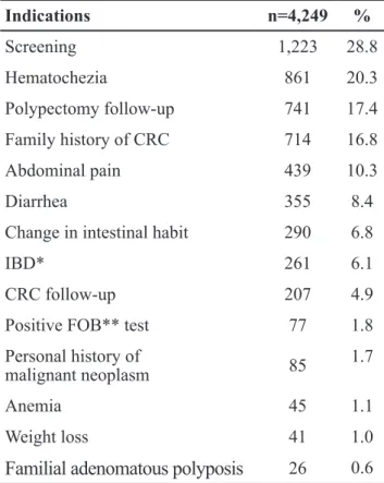
Indications n=4,249 %
Screening 1,223 28.8
Hematochezia 861 20.3
Polypectomy follow-up 741 17.4
Family history of CRC 714 16.8
Abdominal pain 439 10.3
Diarrhea 355 8.4
Change in intestinal habit 290 6.8
IBD* 261 6.1
CRC follow-up 207 4.9
Positive FOB** test 77 1.8
Personal history of
malignant neoplasm 85
1.7
Anemia 45 1.1
Weight loss 41 1.0
Familial adenomatous polyposis
26 0.6Table 1. Indications of colonoscopy.
Source: Clínica Lucano, Curitiba, Paraná. *IBD: inlammatory bowel disease; **FOB: fecal occult blood (CRC). Note: the study had patients with more than one indication of colonoscopy.
was more than 1,000 colonoscopy exams and more than 200 colonoscopy exams per year.
Between 2002 and 2006, the exams were per-formed using a video colonoscope (Fujinon 2200). As of 2006, the device used in the exam was a Fujinon
video colonoscope, model 4400 EC-590ZW/L, with magniication and chromoscopy.
The most frequently used solution was 1,000 mL
of mannitol at 10%, combined or not with sodium pi
-cosulfate. The patients were submitted to endovenous sedation using propofol, assisted by a anesthesiologist.
The patients were from the clinic and other
exter-nal ofices, and they came speciically to be submitted
to colonoscopy.
Deinitions
The study considered an advanced adenoma
le-sions that fulilled one or more of these criteria: diame -ter of min. 1 cm; more than 25% of the area with villous component or with high-grade dysplasia (HGD)23.
Neoplastic lesions include adenomas and CRC. Distal neoplastic lesions were located under the
splen-ic lexure, while the proximal neoplastsplen-ic lesions were above the splenic lexure.
Signiicant indings included: IBD, vascular le -sions, diverticular disease and neoplastic lesions.
statistical analysis
Descriptive parametric statistics with frequency
tables were used in data evaluation. Mean, median and
standard deviation were calculated using SPSS®, version
16.0 (IBM Corporation, SPSS Inc., Chicago IL, USA). The hypotheses were tested using the χ2 test, with evaluation of independent variables. Fisher’s ex
-act test was used in variables with n<5. The level of signiicance (α) was 5%.
resuLts
During the period mentioned above, data were col -lected from 5,000 colonoscopy exams performed in 3,687 patients. In 4,249 colonoscopy exams, the reason for indi-cation was characterized. Hematochezia was the second most frequent indication, with 861 (20.3%) individuals.
Table 1 shows the reason for indicating colonoscopy. The lowchart in Figure 1 illustrates the sample
that followed the inclusion and exclusion criteria. The
861 patients with hematochezia
n=683
178 excluded patients
- 17 patients under 20 years old - 95 cases of family history of CCR
- 20 cases of personal history of malignant neoplasm - 13 cases of prior polypectomy
- 12 cases of weight loss - 09 cases of anemia
- 05 cases of inflammatory bowel disease - 04 incomplete exams
- 03 positive fecal occult blood test
Source: Clínica Lucano.
Gender
n
%
Female
486
71.2Male
197
28.8total 683 100.0
Table 2. Distribution of patients with hematochezia by gender.
Source: Clínica Lucano, Curitiba, Paraná.
Age group n %
<50 years old 372 54.5
≥50 years old 311 45.5
total 683 100.0
Table 3. Distribution of patients with hematochezia by age group.
Source: Clínica Lucano, Curitiba, Paraná. χ2=0.81; p>0.05.
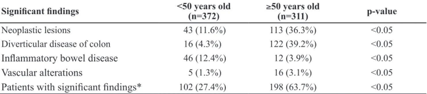
Signiicant indings <50 years old (n=372) ≥50 years old
(n=311) p-value
Neoplastic lesions 43 (11.6%) 113 (36.3%) <0.05
Diverticular disease of colon 16 (4.3%) 122 (39.2%) <0.05
Inlammatory bowel disease
46 (12.4%) 12 (3.9%) <0.05Vascular alterations
5 (1.3%) 16 (3.1%) <0.05Patients with signiicant indings*
102 (27.4%) 198 (63.7%) <0.05Table 4. Distribution of signiicant indings according to patients over and under 50 years old with
hematochezia.
Source: Clínica Lucano, Curitiba, Paraná. The total value is the number of patients with signiicant indings. The study had patients with more than one signiicant inding.
total sample included 683 patients – 197 males and
486 females (Table 2).
Mean age was 49.46 years (SD±15.51 years). The minimum age was 20 years and the maximum
age was 92. The frequency distribution of patients by closed interval of age class is described in Chart 1.
In this study, there was no association of hema-tochezia complaint with age group among the patients
over or under 50 years old (Table 3).
Table 4 describes the frequency of signiicant indings in patients distributed by age group. Neoplas -tic lesions, diver-ticular disease of colon, vascular al-teration and CRC were more prevalent in patients over
50 years old with statistical signiicance (p<0.05). IBD
was more frequent in the group of patients under 50
years old (p<0.05).
In total, 304 polypectomy exams were performed in the 184 (26.9%) patients with polyps. Sixteen polyps were lost in the light. Two patients presented leiomyo-ma and were not included in the analysis. From the 286
polyps, 178 were benign neoplasm (Table 5); 79 ad -vanced adenomas were found in 73 (10.68%) patients.
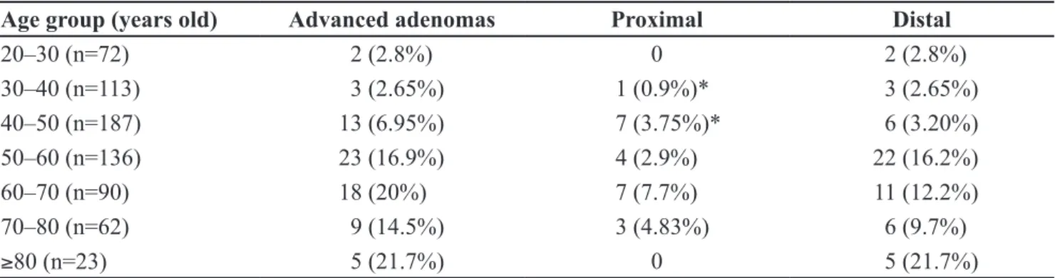
In the group of 30–40 years old, ive advanced
adenomas were detected in three (2.65%) patients.
Only one (0.9%) of them was proximal, tubular, with -out HGD, of 10 mm diameter and located in the as-cending colon.
In the group of 40–50 years old, 14 advanced ad-enomas were detected in 13 patients (6.95%). Seven patients (3.75%) had proximal adenomas. Five of these seven patients with proximal adenomas were over 45
years old. Four tubular adenomas presented HGD and one adenoma was classiied as serrated, with diameter
over 10 mm. The two advanced adenomas found in
20–29 72 (10.5%)
113 (16.5%)
187 (27.4%)
136 (19.9%)
90 (13.2%) 62
(9.1%) 23 (3.4%)
0 20 40 60 80 100 120 140 160 180 200
30–39 40– 49 50–59 60–69 70–79
Age (years old)
Number
of patients
80
patients of 40–45 years old had 10 mm diameter, one
was tubular with HGD and one was serrated without HGD (Table 6).
Table 7 shows the classiication of malignant col -orectal lesions. No malignant lesion was proximally located in patients under 50 years of age.
DIsCussION
International series of studies have described
that around 20% of colonoscopy exams are indicated due to hematochezia, which is the second most fre-quent indication24,25. Some Brazilian series of studies
show hematochezia as the most frequent indication of colonoscopy26,27. In this study, it was the second most
frequent indication of colonoscopy.
There is no doubt that hematochezia – of any
size – is an indication of colonoscopy for patients over 50 years old. In patients under this age group,
the incidence of neoplastic lesions is lower and be
-nign oriicial causes are frequent reasons for search -ing medical attention. For this group, there is no
established consensus regarding the indication of
colonoscopy to investigate hematochezia in the dif-ferent groups of medical specialties7,10. The assistant
physician should decide on how to start the
investi-Histology of polyps
<50 years old (n=372) ≥
50 years old (n=311)
Tubular 29 88
Tubulovillous 05 31
Villous 00 02
Serrated 17 06
total 51 127
Table 5. Distribution of benign neoplastic polyps according to the histological type by age group.
Source: Clínica Lucano, Curitiba, Paraná.
Age group (years old) Advanced adenomas Proximal Distal
20–30 (n=72) 2 (2.8%) 0 2 (2.8%)
30–40 (n=113) 3 (2.65%) 1 (0.9%)* 3 (2.65%)
40–50 (n=187) 13 (6.95%) 7 (3.75%)* 6 (3.20%)
50–60 (n=136) 23 (16.9%) 4 (2.9%) 22 (16.2%)
60–70 (n=90) 18 (20%) 7 (7.7%) 11 (12.2%)
70–80 (n=62) 9 (14.5%) 3 (4.83%) 6 (9.7%)
≥80 (n=23) 5 (21.7%) 0 5 (21.7%)
Table 6. Distribution of patients with advanced adenomas according to the age group and location.
Source: Clínica Lucano, Curitiba, Paraná. *Fisher’s exact test, comparing the frequencies of patients with proximal advanced adenomas, between the patients of 30–40 and 40–50 years old (p=0.266). The study had patients with proximal and distal advanced adenomas.
Age group Malignant colorectal
lesions n (%) Proximal Distal
20–30 (n=72) 0 (0) 0 0
30–40 (n=113) 4 (3.5) 0 04
40–50 (n=187) 8 (4.7) 0 08
50–60 (n=136) 5 (3.7) 0 05
60–70 (n=90) 12 (13.3) 0 12
70–80 (n=62) 6 (9.7) 0 06
≥80 (n=23) 3 (13) 02 01
Table 7. Distribution of patients with malignant lesions by age group and location.
gation and whether to use lexible rectosigmoidos -copy or colonos-copy.
The fear of not diagnosing potentially healable
colorectal neoplastic lesions favors the indication of colonoscopy in this population. However, the costs, risks and discomfort in the preparation for this exam
may not be higher than the beneits to patients with
hematochezia.
Polyps bleed at low frequency and seem to be randomly identiied in the bleeding investigation28. For this reason, it may be dificult to establish a direct relation between hematochezia and polyp as a cause.
The use of colonoscopy has increased in the last years, particularly in young adults, while the use of
lexible rectosigmoidoscopy has decreased29.
The literature clearly shows the best initial meth -od to investigate this population. Investigators suggest colonoscopy to patients with hematochezia. However, most of these studied did not analyze the patients in terms of age group8,12-14,30,31.
From another standpoint, other authors observed that most signiicant lesions are distally located, espe
-cially CRC. They concluded that, in patients between 30 and 39 years old, lexible rectosigmoidoscopy would be suficient. For the group between 40 and 49 years old, with hematochezia, the patients should be considered on a case-by-case basis, with colonoscopy or lexible rectosigmoidoscopy17-20,22,32.
Neoplastic lesions, diverticular disease of colon and vascular alterations were most frequent in patients over 50 years old with hematochezia. IBDs were more prevalent in patients under 50 years old. These data
were statistically signiicant and agree with the epide -miology of these disorders.
This study did not classify the type of hemato-chezia in terms of time to manifestation. Guillem,
Forde and Treat did not detect signiicant differences between the indings and the form of bleeding (acute
and chronic)14. Fine et al., in a prospective study, used a board of colors to help the patient determine the type of bleeding. They concluded that the color of stool is
not a good predictor of disease location and severity33. It should be noted that the information about bleeding is more subjective and the patients is not always able
to characterize it.
Proximal advanced adenomas were found in a much lower proportion in patients of 30–50 years old.
No proximal CRC was detected in patients with hema-tochezia under 50 years old.
Fine et al., in a prospective study with 58 patients under 40 years old with hematochezia, detected three patients with proximal CRC. They suggested that
colonoscopy should be performed with this group.
These authors were the only that detected proximal CRC in this type of population33.
Wong et al. detected adenoma and CRC in 11.6% of the 223 patients under 40 years old with hematochezia. Twenty-six (9.9%) patients had ad-enomas, 6 of them were proximally located. Four (1.8%) had CRC, all distally located34. These results
are similar to those found in this study. The authors
concluded that colonoscopy should be performed in
patients under 50 years old with hematochezia. This
conclusion can be considered controversial, as ma
-lignant lesions were distal and most benign polyps
were also distal.
Van Rosendaal et al. found a malignant lesion
proximal to splenic lexure in a 44-year-old patient, among total 61 patients analyzed by hematochez -ia, under 55 years of age. These authors concluded that, in young patients with hematochezia, the
in-vestigation can be started using lexible rectosig -moidoscopy35. Nikpour and Ali Asgari came to the
same conclusion in a series of studies that analyzed 402 patients32.
David Lieberman concluded, in a literature
review until 2002, that patients with hematoche-zia during or after evacuation, without family
his-tory of CRC, would be properly evaluated by flex
-ible rectosigmoidoscopy. From 40 to 49 years of age, each case would be individually considered,
with the option to use either rectosigmoidoscopy or colonoscopy36.
Carlo et al., in a prospective study that analyzed 417 patients with hematochezia without risk
fac-tors for CRC, evaluated the colonoscopic indings of
this population. They grouped the patients into two groups: over and under 45 years old. They detected two (1.1%) polyps over 10 mm, 29 (16.1%) patients with IBD and no CRC in the younger group. No proxi-mal neoplastic lesion was found. They concluded that
lexible rectosigmoidoscopy can be used as the initial
Spinzi et al., in a multicenter prospective study
conducted in Italy, analyzed 622 patients between 30
and 50 years old, with hematochezia, using similar exclusion criteria to those considered in this study.
The incidence of CRC was 0.6% in both groups of
30–39 and 40–49 years old. Seven (2.2%) patients of 30–39 years old presented advanced adenomas, all distally located. In patients of 40–49 years old, 11 (3.5%) patients presented advanced adenomas, only 3 (0.96%) with proximal adenomas. They concluded
that lexible rectosigmoidoscopy would be suficient
in patients under 40 years old with hematochezia. In
patients between 41 and 50 years of age with hemato
-chezia, the probability of inding proximal adenomas is rare, as colonoscopy should not be habitually used
in this group20.
The last study in evidence was the investigation conducted James Church, in 199121. In the irst study, Church, in a prospective analysis, described 115 pa -tients with hematochezia without risk factors for CRC
and who had been submitted to colonoscopy. He ob -served a proximal adenoma proximal and no CRC.
In 2008, Marderstein and Church published
a similar prospective study that analyzed 703
pa-tients submitted to colonoscopy with bright bleed -ing after or dur-ing evacuation. Among the 183 patients under 50 years old, only 3 (1.6%) had ad-vanced adenoma and no patient presented CRC.
They concluded that colonoscopy would be unnec -essary in this group22.
Studies that analyzed autopsies detected 0.03%
of proximal adenoma or CRC in patients between 30
and 39 years old37. Nelson et al. estimated that the risk of CRC in patients under 40 years old is 0.06%. On
the other hand, the risk of serious complications in colonoscopies can reach 0.3%38.
Flexible rectosigmoidoscopy is less costly when
compared to colonoscopy and is usually not performed
under sedation. Although undesirable side effects are
known related to anesthesia drugs used in endoscopy, more recent studies do not show serious complications
involving signiicant clinical impacts39.Not using, or using, anesthesia would be more related to socioeco -nomic than to medical aspects.
Around 30% of lexible rectosigmoidoscopy ex
-ams should be complemented with colonoscopy, es -pecially due to distal adenomas. Lyra Jr. et al., when
analyzing 74 patients showing rectal adenomas in rigid rectosigmoidoscopy, found proximal neoplasm in 42.5% of the patients40. These patients will be sub
-mitted to two procedures, which could cause incon-veniences.
The ability to reach the splenic lexure is vari
-able. Studies show success rats up to 84.8%41. Then, not all exams will be complete.
This theme is polemical and involves causes
af-liction to both physician and patient when the meth
-od has to be selected. The medical resources are lim -ited in developing countries like Brazil. Colonoscopy
is not available to all patients served by the Uniied Health System (SUS – the public health facilities in
Brazil). The indiscriminate indication of colonosco-py increases lines of patients waiting for the service
at public health facilities. Then, the rationalization of
colonoscopy indication to young adults with hemato-chezia, without risk factors for CRC, would prevent the indiscriminate access to lines for this exam.
Con-sequently, exams of higher priority would be per -formed more rapidly.
For patients with risk factors for CRC with he-matochezia, regardless of their age, colonoscopy re-mains as the most effective method of diagnosis.
Despite the limitations of this study – prospec-tive analysis and considering a population from a
specialized clinic –, it agrees with previously pub -lished results.
It should be noted that the recommendation of the most adequate diagnostic method should be based on studies with proper methodology. The indings of this study should be interpreted within the context of its
limitations and can guide the indication of colonosco-py more rationally, in the population of young adults, with hematochezia and without risk factors for CRC.
CONCLusIONs
The rationalization of colonoscopy indication is required, considering the increasing demand for
this exam, especially at public health services. He -matochezia was the second most frequent indication
reFereNCes
1. Rockey DC, Paulson E, Niedzwiecki D, Davis W, Bosworth HB, Sanders L, et al. Analysis of air contrast barium enema, computed tomographic colonography, and colonoscopy: prospective comparison. Lancet 2005;365(9456):305-11. 2. Winawer SJ, Zauber AG, Ho MN, O’Brien MJ, Gottlieb
LS, Sternberg SS, et al. Prevention of colorectal cancer by colonoscopic polypectomy. The National Polyp Study Workgroup. N Engl J Med 1993;329(27):1977-81.
3. Winawer SJ. The achievements, impact, and future of the National Polyp Study. Gastrointest Endosc 2006;64(6):975-8.
4. Morini S, Hassan C, Meucci G, Toldi A, Zullo A, Minoli G. Diagnostic yield of open access colonoscopy according to appropriateness. Gastrointest Endosc 2001;54(2):175-9. 5. Farley DR, Bannon MP, Zietlow SP, Pemberton JH, Ilstrup
DM, Larson DR. Management of colonoscopic perforations. Mayo Clin Proc 1997;72(8):729-33.
6. Davila RE, Rajan E, Baron TH, Adler DG, Egan JV, Faigel DO, et al. ASGE guideline: colorectal cancer screening and surveillance. Gastrointest Endosc 2006;63(4):546-57. 7. Peytremann-Bridevaux I, Arditi C, Froehlich F, O’Malley
J, Fairclough P, Le Moine O, et al. Appropriateness of colonoscopy in Europe (EPAGE II). Iron-deiciency anemia and hematochezia. Endoscopy 2009;41(3):227-33.
8. Helfand M, Marton KI, Zimmer-Gembeck MJ, Sox HC Jr. History of visible rectal bleeding in a primary care population. Initial assessment and 10-year follow-up. JAMA 1997;277(1):44-8.
9. Talley NJ, Jones M. Self-reported rectal bleeding in a United States community: prevalence, risk factors, and health care seeking. Am J Gastroenterol 1998;93(11):2179-83.
10. Davila RE, Rajan E, Adler DG, Egan J, Hirota WK, Leighton JA, et al. ASGE Guideline: the role of endoscopy in the patient with lower-GI bleeding. Gastrointest Endosc 2005;62(5):656-60. 11. ANAES - Agence Nationale d’Accréditation et d’Evaluation
en Santé. Endoscopie digestive basse : indications en dehors du dépistage en population (excluding population screening). 2004 [cited 2010 May 10]; Available from: http://www. has-sante.fr/portail/upload/docs/application/pdf/Endoscopy_ guidelines.pdf
12. Tedesco FJ, Waye JD, Raskin JB, Morris SJ, Greenwald RA. Colonoscopic evaluation of rectal bleeding: a study of 304 patients. Ann Intern Med 1978;89(6):907-9.
13. Shinya H, Cwern M, Wolf G. Colonoscopic diagnosis and management of rectal bleeding. Surg Clin North Am 1982;62(5):897-903.
14. Guillem JG, Forde KA, Treat MR. The impact of colonoscopy on the early detection of colonic neoplasms in patients with rectal bleeding. Ann Surg 1987;206:606-11.
15. Acosta JA, Fournier TK, Knutson CO, Ragland JJ. Colonoscopic evaluation of rectal bleeding in young adults. Am Surg 1994;60(11):903-6.
16. Lewis JD, Shih CE, Blecker D. Endoscopy for hematochezia in patients under 50 years of age. Dig Dis Sci 2001;46(12):2660-5.
17. Mulcahy HE, Patel RS, Postic G, Eloubeidi MA, Vaughan JA, Wallace M, et al. Yield of colonoscopy in patients with nonacute rectal bleeding: a multicenter database study of 1766 patients. Am J Gastroenterol 2002;97(2):328-33. 18. Eckardt VF, Schmitt T, Kanzler G, Eckardt AJ, Bernhard G.
Does scant hematochezia necessitate the performance of total colonoscopy? Endoscopy 2002;34(8):599-603.
19. Carlo P, Paolo RF, Carmelo B, Salvatore I, Giuseppe A, Giacomo B, et al. Colonoscopic evaluation of hematochezia in low and average risk patients for colorectal cancer: a prospective study. World J Gastroenterol 2006;12(45):7304-8.
20. Spinzi G, Fante MD, Masci E, Buffoli F, Colombo E, Fiori G, et al. Lack of colonic neoplastic lesions in patients under 50 yr of age with hematochezia: a multicenter prospective study. Am J Gastroenterol 2007;102(9):2011-5.
21. Church JM. Analysis of the colonoscopic indings in patients with rectal bleeding according to the pattern of their presenting symptoms. Dis Colon Rectum 1991;34(5):391-5. 22. Marderstein EL, Church JM. Classic “outlet” rectal bleeding
does not require full colonoscopy to exclude signiicant pathology. Dis Colon Rectum 2008;51(2):202-6.
23. Winawer SJ, Zauber AG. The advanced adenoma as the primary target of screening. Gastrointest Endosc Clin N Am 2002;12(1):1-9.
24. Lieberman DA, De Garmo PL, Fleischer DE, Eisen GM, Helfand M. Patterns of endoscopy use in the United States. Gastroenterology 2000;118(3):619-24.
25. Fasoli R, Repaci G, Comin U, Minoli G. A multi-centre North Italian prospective survey on some quality parameters in lower gastrointestinal endoscopy. Dig Liver Dis 2002;34(12):833-41. 26. Santos JM, Felicio F, Lyra Jr HF, Martins MRC, Cardoso FB.
Análise dos Pólipos Colorretais em 3.491 Videocolonoscopias. Rev bras Coloproct 2008;28(3):299-305.
In patients under 50 years old, with hematoche-zia, without risk factor for CRC, the prevalence of ad-vanced adenomas and proximal CRC to the splenic
lexure was very low.
No malignant neoplasm was proximally located in patients under 50 years of age. No statistical
sig-niicance was observed in indings from proximal
advanced adenomas when comparing the patients of 30–40 and 40–50 years old.
Flexible rectosigmoidoscopy seems to be a suf
-icient initial diagnostic method to evaluate neoplastic
27. Nahas SC, Marques CF, Araujo SA, Aisaka AA, Nahas CS, Pinto RA, et al. [Colonoscopy as a diagnostic and therapeutic method of the large bowel diseases: analysis of 2,567 exams]. Arq Gastroenterol 2005;42(2):77-82.
28. Ahlquist DA, Wieand HS, Moertel CG, McGill DB, Loprinzi CL, O’Connell MJ, et al. Accuracy of fecal occult blood screening for colorectal neoplasia. A prospective study using Hemoccult and HemoQuant tests. JAMA 1993;269(10):1262-7.
29. Karasick S, Ehrlich SM, Levin DC, Harford RJ, Rosetti EF, Ricci JA, et al. Trends in use of barium enema examination, colonoscopy, and sigmoidoscopy: is use commensurate with risk of disease? Radiology 1995;195(3):777-84.
30. Brenna E, Skreden K, Waldum HL, Marvik R, Dybdahl JH, Kleveland PM, et al. The beneit of colonoscopy. Scand J Gastroenterol 1990;25(1):81-8.
31. Graham DJ, Pritchard TJ, Bloom AD. Colonoscopy for intermittent rectal bleeding: impact on patient management. J Surg Res 1993;54(2):136-9.
32. Nikpour S, Ali Asgari A. Colonoscopic evaluation of minimal rectal bleeding in average-risk patients for colorectal cancer. World J Gastroenterol 2008;14(42):6536-40.
33. Fine KD, Nelson AC, Ellington RT, Mossburg A. Comparison of the color of fecal blood with the anatomical location of gastrointestinal bleeding lesions: potential misdiagnosis using only lexible sigmoidoscopy for bright red blood per rectum. Am J Gastroenterol 1999;94(11):3202-10.
34. Wong RF, Khosla R, Moore JH, Kuwada SK. Consider colonoscopy for young patients with hematochezia. J Fam Pract 2004;53(11):879-84.
35. Van Rosendaal GM, Sutherland LR, Verhoef MJ, Bailey RJ, Blustein PK, Lalor EA, et al. Deining the role of iberoptic sigmoidoscopy in the investigation of patients presenting with bright red rectal bleeding. Am J Gastroenterol 2000;95(5):1184-7.
36. Lieberman D. Rectal bleeding and diminutive colon polyps. Gastroenterology 2004;126(4):1167-74.
37. Koretz RL. Malignant polyps: are they sheep in wolves’ clothing? Ann Intern Med 1993;118(1):63-8.
38. Nelson DB, McQuaid KR, Bond JH, Lieberman DA, Weiss DG, Johnston TK. Procedural success and complications of large-scale screening colonoscopy. Gastrointest Endosc 2002;55(3):307-14.
39. Ivano FH, Romeiro PCM, Matias JEF, Baretta GAP, Kays AK, Sasakis CA, et al. Estudo comparativo de eicácia e segurança entre propofol e midazolam durante sedação para colonoscopia. Rev bras Cir 2010;37(1):10-6.
40. Lyra JR HF, Bonardi MA, Schiochet VJC, Baldin JR AB, Carmes ER, Sartor MC, et al. Importância da colonoscopia no rastreamento de pólipos e câncer em pacientes portadores de pólipos retais. Rev Bras Coloproct 2005;25(3):226-34. 41. Weissfeld JL, Schoen RE, Pinsky PF, Bresalier RS, Church
T, Yurgalevitch S, et al. Flexible sigmoidoscopy in the PLCO cancer screening trial: results from the baseline screening examination of a randomized trial. J Natl Cancer Inst 2005;97(13):989-97.
Correspondence to: Cristiano Denoni Freitas