w w w . r b h h . o r g
Revista
Brasileira
de
Hematologia
e
Hemoterapia
Brazilian
Journal
of
Hematology
and
Hemotherapy
Original
article
CD144,
CD146
and
VEGFR-2
properly
identify
circulating
endothelial
cell
Mariane
Cristina
Flores-Nascimento
∗,
Aline
Morandi
Alessio,
Fernanda
Loureiro
de
Andrade
Orsi,
Joyce
Maria
Annichino-Bizzacchi
UniversidadeEstadualdeCampinas(UNICAMP),Campinas,SP,Brazil
a
r
t
i
c
l
e
i
n
f
o
Articlehistory: Received15July2014 Accepted28November2014 Availableonline31January2015
Keywords: Endothelialcells CD146antigens
a
b
s
t
r
a
c
t
Studiesevaluatingcirculatingendothelialcellsbyflowcytometryarefacedbyalackof con-sensusaboutthebestcombinationofmonoclonalantibodiestobeused.Therarityofthese cellsinperipheralblood,whichrepresent0.01%ofmononuclearcells,drasticallyincreases thischallenge.
Objective:Theaimofthisstudyistosuggestsomecombinationsofmarkersthatwould safelyandproperlyidentifythesecells.
Methods:Flowcytometryanalysisofcirculatingendothelialcellswasperformedapplying threedifferentpanelscomposed ofdifferentcombinationsof theCD144,CD146,CD31, CD133,CD45andanti-Vascularendothelialgrowthfactorreceptor-2antibodies.
Results:Inspiteoftherarityoftheevents,theyweredetectableandpresentedsimilar inter-personnumbersofcirculatingendothelialcells.
Conclusion:Thecombinationofmarkerssuccessfullyidentifiedthecirculatingendothelial cellsinhealthyindividuals,withtheuseofthreedifferentpanelsconfirmingtheobtained dataasreliable.
©2015Associac¸ãoBrasileiradeHematologia,HemoterapiaeTerapiaCelular.Published byElsevierEditoraLtda.Allrightsreserved.
Introduction
Endothelialcells,locatedintheintimalayerofbloodvessels, evolveduring thevasculogenesis processinthe embryonic period.Thecirculatingformofthesecellswasfirstdescribed in1970andchallengedthetraditionalconceptthat endothe-lial regenerationand angiogenesis occurred exclusively via the proliferation ofthe pre-existing residentvessel wallof
∗ Correspondingauthorat:LaboratóriodeHemostasia,HemocentrodeCampinas,UniversidadeEstadualdeCampinas(UNICAMP),Rua
CarlosChagas,480,13083-970Campinas,SP,Brazil.
E-mailaddress:[email protected](M.C.Flores-Nascimento).
endothelialcells.1,2Thefirststudies,performedbytwogroups,
reported that humanCD34+ cells,isolated from circulating
peripheralblood,umbilicalcordbloodandbonemarrow,could differentiateintoendothelialcellsinvitroandinvivoinmouse models, thereby contributing to neoendothelialization and neovascularizationintheadultorganism.3,4
Nowadaysthesecirculatingendothelialcells(CEC)arewell described asoriginatingfromthevascularwallorrecruited from the bone marrow (progenitor endothelial cells).3
http://dx.doi.org/10.1016/j.bjhh.2014.11.014
Previousstudiesdescribedproliferatingclustersof endothe-lialcells invesselswithno sign ofvasculardenudationor injury,whichsupportsthetheoryofendogenousendothelial replacement.5–7Indifferentischemicmodels,therateof
incor-porationofbonemarrow-derivedcellsrangesfrom0%to57% butachieves80%invasculargrafts.8–10
Increased numbers of these cells have been identified inresponse to ischemia and vascular trauma11,12 inacute
myocardial infarction,13 sickle cell anemia,14 vasculitis,15
pulmonary hypertension16 and these cells have also been
attributed angiogenic potential.17 Some authors have also
postulatedthatCECmayactasanovelmarkertodistinguish betweenquiescentandactivediseasestates,suchasinsickle cell anemia, thalassemia, Kawasaki’s disease, and various cancers.14,18–20CECseemtoplayanactiverolein
hemosta-sis,bloodcoagulationandfibrinolysis,plateletandleukocyte interactionswiththevesselwall,lipoproteinmetabolism, his-tocompatibilityantigenpresentation,muscletoneregulation andarterialpressure.21
Althoughthegold-standardmethodtoevaluateCECisflow cytometry,thedeterminationofCECshasprovedtobe diffi-cultduethelackofaspecificmonoclonalantibodyagainst thecells22–24 and theabsenceofaconsensusregardingthe
bestcombinationofmarkers.Consideringthat,noconsensus hasbeenreacheduntilthismomentastowhichisthebest paneltoaccuratelyidentifyendothelialcellsandthe under-standingoftheimportanceofaccuratelyanalyzingthesecells, theaimofthispaperistoproposeacombinationof mark-ersthattogethermayperformthisanalysis.Thedefinitionof anappropriatepaneltostudythesecellsiscrucialtomakeit possibletocomparetheresultsofdifferentresearchgroups.
Methods
Inthis study,CEC were analyzedbyflow cytometry apply-ingthreedifferentpanelscomposedoftheantibodiesCD144, CD146, CD31, CD133, CD45 and anti-Vascular endothelial growthfactor receptor-2(VEGFR2),remembering thatthese cellscanpresentmorethanonephenotype.
ThisstudywasapprovedbythelocalResearchEthics Com-mitteeandwasinaccordancewiththeDeclarationofHelsinki. Aftersigningwritteninformedconsentforms,8mLof periph-eralbloodwerecollectedfromtheantecubitalveinof20blood donors (10 male, 10 female;mean age: 34.4±2.2 years) at theHemocentroinCampinas/UNICAMP.Participantswerenot takinganymedications.Thecollectionwasperformedusing twovacuumtubes(GreinerBio-One,Kremsmunster,Austria) containingEthylenediaminetetraaceticacid(EDTA),withthe firsttubebeingusedexclusivelyforbloodcountsdueto pos-siblecontaminationwithtracesofcollagen,thrombin25and
endothelialcellsduringvenipuncture.26Thesecondtubewas
usedforflowcytometryanalysis.Preparationofthesamples wascarriedoutimmediatelyaftercollection,andwere subse-quentlystoredat4◦Cuntilflowcytometry.
Absolute CEC number was derived from the white blood cell count, and defined as positive forCD31, CD144, CD146, VEGFR2 and negative for CD45 and CD133.3,23
The mouse anti-human conjugated antibodies used were fluorescein isothiocyanate (FITC)-labeled anti-CD31 (clone
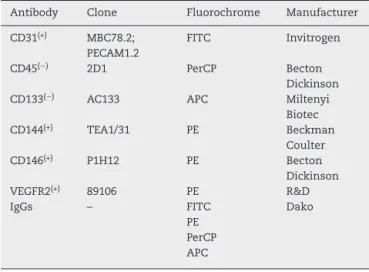
Table1–Monoclonalantibodiesemployedincirculating endothelialcellsanalyses.
Antibody Clone Fluorochrome Manufacturer
CD31(+) MBC78.2;
PECAM1.2
FITC Invitrogen
CD45(−) 2D1 PerCP Becton
Dickinson CD133(−) AC133 APC Miltenyi
Biotec CD144(+) TEA1/31 PE Beckman
Coulter CD146(+) P1H12 PE Becton
Dickinson VEGFR2(+) 89106 PE R&D
IgGs – FITC
PE PerCP APC
Dako
MBC78.2;PECAM1.2,Invitrogen),anti-CD34(clone8G12; Bec-tonDickinson,Bioscences),phycoerthrin(PE)-labeledCD144 (cloneTEA1/31,BeckmanCoulter),anti-CD146(cloneP1H12, BD Bioscences), anti-VEGFR2 (clone 89106, R&D), peri-dinim chlorophyll (PerCP)-labeledanti-CD45(clone 2D1,BD Bioscences), and allophycocianin (APC)-labeled anti-CD133 (cloneAC133,MiltenyiBiotecGmbH,BergischGladbach, Ger-many)(Table1).Threedifferentpanelswerecreatedinthree tubesinanattempttocharacterizeCECwithdifferent pheno-typesasshowninTable2.
A quantityof100Lofblood (withaleukocyte
concen-tration between5and 10×103/
L) wasincubated with the
fluorochrome-labeledmonoclonalanti-humanantibodiesfor 20minat4◦Cinthedarkforthestainingprocedure.Theblood
count was performed using ahematological analyzer (Cell Dyn®;AbbottLaboratories,IL,USA).Redbloodcellswerelysed
byadding2mLofFACSlysingsolution(dilutedat1:10;Becton Dickinson)for10minat4◦C.Theremainingleucocyteswere
washedwith2mL2%phosphatebufferedsaline/bovineserum albuminbuffer(PBS/BSA)atpH=7.4,centrifugedat600×gfor 5minandresuspendedin500Lofwashbuffer.The
acqui-sition of500,000 cells or the totalvolume ofthe tube was performedusingaFACScalibur®flowcytometer(Becton
Dick-inson,SanJose, CA,USA)andanalyzedbyCell-Quest® and
Paint-a-Gate®computerprograms(BD,Bioscences).
The threshold was defined by a forward scatter (FSC) detectorwhichwasloweredinordertoincludelymphocytes. Platelets,debris andleucocyteswere excludedaccordingto theirFCS×SSCandSSC×CD45positions.CECwereanalyzed
Table2–Monoclonalantibodiesappliedinthe identificationandquantificationofmaturecirculating endothelialcells.
Panel Monoclonalantibodies
1 CD31FITC(+),CD144PE(+),CD45PerCP(−)
andCD133APC(−)
2 CD31FITC(+),CD146PE(+),CD45PerCP(−)
andCD133APC(−)
3 CD31FITC(+),VEGFR2PE(+),CD45PerCP(−)
0 256 512 768 1024
FSC 0
256 512 768 1024
SSC SSC
0 256 512 768 1024
CD 45
CD 45
100 101 102 103 104
CD45
100 101 102 103 104
100 101
102 103
104
100 101 102 103 104
CD 31 CD 31
CEC
CD 144 SSC
100
101
102 103
104
100 101 102 103 104 100
101 102
103 104
0 256 512 768 1024
100
101
102
103
104
CD 31 CD 144
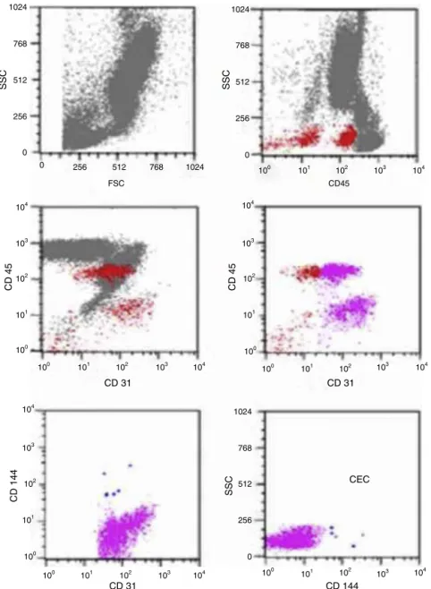
Figure1–Analysisstrategyfortheidentificationofmaturecirculatingendothelialcells(CEC)byflowcytometry.The negativepopulationforCD45wasselectedandanalyzedforthepositivityofendothelialmarkers(CD144,CD146,CD31and
CD133anti-VEGFR2).
according to CD144×CD31×CD133, CD146×CD31×CD133 andanti-VEGFR2×CD31×CD133characteristics.Thestrategy appliedintheseCECanalyzesisshowninFigure1.
Statistical
analysis
Data are presented as mean±standard error of the mean (SEM). TheMann–Whitney test was used to compare con-tinuous variables. Analyses were performed using the R DevelopmentCoreTeam2010Software(Vienna,Austria)and p-values≤0.05wereconsideredstatisticallysignificant.
Results
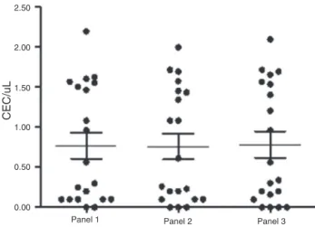
Aspredicted,theseeventswereveryrare,althoughdetectable by flow cytometry using the aforementioned panels,
which gave similar inter-person numbers of CEC (Panel 1: 0.76±0.16cells/L; Panel 2: 0.75±0.15cells/L; Panel 3:
0.78±0.16cells/L). There was no significant difference
regarding their quantification (p-value=0.9; Mann–Whitney test),indicatingthatthesemarkerspresentedsimilarpatterns ofCECexpressioninhealthyindividuals(Figure2).However, differentclinicalconditionsmodifythisbehavior.
Discussion
Inthis study,CECnumbers wereevaluatedinhealthy indi-viduals.Aspreviouslydescribedintheliterature,theseevents howeverrare,17,27weredetectablebyflowcytometryanalysis.
Panel 1 Panel 2 Panel 3 0.00
0.50 1.00 1.50 2.00 2.50
CEC/uL
Figure2–Quantificationbyflowcytometryofcirculating endothelialcells(CEC)withdifferentendothelialmarkers
(CD144,CD146andVEGFR-2)fromhealthysubjects.
thebestcombinationofsurfacemarkersforthistask.Thus, inmany studies there is no certaintyas to whether CECs havebeencorrectlyidentified.However,CECscanbeidentified asthecells expressingendothelialmarkers(CD146, CD144, vWF,VEGFR2)intheabsenceofhematopoietic(CD45,CD14) andprogenitormarkers(CD34,CD133).15,17,29,30Several
proto-colshaveproposedtheuseofwholebloodoramononuclear concentrateobtainedafterenrichmentwithficoll paque to identifythese cells byflowcytometry. Sorting or magnetic beadscanalsobeused;however, thismethodpresentsthe samelimitationsasflowcytometry.Furthermore,theuseof magneticbeadsrarelyprovidestheprecisepurityofthe elu-triatedcellsasthemethodisgenerallyperformedwithone marker,suchasCD146.Therefore,asecondmethod,suchas fluorescencemicroscopy,isusuallyrequiredtoconfirmand quantify the CECs.17 Immunohistochemistry is not a good
optionforthesamereasonsaggravatedbytheextremerarity ofthesecellsinperipheralblood,about0.01%ofmononuclear bloodcells,17,27 and alackofstainingcould beerroneously
interpretedasafalsenegativeresult.Therefore,noneofthese possibilitieshaveemergedasthebestchoice,andaneffective comparisonofresultsbetweenlaboratoriesisdifficult.31
Furthermore,severaltechnicalissuesmustbetakeninto account inorder to truly analyze rare cells such as CECs. Thefirst stepinthe techniqueinvolves extensive cleaning andwashingprocedurestoremoveresidualcellsand parti-cles.Fluorochrome-matched isotypecontrols,currently not favoredforcommon assays,are fairlycrucialinrare event analysis,wheretheyprovideagoodestimateofnonspecific bindingofantibodiestocells.Khanetal.17mentionedthat,
evenwithfreshlydrawnperipheralblood,nonspecificbinding ofisotypecontrolsmaybedetectedin0.1–0.5%ofanalyzed cells.Inmostclinicalassays,thesenonspecific-bindings do not significantly affect data, but in the evaluation of rare cellstheydo.InCECanalysis,thesebindingscanrepresent abackground higherthan the specificcell events.Another pointis thelarge number ofcells(over 500,000) thatmust becountedtoobtainstatisticallymeaningfulnumbersofrare cells.Inthecurrentanalyses,thefirststepwastheexclusion ofCD45(+)cellsbytheSSC×CD45gate;however,asinsome
individualsthelimitbetweenpositiveandnegative popula-tionsisnotveryclear,theSSC×FSCgatewasalsoutilizedto excludediscrepantevents.Furthermore,theantigen expres-sionmay bevariableand mayinvolve othercell lineswith overlappingexpressionofantigens.Forinstance,CD146 rec-ognizeMUC18/S-endo,whichisalsoexpressedinactivatedT cells.Thus,asecondmarker, suchasCD45,wasneededto distinguishthesecells.17Thesameapproachwasadoptedfor
CD31,whichrecognizesPECAM-1presentinendothelialcells, platelets, monocytes,granulocytesand Bcells,whichwere alsoexcludedbyCD45.Anti-VEGFR2andCD144are endothe-lialcellmarkers,astheybindtotheVEGFandVE-Cadherin receptors,respectively.Inaddition,bydifferentiatingbetween mature and precursor endothelial cells, CD133 helped the identificationofCECsasastemcellmarker.CD34expression onendothelialcellsrepresentsaproblemforCECevaluationas itsexpressionisalsofoundinhematopoieticstemcells,17and
thismarkercanbeshowninmatureandimmature endothe-lialcells.32
Anotherstudyperformedwithdeepveinthrombosis(DVT) patientsandcontrolsusingthesamethreepanelsasthisstudy suggestedthattheuseofonlyonepanelmaynotbesufficient toaccuratelyanalyzeCECs.Inthisstudy,ahighersensitivity forCECdetectionwasobservedforoneofthepanels(Panel1) ratherthantheothertwo(unpublisheddata).Regardingthe resultsobtainedwithDVTpatients,wehypothesizedthatthe useoftwoormorepanelscouldincreasetheaccuracyofthe analysisundercertainclinicalconditions.Webelievethatthe expressionofsomeepitopesmaybealteredbysomediseases.
Conclusions
Anyofthesecombinationsofmarkerscanbeusedto success-fullydetermineCECsinhealthyindividualswiththeuseoftwo ormorepanelstoconfirmtheresults.Moreaccuratestudies performedwiththesecellsshouldincreaseour understand-ingregardingtheirphysiologyandinvolvementinreparative processesfavoringtheirpotentialapplicationintheclinical practice.
Conflicts
of
interest
Theauthorsdeclarenoconflictsofinterest.
Funding
FAPESPandCNPq.
Acknowledgments
r
e
f
e
r
e
n
c
e
s
1. BouvierCAGE,CintronJR,BernhardtB,SpaetTH.Circulating endotheliumasanindicatorofvascularinjury.ThrombDiath Haemorrh.1970;40:163–8.
2. HladovecJR.Circulatingendothelialcellsisolatedtogether withplateletsandtheexperimentalmodificationoftheir countsinrats.ThrombRes.1973;3(6):665–74.
3. AsaharaT,MuroharaT,SullivanA,SilverM,vanderZeeR,Li T,etal.Isolationofputativeprogenitorendothelialcellsfor angiogenesis.Science.1997;275(5302):964–7.
4. ShiQ,RafiiS,WuMH,WijelathES,YuC,IshidaA,etal. Evidenceforcirculatingbonemarrow-derivedendothelial cells.Blood.1998;92(2):362–7.
5. PrescottMF,MullerKR.Endothelialregenerationin hypertensiveandgeneticallyhypercholesterolemicrats. Arteriosclerosis.1983;3(3):206–14.
6. TaylorRG,LewisJC.Endothelialcellproliferationand monocyteadhesiontoatheroscleroticlesionsofwhite carneaupigeons.AmJPathol.1986;125(1):152–60.
7. RafiiS,OzMC,SeldomridgeJA,FerrisB,AschAS,Nachman RL,etal.Characterizationofhematopoieticcellsarisingon thetexturedsurfaceofleftventricularassistdevices.Ann ThoracSurg.1995;60(6):1627–32.
8. KalkaC,MasudaH,TakahashiT,Kalka-MollWM,SilverM, KearneyM,etal.Transplantationofexvivoexpanded endothelialprogenitorcellsfortherapeutic
neovascularization.ProcNatlAcadSciUSA.2000;97(7):3422–7.
9. ZiegelhoefferT,FernandezB,KostinS,HeilM,VoswinckelR, HelischA,etal.Bonemarrow-derivedcellsdonotincorporate intotheadultgrowingvasculature.CircRes.2004;94(2):230–8.
10.KaushalS,AmielGE,GuleserianKJ,ShapiraOM,PerryT, SutherlandFW,etal.Functionalsmall-diameterneovessels createdusingendothelialprogenitorcellsexpandedexvivo. NatMed.2001;7(9):1035–40.
11.Dignat-GeorgeF,SampolJ.Circulatingendothelialcellsin vasculardisorders:newinsightsintoanoldconcept.EurJ Haematol.2000;65(4):215–20.
12.RafiiS,LydenD,BenezraR,HattoriK,HeissigB.Vascularand haematopoieticstemcells:noveltargetsfor
anti-angiogenesistherapy?NatRevCancer.2002;2(11):826–35.
13.DamaniS,BacconiA,LibigerO,ChourasiaAH,SerryR, GollapudiR,etal.Characterizationofcirculatingendothelial cellsinacutemyocardialinfarction.SciTranslMed. 2012;4(126):126ra33.
14.SoloveyA,LinY,BrowneP,ChoongS,WaynerE,HebbelRP. Circulatingactivatedendothelialcellsinsicklecellanemia.N EnglJMed.1997;337(22):1584–90.
15.WoywodtA,StreiberF,deGrootK,RegelsbergerH,HallerH, HaubitzM.Circulatingendothelialcellsasmarkersfor ANCA-associatedsmall-vesselvasculitis.Lancet. 2003;361(9353):206–10.
16.BullTM,GolponH,HebbelRP,SoloveyA,CoolCD,TuderRM, etal.Circulatingendothelialcellsinpulmonary
hypertension.ThrombHaemost.2003;90(4):698–703.
17.KhanSS,SolomonMA,McCoyJPJr.Detectionofcirculating endothelialcellsandendothelialprogenitorcellsbyflow cytometry.CytometryBClinCytom.2005;64(1):1–8.
18.NakataniK1,TakeshitaS,TsujimotoH,KawamuraY, TokutomiT,SekineI.Circulatingendothelialcellsin Kawasakidisease.ClinExpImmunol.2003;131(3): 536–40.
19.ButthepP,RummavasS,WisedpanichkijR,
JindadamrongwechS,FucharoenS,BunyaratvejA.Increased circulatingactivatedendothelialcells,vascularendothelial growthfactor,andtumornecrosisfactorinthalassemia.AmJ Hematol.2002;70(2):100–6.
20.BeerepootLV,MehraN,VermaatJS,ZonnenbergBA,Gebbink MF,VoestEE.Increasedlevelsofviablecirculatingendothelial cellsareanindicatorofprogressivediseaseincancer patients.AnnOncol.2004;15(1):139–45.
21.VapaataloH,MervaalaE.Clinicallyimportantfactors influencingendothelialfunction.MedSciMonit. 2001;7(5):1075–85.
22.KörblingM,ReubenJM,GaoH,LeeBN,HarrisDM,CogdellD, etal.Recombinanthumangranulocyte-colony-stimulating factor-mobilizedandapheresis-collectedendothelial progenitorcells:anovelbloodcellcomponentfortherapeutic vasculogenesis.Transfusion(Paris).2006;46(10):1795–802.
23.MancusoP,BurliniA,PruneriG,GoldhirschA,MartinelliG, BertoliniF.Restingandactivatedendothelialcellsare increasedintheperipheralbloodofcancerpatients.Blood. 2001;97(11):3658–61.
24.MancusoP,CalleriA,CassiC,GobbiA,CapilloM,PruneriG, etal.Circulatingendothelialcellsasanovelmarkerof angiogenesis.AdvExpMedBiol.2003;522:83–97.
25.GoonPK,BoosCJ,StonelakePS,BlannAD,LipGY.Detection andquantificationofmaturecirculatingendothelialcells usingflowcytometryandimmunomagneticbeads:a methodologicalcomparison.ThrombHaemost. 2006;96(1):45–52.
26.GoonPK,BoosCJ,LipGY.Circulatingendothelialcells: markersofvasculardysfunction.ClinLab.
2005;51(9–10):531–8.
27.Dignat-GeorgeF,SampolJ,LipG,BlannAD.Circulating endothelialcells:realitiesandpromisesinvasculardisorders. PathophysiolHaemostThromb.2004;33(5–6):495–9.
28.TimmermansF,PlumJ,YöderMC,IngramDA, VandekerckhoveB,CaseJ.Endothelialprogenitorcells: identitydefined?JCellMolMed.2009;13(1):87–102.
29.PeichevM,NaiyerAJ,PereiraD,ZhuZ,LaneWJ,WilliamsM, etal.ExpressionofVEGFR-2andAC133bycirculatinghuman CD34(+)cellsidentifiesapopulationoffunctionalendothelial precursors.Blood.2000;95(February(3)):952–8.
30.JacquesN,VimondN,ConfortiR,GriscelliF,LecluseY, LaplancheA,etal.Quantificationofcirculatingmature endothelialcellsusingawholebloodfour-colorflow cytometricassay.JImmunolMethods.2008;337(2): 132–43.
31.MancusoP,BertoliniF.Circulatingendothelialcellsas biomarkersinclinicaloncology.MicrovascRes. 2010;79(3):224–8.
32.HarhouriK,KebirA,GuilletB,Foucault-BertaudA,Voytenko S,Piercecchi-MartiMD,etal.SolubleCD146displays angiogenicpropertiesandpromotesneovascularizationin experimentalhind-limbischemia.Blood.