DOI: 10.1590/0004-282X20160058
ARTICLE
The extended pterional approach allows
excellent results for removal of anterior
cranial fossa meningiomas
O acesso pterional extendido permite excelente resultado para a ressecção dos
meningiomas da fossa craniana anterior
Jose Carlos Lynch1, Mariangela Barbi Gonçalves1, Celestino Esteves Pereira2, Wladimir Melo2, Gianni
Ferraz Temponi2
Olfactory groove meningiomas (OGMs) and tuberculum sellae meningiomas (TSMs) arise both in the anterior crani-al fossa. OGMs are a subset of meningiomas that grows over
the cribiform plate and the frontosphenoidal suture. hey
represent 8–18% of all intracranial meningiomas1,2,3,4,5. OGMs
displace the optic nerve (ON) and the chiasm inferiorly and posteriorly. TSMs originate from the limbus sphenoidale,
chi-asmatic sulcus and frontosphenoidal suture. hey comprise
3 to 12% of all intracranial meningiomas1,6,7,8,9,10,11. TSMs
dis-place the ON superiorly and laterally and the chiasm superi-orly and posterisuperi-orly6,12,13,14,15.
he distance between the tuberculum sellae and the cri -biform plate is a scanty 2 cm1.Nonetheless, OGMs and TSMs
have features in common, mainly in cases of large meningio-mas with invasion of the surrounding structures. On the ba-sis of preoperative imaging studies and intraoperative
inspec-tion, it is often diicult to determine the exact origin of the
meningioma1,3,6,8,10,11,13,14,15,16(Figures 1, 2, 3, 4).
Despite the advances of microsurgery, the management of anterior fossa meningiomas remains a great challenge. Since these tumors grew in close contact with neural and
vascu-lar structures that cannot be sacriiced or retracted, they still
1Rede D’Or São Luiz, Rio de Janeiro RJ, Brasil;
2Hospital Federal dos Servidores do Estado do Rio de Janeiro, Serviço de Neurocirugia, Rio de Janeiro RJ, Brasil.
Correspondence: José Carlos Lynch; Rua Jardim Botânico, 600/605; 22461-000 Rio de Janeiro RJ, Brasil; E-mail: [email protected]
Conflict of interest: There is no conflict of interest to declare.
Received 21 October 2015; Accepted 09 December 2015.
ABSTRACT
Objective: To describe a unique operative strategy, instead the classical pterional approach, and to analyses it safety and effectiveness for removal of anterior cranial fossa meningiomas. Method: We identify 38 patients with tuberculum sellae and olphactory groove meningiomas operated between 1986 and 2013.Medical charts, operative reports, imaging studies and clinical follow-up evaluations were reviewed and analyzed retrospectively. The pterional craniotomy is extended toward the frontal bone providing access through the subfrontal route, besides the usual anterolateral view provided by the classical pterional approach. Results: Surgical mortality occurred in one patient (2.6%). Gross total resection was achieved in 27 patients (86.8%). Median time of follow-up was 69.4 months. Conclusion: The extended pterional approach allows excellent results. Total removal of meningiomas of the anterior cranial fossa was obtained in 86.8 % of patients, with low morbidity and mortality.
Keywords: meningiomas; cranal fossa, anterior; microsurgery.
RESUMO
Objetivo: Descrever a craniotomia pterional estendida, ao invés da abordagem pterional clássica, e analisar sua segurança e eficácia para a remoção dos meningiomas da fossa anterior. Método: Identificamos 38 pacientes com meningiomas do tubérculo da sela e da goteira olfatória operados entre 1986 e 2013. Os prontuários, relatórios cirúrgicos, exames de imagem e acompanhamento pós-operatório foram analisados retrospectivamente. A craniotomia pterional com extensão para o osso frontal permite acesso pela via subfrontal além da via anterolateral do acesso pterional clássico. Resultados: A mortalidade cirúrgica foi de 2,6% (um paciente). A remoção total foi alcançada em 86,8% (27 pacientes) com um tempo médio de seguimento de 69,4 meses. Conclusão: A abordagem pterional estendida permite excelentes resultados. A remoção total dos meningiomas da fossa craniana anterior foi obtida em 86,8% dos pacientes, com baixa morbi-mortalidade.
Figure 2. Preoperative coronal (A) and sagittal (B) gadolinium-enhanced T1-weighted MRI revealed a large bilateral OGM in a 71-years old man with mood alterations and progressive apathy. Postoperative sagittal (C) gadolinium-enhance T1- weighted MRI, confirming GTR obtained through a right pterional approach.
A
B
C
Figure 1. Preoperative gadolinium-enhanced T1-weighted coronal (A) and sagittal (B) MRI demonstrating a large OGM in a 44-year-old woman with progressive decrease of visual function. Postoperative coronal (C) and sagittal (D) T1-weighted contrast-enhanced MRI demonstrating GTR.
A
B
C
D
Figure 4. T1-weighted MRI with contrast enhancement in cases of A: small, B: medium, C: large: and D: giant OGMs and TSMs.
A
B
C
D
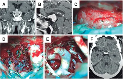
Figure 3. Preoperative coronal (A) and sagittal (B) gadolinium-enhanced T1-weighted MRI revealed a TSM. Intraoperative images (C). Following dissection of the arachnoid, the tumor is clearly seen in the tuberculum sellae. After removal of the suprasellar portion, the meningioma is identified medially to the right ON (D). After GTR, the opposite internal carotid artery, posterior communicating artery and carotid artery bifurcation are completely exposed (E). Early postoperative images showing GTR (F).
A
B
C
raise controversies related to the best surgical approaches to deal with these lesions1,5,8,10,11,12,13,14,15,16,17,18,19,20,21,22,23,24,25.
In our study, we describe a unique operative strategy and personal nuances of the microsurgical technique. We also emphasize the surgical results and discuss the best operative approach in dealing with such challenging lesions.
METHOD
Data collection
hirty-eight patients with anterior fossa meningiomas underwent surgery through the extended pterional trans-syl -vian approach, performed mainly by the senior author (JCL),
between 1986 and 2013. he demographic and clinical pro
-iles of the patients are summarized in Tables 1 and 2 respec -tively. Duration of symptoms ranged from 3 to 120 months
(mean, 12 months). he intraoperative videos and photos
of 27 patients were retrospectively analyzed for nuances of
the microsurgical technique. he Simpson grade of menin -gioma resection was determined through review of the op-erative report, surgeon’s assessment and postopop-erative im-ages. Pathological review was performed based on the World
Health Organization (WHO) guidelines. he need for in -formed consent was waived due to the retrospective charac-ter of the study.
Tumor characteristics
As depicted from preoperative MRI scans and conirmed
during surgery, meningiomas located in the anterior fossa
were classiied into two subgroups according to their dural at -tachment. Tumors attached to the cribiform plate and to the
frontosphenoidal suture were classiied as olfactory groove meningiomas. hose attached to the limbus sphenoidale,
chiasmatic sulcus and frontosphenoidal suture were
classi-ied as tubercullum sellae meningiomas. We also classiclassi-ied
anterior fossa meningiomas according to their size as giant (> 6 cm), large (4-6 cm), medium (2-4 cm) and small (< 2 cm).
Surgical approach and microsurgical technique
Patients underwent surgery according to the tech-nique described by Yasargil in 1975 with the following
modifications: a pterional craniotomy with extension to -ward the frontal bone providing access through the sub-frontal route, besides the usual anterolateral view pro-vided by the classical pterional approach. Craniotomy is performed from the frontal bone to the ipsilateral supra-orbital prominence, avoiding the frontal sinus. The fron-totemporal duramater is opened in a curvilinear fashion over the Sylvian fissure and a second incision is directed
toward the falciform ligament. The extended pterional ap -proach described above was used in all cases.
OGMs
he progressive elevation of the tumor from the cribiform
plate and planum sphenoidale reveals small arteries that ir-rigate the meningioma. In order to decrease intraoperative bleeding, early interception of these feeding arteries is
essen-tial. he tumor capsule is bipolar coagulated medial to the
ipsilateral ON and the tumor is debulked. If dissection of the tumor from the perforators is tenacious, it is better to leave a shell of the tumor on the vessel wall than to risk the rupture
of the artery. he basal dura with tumor invasion is bipolar coagulated and striped of, and the underline hypertrophic
bone is partially drilled away without entering the sphenoi-dal sinus (Simpson Grade 2).
TSMs
he dissection must start in the planum sphenoidale or
tuberculum sellae, bipolar coagulating the feeding arter-ies. Internal tumor debulking is achieved by piecemeal
re-section. he tumor located posterolaterally to the ON and
medial to the internal carotid artery is carefully dissected through the optic carotid triangle and removed from
be-neath the ON. A diamond ball must be used for drilling the roof of the optic canal when the tumor extends through it.
Copious irrigation is mandatory to prevent damage to the
ON by the heat. he meningioma is exposed and careful -ly dissected from the inferior and medial aspect of the ON with minimal manipulation.
Patients Follow-up
In the immediate postoperative period, patients were submitted to contrast-enhanced computed tomogra-phy scans. All patients were followed up with Magnetic Resonance Imaging (MRI) studies 3 and 12 months after sur-gery. he mean follow-up period was69,4 months (range,
4-324 months). hereafter, patients were reexamined or at least interviewed by telephone. he Glasgow Outcome Scale (GOS) deined the outcome (Table 3).
Table 2. Clinical presentation.
Symptoms Number Percentage
Personality changes 28 73.4
Headache 14 36.8
Seizure 6 18.1
Visual deficit 9 13.2
Obesity 3 7.8
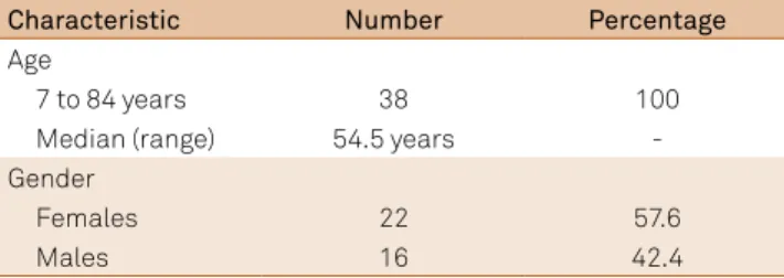
Table 1. Demographic characteristics of the study population.
Characteristic Number Percentage
Age
7 to 84 years 38 100
Median (range) 54.5 years
-Gender
Females 22 57.6
RESULTS
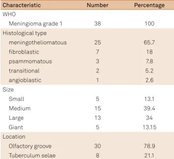
Tumor size, histological subtype and location are sum-marized in Table 4.
Operative mortality was 2.6% (one patient) due to a pul-monary thromboembolism 15 days after discharged from hospital. A 77 years-old female died 3 months after sur-gery due to an acute subdural hematoma. One patient died 18 months after discharged from hospital because of a
pul-monary carcinoma. Other patient afected by obesity and cardiac failure died 2 years after the operation. Excellent
or good outcome (GOS 4 or 5) was achieved in 31 patients
(81,5%). here were 2 patients with postoperative Cerebro
Spinal Fluid (CSF) leakage who returned to the operating room for repair. Surgical outcome and follow up are sum-marized in Table 3.
DISCUSSION
Surgical approaches and microsurgical techniques
Various approaches have been described in the literature to treat anterior fossa meningiomas, including the pterion-al1,7,10,11,13,17,20,22,26, uni3,5,17,23,24 or bilateral subfrontal8,11,21,27 and
cranial base approaches9,2,5,16,25,,27. he surgical technique pre
-sented here is modiied from one described earlier by Yasargil
and others1,7,10,11.
he extended pterional approach has a number of ad -vantages over the bifrontal craniotomy: provides the short-est distance to the tuberculum sellae, the early release of CSF
from the basal cisterns, allows brain relaxation and mini
-mizes frontal lobe retraction. Early exposure of the ON and chiasm provides protection of the visual system. he identi
-ication of the internal carotid artery improves the ability to
dissect the anterior cerebral artery and its branches, allowing protection of these vessels 1,7,10,11,13,14,17,19,20,22,26,28,29,30.
he disadvantage of the pterional approach is the inad -equate visualization of the undersurface of the ipsilateral ON and chiasm. However we can circumvent this problem by moving the surgical microscope medially, associated with
lateral tilt of the operative table. his surgical nuances tech -nique was not published before.
Nakamura et al.11 compared the results of their patients
operated via bifrontal and frontolateral approaches. hey
claimed that the frontolateral and pterional approaches provide remarkable improvement compared with the bi-frontal approach. To decrease tumor recurrence, some au-thors recommend cranial base approaches such as uni or bilateral orbital osteotomy and cranial base drilling and reconstruction2,5,8,16,25.
he demographic characteristics of the patients in this series did not inluence the results.
Extend of resection and recurrence
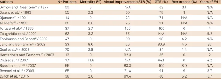
Gross total resection (GTR) was accomplished by a complete macroscopic lesion removal and coagulation of its dural attachment (Simpson grade II) in 33 (86,8%) pa-tients and subtotal resection in 5 (13.1%).Published pa-pers on TSMs and OGMs have reported GTR rates rang-ing from 71% to 100% (Table 2). Some surgeons stated that Simpson Grade I resection including dural attachment and underlying tumor-infiltrated bone is critical in preventing future recurrence.25 Nevertheless, several surgeons,
includ-ing the authors of the current paper, have preferred a more conservative approach, not entering the paranasal sinus-es, because of the risk of postoperative CSF leakage and infection, especially in elderly patients4,9,10,11,13,17,18,20,21,22,23.
In published microsurgical series, the recurrence rate for TSMs and OGMs, with a follow-up period ranging from 2 to 9.3 years, varies from 0 to 41%.5,10,13,16 The recurrence
rate of this sample, with a median follow-up of 5.7 years (range 4–324 months), was 5.2%.
Table 4. Tumor characteristics.
Characteristic Number Percentage
WHO
Meningioma grade 1 38 100
Histological type
meningotheliomatous 25 65.7
fibroblastic 7 18
psammomatous 3 7.8
transitional 2 5.2
angioblastic 1 2.6
Size
Small 5 13.1
Medium 15 39.4
Large 13 34
Giant 5 13.15
Location
Olfactory groove 30 78.9
Tuberculum selae 8 21.1
WHO: World Health Organization.
Table 3. Surgical outcome and follow-up.
Number Percentage
GTR 33 86
Mortality 1 2.6
Complications
Seizures 3 7.8
CSF leak 2 5.2
Meningitis 2 5.2
Recurrence 2 5.2
GOS
1 4 10.5
3 3 7.9
4 8 21.1
5 23 60.5
Table 5. Microsurgical series for TSM and OGM.
Authors Nº Patients Mortality (%) Visual Improvement/STB (%) GTR (%) Recurrence (%) Years of F/U
Symon and Rosentein18 / 1977 33 3 N/A 82 3.1 N/A
Solero et al.4 / 1983 55 2.3 60 78 3 N/A
Ojemann21 / 1991 14 0 73 71 N/A N/A
Al-Mefty25 / 1993 35 8.6 25 91 N/A N/A
Turazzi et al.20 / 1999 37 2.7 100 100 0 4
Zeugaridis et al. / 2001 62 3.2 65 N/A N/A 5.2
Fahlbusch and Schott6 / 2002 47 0 80 98 4.2 N/A
Jallo and Benjamim13 / 2002 23 8.6 55 86.9 4.5 93
Goel et al.23 / 2002 70 2.8 N/A 84 1.4 N/A
Hentschels and Demonte16 / 2003 13 0 92.3 85 0 2
Colli et al.8 / 2007 17 11.8 N/A 94.1 0 4.2
Bassioni et al.29 / 2007 55 0 83.3 100 8.9 N/A
Romani et al.19 / 2009 65 0 21.4 91 9 3.7
Lynch et al. / 2015 38 2.6 89.4 86 5.2 5.7
TSM: Tuberculum Sellae meningiomas; OGM: Olfactory groove meningiomas. Mortality and clinical outcome
Cushing6 reported an operative mortality of 27.5%. As a
result of the reinements of microsurgical techniques, death
rates had declined11,18,20,23,26,29. In this present series, the
surgi-cal mortality occured in one patient (2.6%), with 31(81.5%) patients obtaining GOS 4 or 5. Al-Mefty25 and Solero et al.1
observed higher mortality rates in patients with tumors ex -ceeding 3 cm in diameter, compared with mortality rates in patients with smaller tumors. In our series, 18 individuals (47.36%) harbored large or giant tumors, but we did not ob-served increase in mortality in this group of patients.
Nowadays, the preservation of vision is an important goal of treatment. Fahlbusch and Schott10 and Symon18
found tumors smaller than 3 cm to be associated with better visual outcomes than tumors larger than 3 cm in
diameter. In this sample, improvement of vision occured in 10.5% of patients and preserved vision with no further de-terioration in 89.4%. Improvement or stabilization of vision has been reported in 48.8 to 100% of patients undergoing surgery (Table 5).
We can conclude that the extended pterional transsyl -vian approach has many advantages. It is simple and fast,
while preserving normal anatomy. he early exposure of the
ON and chiasm provides protection of the visual system. In this paper we show that we can achieve a low mortality and morbidity, with a high rate of GTR, fewer complications,
and low recurrence rates with the extended pterional trans
-sylvian approach. Our study conirms that the pterional ap
-proach and its variants are efective to remove anterior fossa
meningiomas.
References
1. Solero CL, Giombini S, Morello G. Suprasellar and olfactory meningiomas: report on a series of 153 personal cases. Acta Neurochir (Wien). 1983;67(3-4):181-94. doi:10.1007/BF01401420
2. Colli BO, Carlotti CG Jr, Assirati JA Jr, Santos MB, Neder L, Santos AC et al. Olfactory groove meningiomas: Surgical technique and follow-up review. Arq Neuropsquiatr. 2007;65(3B):795-9. doi:10.1590/S0004-282X2007000500012
3. Tella Jr OI, Paiva Neto MA, Herculano MA, Faedo Neto A. [Olfactory groove meningioma]. Arq Neuropsiquiatr. 2006;64(1):83-7. Portuguese. doi:10.1590/S0004-282X2006000100017.
4. Coppens JR, Couldwell WT. Olfactory groove meningiomas. In: Pamir MN, Black PM, Fahlbush R, editors. Meningiomas: a comprehensive text. New York: Saunders; 2009. p. 373-86.
5. Fox F, Khurana VG, Spetzler RF. Olfactory groove/planum sphenoidale meningiomas. In: Lee JH, editor. Meningiomas: diagnosis, treatment, and outcome, New York: Springer; 2008. p. 327-32.
6. Cushing H, Eisenhardt L. Meningiomas: their classification, regional behavior, life history, and surgical end results. Baltimore: Charles C. Thomas; 1938. p. 224-283.
7. Yasargil MG: General operative techniques. In: Yasargil MG, editor. Microneurosurgery. Vol. 1: Microsurgical Anatomy of the basal
cisterns and vessels of the brain, diagnostic studies, general operative techniques and pathological considerations of the intracranial aneurysms. New York: Georg Thieme/Thieme-Stratton; 1984. p. 208-33.
8. McDermott MW, Rootman J, Durity FA. Subperiosteal, subperiorbital dissection and division of the anterior and posterior ethmoid arteries for meningiomas of the cribriform plate and planum sphenoidale: technical note. Neurosurgery. 1995;36(6):1215-9. doi:10.1227/00006123-199506000-00027
9. Mathieson T, Lindqvist C, Kihlström, Karlsson B. Recurrence of cranial base meningiomas. Neurosurgery. 1996;39(1):2-7. doi:10.1097/00006123-199607000-00002
10. Fahlbusch R, Schott W. Pterional surgery of meningiomas of the tuberculum sellae and planum sphenoidale: surgical results with special consideration of ophthalmological and endocrinological outcomes. J Neurosurg. 2002;96(2):235-43. doi:10.3171/jns.2002.96.2.0235
12. Kempe LG: Olfactory groove meningioma. In: Kempe LG, editor. Operative neurosurgery. Vol 1: Cranial, cerebral, and intracranial vascular disease. New York:, Springer; 1968. p. 104-8.
13. Jallo GI, Benjamin V. Tuberculum sellae meningiomas: microsurgical anatomy and surgical technique. Neurosurgery. 2002;51(6):1432-40. doi:10.1097/00006123-200212000-00013
14. Spektor S, Valarezo J, Fliss DM, Gil Z, Cohen J, Goldman J et al. Olfactory groove meningiomas from neurosurgical and ear, nose, and throat perspectives: approaches, techniques, and outcomes. Neurosurgery. 2005;57(4 Suppl): 268-80. doi:10.1227/01.NEU.0000176409.70668.EB
15. Zevgaridis D, Medele RJ, Müller A, Hischa AC, Steiger HJ. Meningiomas of the sellar region presenting with visual impairment: impact of various prognostic factors on surgical outcome in 62 patients. Acta Neurochir (Wien). 2001;143(5):471-6. doi:10.1007/s007010170076
16. Hentschel SJ, DeMonte F. Olfactory groove meningiomas. Neurosurg Focus. 2003;14(6):e4.
17. Aguiar PH, Tahara A, Almeida AN, Simm R, Silva AN, Maldaun MV et al. Olfactory groove meningiomas: approaches and complications. J Clin Neurosci. 2009;16(9):1168-73. doi: 10.1016/j.jocn.2008.12.013
18. Symon L, Olfactory groove and suprasellar meningiomas. In: Krayonbühl H, editor. Advances and technical standards in neurosurgery. Wien: Springer; 1977. p. 67-91.
19. Romani R, Lehecka M, Gaal E, Toninelli S, Celik O, Niemelä M et al. Lateral supraorbital approach applied to olfactory groove meningiomas: experience with 66 consecutive patients. Neurosurgery. 2009;65(1):39-52. doi:10.1227/01.NEU.0000346266.69493.88
20. Turazzi S, Cristofori L, Gambin R, Bricolo A. The pterional approach for the microsurgical removal of olfactory groove meningiomas. Neurosurgery. 1999;45(4):821-6. doi:10.1097/00006123-199910000-00016
21. Ojemann RG. Olfactory groove meningiomas. In: Al-Mefty O, editor. Meningiomas. New York: Raven; 1991. p. 383-93.
22. Hassler W, Zentner J. Pterional approach for surgical treatment of olfactory groove meningiomas. Neurosurgery. 1989;25(6):942-7. doi:10.1227/00006123-198912000-00014
23. Goel A, Muzumdar D, Desai KI. Tuberculum sellae meningioma: a report on management on the basis of a surgical experience with 70 patients. Neurosurgery. 2002;51(6):1358-64.
24. Poppen JL. Operative techniques for removal of olfactory groove and suprasellar meningiomas. Clin Neurosurg. 1964;11:1-7.
25. Al-Mefty O. Tuberculum sellae and olfactory groove meningiomas. In: Sekhar LN, Janecka IP, editors. Surgery of cranial base tumors. New York: Raven; 1993. p. 507-19.
26. Pamir MN, Ozduman K, Belirgen M, Kilic T, Ozek MM. Outcome determinants of pterional surgery for tuberculum sellae meningiomas. Acta Neurochir (Wien). 2005;147(11):1121-30. doi:10.1007/s00701-005-0625-0
27. Bogaev CA, Sekhar LN. Olfactory groove and planum sphenoidale meningiomas. In: Sekhar LN, Fessler RG. Atlas of neurosurgical techniques. New York Brain Thieme; 2006. p. 608-17.
28. Park CK, Jung HW, Yang SY, Seol HJ, Paek SH, Kim DG. Surgically treated tuberculum sellae and diaphragm sellae meningiomas: the importance of short-term visual outcome. Neurosurgery. 2006;59(2):238-43. doi:10.1227/01.NEU.0000223341.08402.C5
29. Bassiouni, H., Asgari, S., Stolke, D.: Olfactory groove meningiomas: functional outcome in a series treated microsurgically. Acta Neurochir (Wien). 2007;149(2):109-21. doi:10.1007/s00701-006-1075-z