1. Department of Pharmacology, Faculty of Medicine, University of Prishtina, Kosovo
2. Departament of Rheumatology, Faculty of Medicine, University of Prishtina, Kosovo
3. Department of Biochemistry, Faculty of Medicine, University of Prishtina, Kosovo
4. Department of Physiology, Faculty of Medicine, University of Prishtina, Kosovo
5. Department of Pharmacology, Cerrahpasa Medical School, Istanbul University, Turkey
Conclusion: This study shows that Hcy status, but not
vitamin B12 status, is associated with BMD in this co-hort of postmenopausal women. We therefore confirm that high Hcy levels are an independent risk factor for osteoporosis. BMD evaluation in women at post menopause with high Hcy levels may be helpful in advi sing precautionary measures.
IntroductIon
Osteoporosis, a major health problem in ageing po pulation, is a chronic skeletal disorder characterized by reduction of bone mineral density (BMD) and compromised bone strength that leads to increased susceptibility to bone fractures1. Results from recent investigations show an association between increased plasma concentrations of homocysteine (Hcy) and reduced BMD and increased risk of bone fractures2-10. Actual ly, hyperhomocysteinemia (HHcy) has been considered as a new independent risk factor for os -teoporosis and fractures, especially in the elderly11,13. Seve ral mechanisms have been proposed to link HHcy with the pathogenesis of osteoporosis: reduction of bone formation by inducing apoptosis in human bone marrow stromal cells12, osteoclastogenesis stimu -lation13,14, bone blood flow reduction and consequen -tly changes on bone biomechanical properties15, de-cline in physical functioning19, alteration of gene ex-pression with reduced methylation capacity20, oxida-tive damage on trabecular structures21, and reduction of bone strength by interfering with collagen cross-links5,22. Despite this, there is continuing inconsisten-cy concerning the relationship between Hinconsisten-cystatus and BMD, with some stu dies showing an association of high plasma concentrations of Hcywith low BMD4,6,16,17,28 while other studies showed no significant asso -ciation3,5,8,30. In general, plasma concentrations of Hcy are age-dependent with higher levels in elderly people,
Relationship of homocysteine levels with lumbar
spine and femur neck BMD in postmenopausal women
Bahtiri E1, Islami H1, Rexhepi S2, Qorraj-Bytyqi H1, Thaçi K3, Thaçi S4, Karakulak Ç5, Hoxha R1
ACTA REUMATOL PORT. 2015;40:355-362
AbstrAct
Objective: The focus of several studies in recent years
has been the association between increased plasma con-centrations of homocysteine (Hcy), reduced bone mine ral density and increased risk of bone fractures. Nevertheless, inconsistencies persist in the literature. Thus, the objective of this study was to investigate the possible relationship between serum Hcy and vitamin B12 status, and bone mineral density, on a group of post-menopausal women.
Materials and methods: One hundred thirty-nine
postmenopausal women were recruited to enter this cross-sectional study. Bone mineral density (BMD) of total hip, femoral neck and lumbar spine was measured by dual-energy X-ray absorptiometry (DXA) and serum Hcy, vitamin B12, parathyroid hormone (PTH), total calcium and magnesium levels were determined. In addi tion, we investigated the relationship of Hcy and vi-tamin B12 and BMD using a meta-analysis approach.
Results: Serum Hcy levels were significantly higher in
osteoporotic women when compared to other BMD groups, and were inversely related to lumbar spine BMD and femur neck BMD. Body mass index and serum Hcy levels were shown to be significant predi -ctors of BMD at lumbar spine, femur neck and total hip. The performed meta-analysis showed that serum Hcy levels were significantly higher in osteoporotic sub-jects compared to normal BMD subsub-jects.
MAtErIAls And MEthods
study dEsIGn And subjEcts
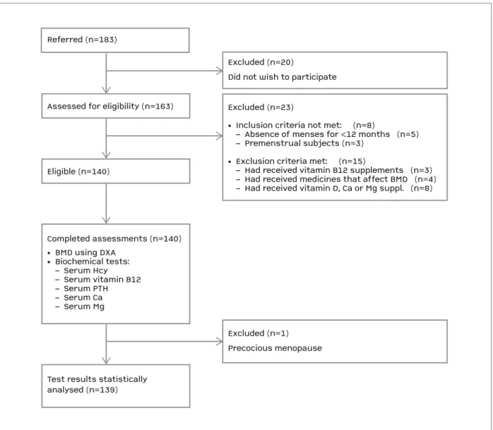
The study was conducted at the University of Prishti-na and Kosovo OccupatioPrishti-nal Health Institute between April 2013 and July 2013. One hundred thirty-nine postmenopausal women, aged between 41 and 65 years, were recruited prospectively to enter this cross--sectional study (Figure 1). The research protocol was in accordance with the Declaration of Helsinki and was approved by the local ethical committee. All the sub-jects provided their written informed consent before inclusion in the study. Postmenopausal women were defined as those whose menses had stopped more than especially in postmenopausal women18.
Vitamin B12 is an important determinant of total Hcy status and may have an indirect impact on BMD via tHcy4,9,28. In addition, vitamin B12 may have direct impact on BMD by affecting osteoblasts activity31. Seve -ral studies have analyzed the relationship between vi-tamin B12 levels and BMD3-5,9,28,33. However, the results of these studies are at odds with each other.
The study main purpose of the study is to investi-gate a possible relationship between serum Hcy and vitamin B12 status and BMD on a group of postme -no pausal women. In addition, we aimed to estimate the relationship of Hcy and vitamin B12 with osteo-porosis risk using a meta-analysis approach.
Referred (n=183)Assessed for eligibility (n=163)Eligible (n=140)Test results statisticallyanalysed (n=139)Excluded (n=1)Precocious menopause
Excluded (n=20)Did not wish to participateExcluded (n=23)• Inclusion criteria not met: (n=8) – Absence of menses for <12 months (n=5) – Premenstrual subjects (n=3)• Exclusion criteria met: (n=15) – Had received vitamin B12 supplements (n=3) – Had received medicines that affect BMD (n=4) – Had received vitamin D, Ca or Mg suppl. (n=8)Completed assessments (n=140)• BMD using DXA• Biochemical tests: – Serum Hcy – Serum vitamin B12 – Serum PTH – Serum Ca – Serum Mg
Referred (n=183)
Assessed for eligibility (n=163)
Eligible (n=140)
Test results statistically analysed (n=139)
Excluded (n=1) Precocious menopause Excluded (n=20)
Did not wish to participate Excluded (n=23)
• Inclusion criteria not met: (n=8)
– Absence of menses for <12 months (n=5) – Premenstrual subjects (n=3)
• Exclusion criteria met: (n=15)
– Had received vitamin B12 supplements (n=3) – Had received medicines that affect BMD (n=4) – Had received vitamin D, Ca or Mg suppl. (n=8)
Completed assessments (n=140) • BMD using DXA • Biochemical tests: – Serum Hcy – Serum vitamin B12 – Serum PTH – Serum Ca – Serum Mg
COBAS Integra 400 Plus (Roche Diagnostics, Switzer-land), while serum vitamin B12 and PTH concentra-tions were determined simultaneously using Electro-chemiluminescence Immunoassay kits on the Elecsys 2010 system (Roche Diagnostics, Switzerland).
stAtIstIcAl AnAlysIs
Results of continuous variables are reported as means ± SD. Chi-square test was used to compare proportions (percentages). One-way ANOVA was used to analyze differences between groups for continuous varia bles followed with post hoc test. Pearson’s correlation ana -lysis was carried out to measure the association of BMD with the study variables.
Multiple regression analysis was performed to deter-mine the relationship between BMD at different regions, age, BMI as well as serum levels of Hcy and vitamin B12. Studies cited in this article were included in the meta-analysis if they met all of the following criteria: (1) used a cross-sectional, cohort or case-control design, (2) study participants were postmenopausal women, (3) evaluated the association between serum Hcy and/or vitamin B12 levels and BMD, (4) provided suf-ficient information on Hcy and/or vitamin B12 levels in osteoporotic patients and normal BMD participants, (5) BMD was measured by DXA, (6) the mean and stan-dard deviation of Hcy and/or vitamin B12 levels were used to calculate the mean difference and 95% CI. Other cited studies in this article that observed or did not observe an association between serum Hcy and/or vitamin B12 and BMD were not included in the meta-analysis because necessary information for meta-analysis was unavailable. Potential between-study heterogeneity of the studies included in the meta-analysis was assessed using Chisquare based Q test and the degree of hetero -geneity was assessed using I2test, which was conside -red significant for I2>50%. When the effects were as-sumed to be homogeneous (I2<50%), the fixed-effects model was used to estimate the pooled mean diffe -rences and their corresponding 95% confidence inter-val (95% CI). Otherwise, the random-effects model was used. Begg’s funnel plots were used to assess potential publication bias. Pooled weighted mean difference of Hcy and vitamin B12 in osteoporotic patients and nor-mal BMD participants was assessed using the Z-test.
Statistical analysis was performed using SPSS soft-ware version 16 (SPSS Inc., Chicago, USA) and Com-prehensive Meta-Analysis (CMA) software version 2 (Biostat, Inc., Englewood, USA). A p value of less than 0.05 was considered as statistically significant. 1 year before inclusion in the study.
The exclusion criteria were: having received vitamin B12 and/or folic acid supplements or other me dicines known to influence levels of Hcy or vitamin B12 or bone mineralization during the preceding one year (glucocorticoids, thiazide diuretics, anticoagulants, an-ticonvulsants, estrogen therapy); having received bis-phosphonates, contraceptives, vitamin D or calcium, magnesium or iron supplements during the preceding six months. Patients were also excluded if they had metabolic bone diseases or endocrine abnormalities (other than diabetes mellitus), malabsorptive disorders, having neoplastic diseases during previous five years, chronic liver or renal diseases, severe cognitive im-pairment or severe psychological problems and if they were drug or alcohol abusers, or current smokers.
Weight and height were measured with a balanced scale and attached stadiometer (Seca model 700-220, Vogel & Halke Gmbh & Co., Hamburg, Germany) in light indoor clothing without shoes before performing DXA, and body mass index (BMI) was calculated as kilograms per square meters (kg/m2).
bMd MEAsurEMEnt
Total hip, femoral neck and lumbar spine (L1-L4) BMD was measured in all subjects included in the study by dual-energy X-ray absorptiometry (DXA) using the same Hologic®DXA scanner (model QDR 4500) and was expressed in absolute values as grams of mineral content per square centimeters of bone area (g/cm2). Quality control of the scanner was performed daily by measuring a phantom’s density that showed stable re-sults at the time of the study. Coefficients of variation of DXA measurements were 1%. Consistent with World Health Organization (WHO) classification system, os-teoporosis is defined as T-score at least -2.5 standard deviations below the mean (≤ -2.5 SD) and osteopenia or low bone mass is defined as a T-score between -2.5 and -1.0 standard deviations below the mean.
blood sAMplInG And sEruM AnAlysEs
Fasting blood samples for measurement of serum Hcy, vitamin B12, total calcium, magnesium, and parathy-roid hormone (PTH) were drawn from the antecubital vein between 8 and 9 AM on the same day of the BMD measurements. Samples were centrifuged within one hour, and the separated serum was immediately stored at -20 ˚C until assayed.
Serum Hcy, total calcium and magnesium concen-trations were determined using automated analyzer,
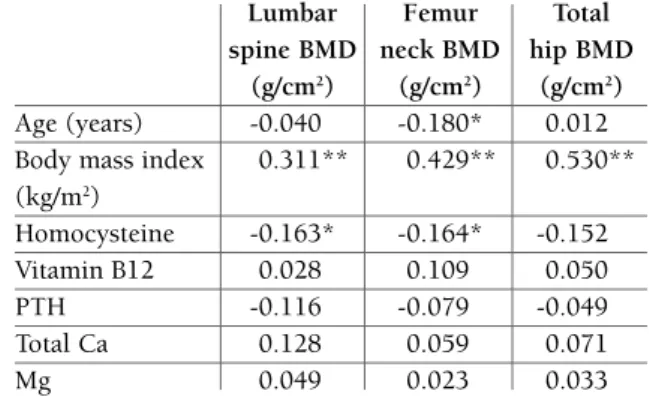
BMD were positively correlated with BMI, while no sig-nificant correlations were found between BMD (at all sites where it was measured) and age. Serum levels of Hcy were inversely related to lumbar spine BMD (r= -0.163, p<0.05) and femur neck BMD (r= -0.164, p<0.05) whereas at total hip the association didn’t reach statistical significance (r = 0.154, p>0.05). No signi -ficant associations were found between serum vitamin B12, PTH, total calcium and magnesium levels and BMD (Table II). Highly significant negative correlation between Hcy and vitamin B12 (r=-0.244, p<0.001) was found (data not shown in the table).
Multiple linear regression analysis in Table III shows the possible influence of age, BMI, serum Hcy and vi-tamin B12 levels in predicting BMD. According to β coefficients of age-adjusted linear regression analysis, main significant predictors of BMD at lumbar spine, fe-mur neck and total hip were BMI and serum Hcy le vels; age itself was a significant predictor at the femoral neck only.
Because there was a significant betweenstudy he -terogeneity in the meta-analysis of the association be-tween Hcy and BMD (c2=42.92; df=5; p<0.001; I2=88.35) the weighted mean difference (WMD) esti-mates of BMD (in g/cm2) were calculated as DerSimo-nian and Laird estimators using random-effects mo del; the WMD for normal BMD versus osteoporosis was 0.826 (95% CI, 0.375-1.276).The forest plot of six rEsults
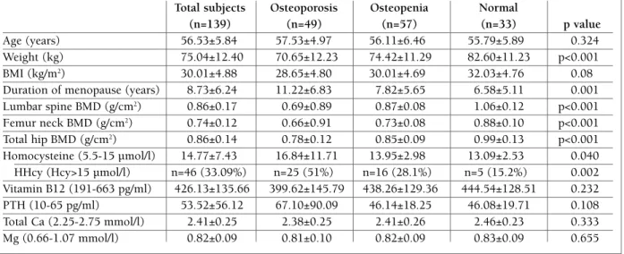
Clinical and biochemical features from the study popu lation expressed in means ± SD with regard to the lo -west T-score are summarized in Table I. According to the lowest T-score in any of the skeletal sites where BMD was measured, 49 subjects had osteoporosis, 57 had low bone mass and 33 had normal BMD values. While there were no differences between BMD groups regarding age (p=0.324), the mean BMI in normal BMD group was statistically higher than in osteoporosis group (p<0.001) and the mean duration of menopause was significantly longer in the osteoporosis group com-pared to the mean duration of menopause in low bone mass and normal BMD groups (p=0.001). The preva-lence of HHcy (Hcy>15 µmol/l) in the study population was 33.09%. The subjects in osteoporosis group had significantly higher serum Hcy levels (16.84±11.71 vs. 13.95±2.98 and 13.09±2.53, respectively, p=0.040) and increased prevalence of HHcy when compared to the subjects in low bone mass and normal BMD groups (51% vs. 28.1% and 15.2%, respectively, p=0.002). There was no statistically significant difference between the groups in serum vitamin B12, PTH, total calcium and magnesium levels (p>0.05).
The results of Pearson’s correlation analysis between BMD and the study variables are shown in Table II. Lumbar spine BMD, femur neck BMD and total hip
tAblE I. clInIcAl And bIochEMIcAl chArActErIstIcs oF 139 postMEnopAusAl woMEn, dIvIdEd AccordInG to thE lowEst t-scorE
Total subjects Osteoporosis Osteopenia Normal
(n=139) (n=49) (n=57) (n=33) p value
Age (years) 56.53±5.84 57.53±4.97 56.11±6.46 55.79±5.89 0.324
Weight (kg) 75.04±12.40 70.65±12.23 74.42±11.29 82.60±11.23 p<0.001 BMI (kg/m2) 30.01±4.88 28.65±4.80 30.01±4.69 32.03±4.76 0.08
Duration of menopause (years) 8.73±6.24 11.22±6.83 7.82±5.65 6.58±5.11 0.001 Lumbar spine BMD (g/cm2) 0.86±0.17 0.69±0.89 0.87±0.08 1.06±0.12 p<0.001
Femur neck BMD (g/cm2) 0.74±0.12 0.66±0.91 0.73±0.08 0.88±0.10 p<0.001
Total hip BMD (g/cm2) 0.86±0.14 0.78±0.12 0.85±0.09 0.99±0.13 p<0.001
Homocysteine (5.5-15 µmol/l) 14.77±7.43 16.84±11.71 13.95±2.98 13.09±2.53 0.040 HHcy (Hcy>15 µmol/l) n=46 (33.09%) n=25 (51%) n=16 (28.1%) n=5 (15.2%) 0.002 Vitamin B12 (191-663 pg/ml) 426.13±135.66 399.62±145.79 438.26±129.36 444.54±128.51 0.232 PTH (10-65 pg/ml) 53.52±56.12 67.10±90.09 46.14±18.25 46.08±19.71 0.108 Total Ca (2.25-2.75 mmol/l) 2.41±0.25 2.38±0.25 2.41±0.26 2.46±0.23 0.333 Mg (0.66-1.07 mmol/l) 0.82±0.09 0.81±0.10 0.82±0.09 0.83±0.09 0.655
studies (Figure 2) with 821 participants shows that serum Hcy levels were significantly higher in osteo-porotic subjects compared to normal BMD subjects (Z=3.590; p=0.0003; 95% CI, 0.375-1.276).
For the vitamin B12 analysis the largest source of
heterogeneity stemmed from the study of Bucciarelli et
al.6After removing this study from the meta-analysis the heterogeneity decreased from 98.65% to 11.99% (c2=5.681; df=5; p=0.338; I2=11.99). The forest plot of six studies with 643 participants shows that serum vitamin B12 levels were lower in osteoporotic subjects (WMD=-0.007) but the difference was not statistically significant under fixed-effect model (Z=-0.082; p=0.935; 95% CI, -0.165-0.152).
dIscussIon
A high serum level of homocysteine has been recently recognized as a risk factor for osteoporosis and osteo-porotic fractures2,7,11,13,19. The role of Hcy is yet not clearly understood, despite the fact that there are se -veral suggested mechanisms for the effect of HHcy in bones5,12-15,19-21. Earlier studies that observed high prevalence of osteoporosis in patients with homocystei -nuria23,24, followed by studies investigating the influen -ce of methylenetetrahydrofolate reductase (MTHFR) polymorphism in BMD25,26,27 were forerunners of more tAblE II. pEArson corrElAtIon coEFFIcIEnts
bEtwEEn bMd And thE study vArIAblEs (r) Lumbar Femur Total spine BMD neck BMD hip BMD
(g/cm2) (g/cm2) (g/cm2) Age (years) -0.040 -0.180* 0.012 Body mass index 0.311** 0.429** 0.530** (kg/m2) Homocysteine -0.163* -0.164* -0.152 Vitamin B12 0.028 0.109 0.050 PTH -0.116 -0.079 -0.049 Total Ca 0.128 0.059 0.071 Mg 0.049 0.023 0.033 *p <0.05; **p<0.01
AStudynameBaines M et al. 2007Cagnacci A et al. 2008Bozkurt N et al. 2009Bucciarelli P et al. 2010Halioglu B et al. 2010Ouzzif Z et al. 20120.23921.21530.42961.47891.14560.50550.8257Std diffin meansStandarderror0.13530.23870.21960.16870.25940.18420.2300Statistics for each studyStd diff in means and 95% CIVariance0.01830.05700.04820.02840.06730.03390.0529Lowerlimit-0.02600.7474-0.00091.14840.63720.14450.3749Upperlimit0.50441.68320.85991.80961.65390.86641.2766Z-Value1.76785.09081.95578.76874.41642.74493.5897p-Value0.07710.00000.05050.00000.00000.00610.0003Relativeweight17.9715.8816.3117.3715.4117.06-2.00-1.000.001.002.00BStudynameBaines M et al. 2007Cagnacci A et al. 2008Bozkurt N et al. 2009Halioglu B et al. 2010Ouzzif Z et al. 2012Kakehasi et al. 2012-0.0770.4630.052-0.188-0.1260.000-0007Std diffin meansStandarderror0.1350.2250.2170.2430.1810.3290.081Statistics for each studyStd diff in means and 95% CIVariance0.0180.0510.0470.0590.0330.1080.007Lowerlimit-0.3420.022-0.374-0.664-0.482-0.645-0.165Upperlimit0.1870.9040.4780.2890.2300.6450.152Z-Value-0.5732.0560.239-0.772-0.6940.000-0.082p-Value0.5670.0400.8110.4400.4881.0000.935Relativeweight36.0612.9513.9111.1019.926.06-2.00-1.000.001.002.00
OsteoporosisNormal BMDOsteoporosisNormal BMD A Studyname Baines M et al. 2007 Cagnacci A et al. 2008 Bozkurt N et al. 2009 Bucciarelli P et al. 2010 Halioglu B et al. 2010 Ouzzif Z et al. 2012 0.2392 1.2153 0.4296 1.4789 1.1456 0.5055 0.8257 Std diff in means Standarderror
0.1353 0.2387 0.2196 0.1687 0.2594 0.1842 0.2300
Statistics for each study Std diff in means and 95% CI
Variance 0.0183 0.0570 0.0482 0.0284 0.0673 0.0339 0.0529 Lower limit -0.0260 0.7474 -0.0009 1.1484 0.6372 0.1445 0.3749 Upper limit 0.5044 1.6832 0.8599 1.8096 1.6539 0.8664 1.2766 Z-Value 1.7678 5.0908 1.9557 8.7687 4.4164 2.7449 3.5897 p-Value 0.0771 0.0000 0.0505 0.0000 0.0000 0.0061 0.0003 Relative weight 17.97 15.88 16.31 17.37 15.41 17.06 -2.00 -1.00 0.00 1.00 2.00 B Studyname Baines M et al. 2007 Cagnacci A et al. 2008 Bozkurt N et al. 2009 Halioglu B et al. 2010 Ouzzif Z et al. 2012 Kakehasi et al. 2012 -0.077 0.463 0.052 -0.188 -0.126 0.000 -0007 Std diff
in means Standarderror 0.135 0.225 0.217 0.243 0.181 0.329 0.081
Statistics for each study Std diff in means and 95% CI
Variance 0.018 0.051 0.047 0.059 0.033 0.108 0.007 Lower limit -0.342 0.022 -0.374 -0.664 -0.482 -0.645 -0.165 Upper limit 0.187 0.904 0.478 0.289 0.230 0.645 0.152 Z-Value -0.573 2.056 0.239 -0.772 -0.694 0.000 -0.082 p-Value 0.567 0.040 0.811 0.440 0.488 1.000 0.935 Relative weight 36.06 12.95 13.91 11.10 19.92 6.06 -2.00 -1.00 0.00 1.00 2.00 Osteoporosis Normal BMD Osteoporosis Normal BMD
FIGurE 2.Forest plots of the meta-analysis of the association between homocysteine (A) and vitamin B12 (B) and bone mineral density
recent studies that investigated the relationship bet -ween serum levels of Hcy and BMD4,6,7,17,27-30. Never-theless, the inconsistencies persist as to whether HHcy is associated with an increased risk of developing os-teoporosis.
In the present crosssectional study of postme no -pausal women, it was demonstrated that serum Hcy levels were significantly higher in osteoporotic women when compared to those with low bone mass or nor-mal BMD. Results of this study have also demonstra ted that high serum Hcy levels were inversely associated to lumbar spine BMD and femur neck BMD and that high Hcy levels were a significant factor to predict BMD at lumbar spine, femur neck and total hip. These fin -dings are in agreement with those of Baines et al.7that showed a significant association of tHcy with BMD as well as significantly higher tHcy levels in the osteo-porosis group. In the same way, Bozkurt et al.4found that plasma Hcy levels were associated with osteo-porosis in a sample of Turkish postmenopausal wo men. Bucciarelli et al.6results also showed a negative associa -tion between Hcy levels and BMD of total femur in a large cohort of postmenopausal women. Similarly, Ouzzif et al.28concluded that Hcy as well as vitamin B12 levels are independent risk factors for osteoporo-sis in Moroccan postmenopausal women. Our results are in accordance with these studies in reporting sig-nificantly higher Hcy levels in osteoporotic subjects as well as in demonstrating a negative association between Hcy levels and BMD. Further supporting our findings are two longitudinal studies; Zhu et al.17found that a high Hcy was associated with greater hip bone loss in elderly women aged 70 to 85. Similarly, Kim et al.29 re-ported detrimental effect of Hcy in femur in men and premenopausal women. Regarding results in these studies and current data, scientific evidence suggests an
association between HHcy and low BMD. The results of the meta-analysis provided additional evidence to suggest that high Hcy levels contribute to low BMD and increase the risk of developing osteoporosis.
On the contrary, there are results from studies that did not find a significant association between Hcy le -vels and BMD. Among earlier studies, Van Meurs et al.30 failed to find evidence of association of Hcy and BMD of femoral neck and lumbar spine, and results from Cagnacci et al.3observed no direct relation between le -vels of Hcy and vertebral BMD in a sample of Italian postmenopausal women. In addition, results from a more recent study of Haliloglu et al.5 showed that serum Hcy and vitamin B12 levels were not related to lumbar spine BMD in postmenopausal women. Final-ly, Rumbak et al.8 results showed that in a population of Croatian postmenopausal women Hcy and vitamin B12 levels were not related to BMD regardless of the mea-surement site. The proposed mechanistic link between HHcy and osteoporosis and osteoporotic fractures is much more complicated and cannot be explained only through BMD5,8. The inconsistencies in these results may be related to different socio-demographic factors, dietary habits, age of participants and BMD measure-ment sites.
The focus of several studies has been also on vitamin B12 levels as far as it is related to the metabolism of Hcy. In a study of Naharci et al.32the protective effect of normal levels of vitamin B12 in femur neck BMD was demonstrated. A non-statistically significant dif-ference in serum vitamin B12 concentrations was found in the current study in the osteoporosis group com-pared to low bone mass and normal BMD groups (399.62±145.79 vs. 438.26±129.36 and 444.54± 128.51, respectively; p=0.232), likewise no significant association with BMD at any of the measurement sites. tAblE III. MultIplE lInEAr rEGrEssIon AnAlysIs oF thE prEdIctors oF luMbAr spInE,
FEMur nEck And totAl hIp bMd
Lumbar spine BMD Femur neck BMD Total hip BMD
(R2=0.131, p=0.001) (R2=0.268, p<0.001) (R2=0.308, p<0.001)
β SE p value β SE p value β SE p value
Age -0.002 0.002 0.333 -0.005 0.002 0.003 -0.001 0.002 0.475
BMI 0.011 0.003 <0.001 0.012 0.002 <0.001 0.015 0.002 <0.001 Homocysteine -0.004 0.002 0.040 -0.003 0.001 0.045 -0.003 0.001 0.035 Vitamin B12 -0.00003 0.0001 0.758 0.0004 0.00006 0.535 -0.000003 0.00007 0.968
These findings are consistent with the results of Kake-hasi et al.33that suggested a lack of relationship between low levels of vitamin B12 and BMD in a sample of se -venty Brazilian postmenopausal women. Similarly, data from the studies of Cagnacci et al.3, Haliloglu et al.5, and Rumbak et al.7, reported no association between vi-tamin B12 status and BMD. In contrast, our findings are inconsistent with the results of Bozkurt et al.4 and of Ouzzif et al.28In addition, the results of El Maghraoui et al.9reported increased bone loss at total femur among elderly women with low serum vitamin B12 levels. Findings of current study and the results of the meta-analysis, which pooled the data on the association be-tween vitamin B12 levels and BMD, suggest that the relation is probably weak; hence vitamin B12 can be considered a less significant contributor to BMD.
The strength of this study is that all the measure-ments are performed with a single DXA scanner and by a single biochemistry lab. In addition, the pooled re-sults of the previous studies in the meta-analysis pro-vided additional evidence that HHcy is associated with an increased risk of osteoporosis. Our study has limi-tations that should be considered. Sample size makes it difficult to generalize the results to the overall popu-lation. The lack of data on vitamin D and folate status in these subjects made it impossible to analyze their potential influence in predicting BMD. Similarly, lack of data on biochemical markers of bone turnover made it impossible to assess potential influence of Hcy on them. Studies with a large sample size are needed to provide definitive evidence for a cause-effect relation of Hcy and vitamin B12 status and BMD.
In conclusion, results from this study show that Hcy status, but not vitamin B12 status, is associated with BMD in this cohort of postmenopausal women. There-fore, our results indicate that high Hcy levels are an in-dependent risk factor for osteoporosis. At the present time there is limited evidence to fully support the hy-pothesis of osteoporosis development as a consequence of low vitamin B12 levels. BMD evaluation in women at post menopause with high Hcy levels may be help-ful in advising precautionary measures and thus give a substantial contribution to low bone mass or osteo-porosis prognosis.
corrEspondEncE to
Rexhep Hoxha
Department of Pharmacology Faculty of Medicine
University of Prishtina, Republic of Kosovo E-mail: rexhephoxha@gmail.com
rEFErEncEs
1. NIH Consensus Development Panel on Osteoporosis Preven-tion, Diagnosis, and Therapy. Osteoporosis prevenPreven-tion, diag-nosis, and therapy. JAMA 2001;285:785-795.
2. Gjesdal CG, Vollset SE, Ueland PM, Refsum H, Drevon CA, Gjessing HK, Tell GS. Plasma total homocysteine level and bone mineral density: the Hordaland Homocysteine Study. Arch In-tern Med 2006;166:88-94.
3. Cagnacci A, Bagni B, Zini A, Cannoleta M, Generali M, Volpe A. Relation of folates, vitamin B12 and homocysteine to vertebral bone mineral density change in postmenopausal women. A five-year longitudinal evaluation. Bone 2008;42:314-320. 4. Bozkurt N, Erdem M, Yilmaz E, Erdem A, Biri A, Kubatova A,
Bozkurt M. The relationship of homocyteine, B12 and folic acid with the bone mineral density of the femur and lumbar spine in Turkish postmenopausal women. Arch Gynecol Obstet 2009;280:381-387.
5. Haliloglu B, Aksungar FB, Ilter E, Peker H, Akin FT, Mutlu N, Ozekici U. Relationship between bone mineral density, bone turnover markers and homocysteine, folate and vitamin B12 le-vels in postmenopausal women. Arch Gynecol Obstet 2010; 281:663-668.
6. Bucciarelli P, Martini G, Martinelli I, et al. The relationship bet-ween plasma homocysteine levels and bone mineral density in post-menopausal women. Eur J Intern Med 2010;21:301-305. 7. Baines M, Kredan MB, Usher J, et al. The association of
homo-cysteine and its determinants MTHFR genotype, folate, vitamin B12 and vitamin B6 with bone mineral density in postmeno-pausal British women. Bone 2007;40:730-736.
8. Rumbak I, Ziži V, Sokoli L, Cvijeti S, Kajfež R, Coli Bari I. Bone mineral density is not associated with homocysteine level, folate and vitamin B12 status. Arch Gynecol Obstet 2012;285: 991-1000.
9. El Maghraoui A, Ghozlani I, Mounach A, et al. Homocysteine, folate, and vitamin B12 levels and vertebral fracture risk in pos-tmenopausal women. J Clin Densitom 2012;15:328-333. 10. van Wijngaarden JP, Doets EL, SzczeciDska A, et al. Vitamin
B12, folate, homocysteine, and bone health in adults and el-derly people: a systematic review with meta-analyses. J Nutr Metab 2013;2013:486186.
11. Herrmann M, Widmann T, Herrmann W. Homocysteine-a new-ly recognized risk factor for osteoporosis. Clin Chem Lab Med 2005;43:1111-1117.
12. Kim DJ, Koh JM, Lee O, et al. Homocysteine enhances apopto-sis in human bone marrow stromal cells. Bone 2006;39:582--590.
13. Herrmann M, Widmann T, Colaianni G, Colucci S, Zallone A, Herrmann W. Increased osteoclast activity in the presence of in-creased homocysteine concentrations. Clin Chem 2005;51: 2348-2353.
14. Vaes BL, Lute C, Blom HJ, et al. Vitamin B(12) deficiency sti-mulates osteoclastogenesis via increased homocysteine and met-hylmalonic acid. Calcif Tissue Int 2009;84:413-422. 15. Tyagi N, Kandel M, Munjal C, et al. Homocysteine mediated
de-crease in bone blood flow and remodeling: role of folic acid. J Orthop Res 2011;29:1511-1516.
16. Gerdhem P, Ivaska KK, Isaksson A, Pettersson K, Väänänen HK, Obrant KJ, Akesson K. Associations between homocysteine, bone turnover, BMD, mortality, and fracture risk in elderly wo-men. J Bone Miner Res 2007;22:127-134.
ef-fects of homocysteine and MTHFR genotype on hip bone loss and fracture risk in elderly women. Osteoporos Int 2009;20: 1183-1191.
18. Zhang H, Tao X, Wu J. Association of homocysteine, vitamin B12, and folate with bone mineral density in postmenopausal women: a meta-analysis. Arch Gynecol Obstet 2014;289:1003--1009.
19. Swart KM, van Schoor NM, Lips P. Vitamin B12, folic acid, and bone. Curr Osteoporos Rep 2013;11:213-218.
20. Enneman AW, van der Velde N, de Jonge R, et al.The associa-tion between plasma homocysteine levels, methylaassocia-tion capaci-ty and incident osteoporotic fractures. Bone 2012;50:1401--1405.
21. Vacek TP, Kalani A, Voor MJ, Tyagi SC, Tyagi N. The role of ho-mocysteine in bone remodeling. Clin Chem Lab Med 2013;51 (3):579-590.
22. Blouin S, Thaler HW, Korninger C, et al. Bone matrix quality and plasma homocysteine levels. Bone 2009;44:959-964. 23. Brenton DP. Skeletal abnormalities in homocystinuria. Postgrad
Med J 1977; 53 (622):488–496
24. Mudd SH, Skovby F, Levy HL, et al. The natural history of ho-mocystinuria due to cystathionine beta-synthase deficiency. Am J Hum Genet 1985;37:1–31
25. Miyao M, Morita H, Hosoi T, et al. Association of methylenete-trahydrofolate reductase (MTHFR) polymorphism with bone mineral density in postmenopausal Japanese women. Calcif Tis-sue Int 2000;66:190–194.
26. Abrahamsen B, Madsen JS, Tofteng CL, et al. A common met-hylenetetrahydrofolate reductase (C677T) polymorphism is as-sociated with low bone mineral density and increased fracture incidence after menopause: longitudinal data from the Danish Osteoporosis Prevention Study. J Bone Miner Res 2003;18: 723--729
27. Golbahar J, Hamidi A, Aminzadeh MA, Omrani GR. Association of plasma folate, plasma total homocysteine, but not methyle-netetrahydrofolate reductase C667T polymorphism, with bone mineral density in postmenopausal Iranian women: a cross-sec-tional study. Bone 2004;35:760-765.
28. Ouzzif Z, Oumghar K, Sbai K, Mounach A, Derouiche el M, El Maghraoui A. Relation of plasma total homocysteine, folate and vitamin B12 levels to bone mineral density in Moroccan healt-hy postmenopausal women. Rheumatol Int 2012;32:123-128. 29. Kim BJ, Koh JM, Ahn SH, et al. High serum total homocysteine
levels accelerate hip bone loss in healthy premenopausal women and men. Bone 2013;52:56-62
30. van Meurs JB, Dhonukshe-Rutten RA, Pluijm SM, et al. Ho-mocysteine levels and the risk of osteoporotic fracture. N Engl J Med 2004; 350:2033–2041
31. Kim GS, Kim CH, Park JY, Lee KU, Park CS. Effects of vitamin B12 on cell proliferation and cellular alkaline phosphatase ac-tivity in human bone marrow stromal osteoprogenitor cells and UMR106 osteoblastic cells. Metabolism 1996;45:1443-1446. 32. Naharci I, Bozoglu E, Karadurmus N, et al. Vitamin B(12) and
folic acid levels as therapeutic target in preserving bone mine-ral density(BMD) of older men. Arch Gerontol Geriatr 2012;54:469-472.
33. Kakehasi AM, Carvalho AV, Maksud FA, Barbosa AJ. Serum le-vels of vitamin B12 are not related to low bone mineral density in postmenopausal Brazilian women. Rev Bras Reumatol 2012;52:863-869.